UNIVERSE
that is made of an infinite amount of
space, matter and time.
[1] note
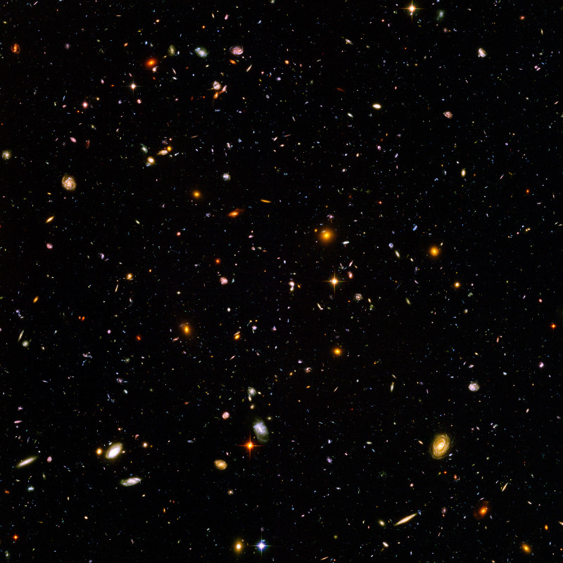
Hubble_ultra_deep_field_high_rez_edit1
is much larger [2] Hubble ultra deep
field high rez
edit1_small.jpg Deutsch: Das Hubble
Ultra Deep Field ist ein Bild einer
kleinen Himmelsregion aufgenommen vom
Hubble-Weltraumteleskop über einen
Zeitraum vom 3. September 2003 bis 16.
Januar 2004. Dabei wurde eine
Himmelsregion ausgewählt, die kaum
störende helle Sterne im Vordergrund
enthält. Man entschied sich für ein
Zielgebiet südwestlich von Orion im
Sternbild Chemischer Ofen. English:
The Hubble Ultra Deep Field, is an
image of a small region of space in the
constellation Fornax, composited from
Hubble Space Telescope data accumulated
over a period from September 3, 2003
through January 16, 2004. The patch of
sky in which the galaxies reside was
chosen because it had a low density of
bright stars in the
near-field. Español: El Campo Ultra
Profundo del Hubble, es una imagen de
una pequeña región del espacio en la
constelación Fornax, compuesta de
datos obtenidos por el telescopio
espacial Hubble durante el período
entre el 3 de Septiembre de 2003 y el
16 de Enero de 2004. Esta parte del
cielo fue escogida por su baja densidad
de estrellas brillantes en sus
proximidades. Français : Le champ
ultra profond de Hubble, une image
d'une petite portion du ciel dans la
constellation du Fourneau, prise par le
télescope spatial Hubble du 3
septembre 2003 au 16 juillet 2004. La
portion de ciel a été choisie car
elle possède peu d'étoiles brillantes
proches. Date 2003-09-03 -
2004-01-16 Source
http://hubblesite.org/newscenter/ar
chive/releases/2004/07/image/a/warn/ Au
thor NASA and the European Space
Agency. Edited by Noodle snacks PD
source: http://upload.wikimedia.org/wiki
pedia/commons/0/0d/Hubble_ultra_deep_fie
ld_high_rez_edit1.jpg
[1] note
Hubble_ultra_deep_field_high_rez_edit1
is much larger [2] Hubble ultra deep
field high rez
edit1_small.jpg Deutsch: Das Hubble
Ultra Deep Field ist ein Bild einer
kleinen Himmelsregion aufgenommen vom
Hubble-Weltraumteleskop über einen
Zeitraum vom 3. September 2003 bis 16.
Januar 2004. Dabei wurde eine
Himmelsregion ausgewählt, die kaum
störende helle Sterne im Vordergrund
enthält. Man entschied sich für ein
Zielgebiet südwestlich von Orion im
Sternbild Chemischer Ofen. English:
The Hubble Ultra Deep Field, is an
image of a small region of space in the
constellation Fornax, composited from
Hubble Space Telescope data accumulated
over a period from September 3, 2003
through January 16, 2004. The patch of
sky in which the galaxies reside was
chosen because it had a low density of
bright stars in the
near-field. Español: El Campo Ultra
Profundo del Hubble, es una imagen de
una pequeña región del espacio en la
constelación Fornax, compuesta de
datos obtenidos por el telescopio
espacial Hubble durante el período
entre el 3 de Septiembre de 2003 y el
16 de Enero de 2004. Esta parte del
cielo fue escogida por su baja densidad
de estrellas brillantes en sus
proximidades. Français : Le champ
ultra profond de Hubble, une image
d'une petite portion du ciel dans la
constellation du Fourneau, prise par le
télescope spatial Hubble du 3
septembre 2003 au 16 juillet 2004. La
portion de ciel a été choisie car
elle possède peu d'étoiles brillantes
proches. Date 2003-09-03 -
2004-01-16 Source
http://hubblesite.org/newscenter/ar
chive/releases/2004/07/image/a/warn/ Au
thor NASA and the European Space
Agency. Edited by Noodle snacks PD
source: http://upload.wikimedia.org/wiki
pedia/commons/0/0d/Hubble_ultra_deep_fie
ld_high_rez_edit1.jpg
light.
[1] note
Hubble_ultra_deep_field_high_rez_edit1
is much larger [2] Hubble ultra deep
field high rez
edit1_small.jpg Deutsch: Das Hubble
Ultra Deep Field ist ein Bild einer
kleinen Himmelsregion aufgenommen vom
Hubble-Weltraumteleskop über einen
Zeitraum vom 3. September 2003 bis 16.
Januar 2004. Dabei wurde eine
Himmelsregion ausgewählt, die kaum
störende helle Sterne im Vordergrund
enthält. Man entschied sich für ein
Zielgebiet südwestlich von Orion im
Sternbild Chemischer Ofen. English:
The Hubble Ultra Deep Field, is an
image of a small region of space in the
constellation Fornax, composited from
Hubble Space Telescope data accumulated
over a period from September 3, 2003
through January 16, 2004. The patch of
sky in which the galaxies reside was
chosen because it had a low density of
bright stars in the
near-field. Español: El Campo Ultra
Profundo del Hubble, es una imagen de
una pequeña región del espacio en la
constelación Fornax, compuesta de
datos obtenidos por el telescopio
espacial Hubble durante el período
entre el 3 de Septiembre de 2003 y el
16 de Enero de 2004. Esta parte del
cielo fue escogida por su baja densidad
de estrellas brillantes en sus
proximidades. Français : Le champ
ultra profond de Hubble, une image
d'une petite portion du ciel dans la
constellation du Fourneau, prise par le
télescope spatial Hubble du 3
septembre 2003 au 16 juillet 2004. La
portion de ciel a été choisie car
elle possède peu d'étoiles brillantes
proches. Date 2003-09-03 -
2004-01-16 Source
http://hubblesite.org/newscenter/ar
chive/releases/2004/07/image/a/warn/ Au
thor NASA and the European Space
Agency. Edited by Noodle snacks PD
source: http://upload.wikimedia.org/wiki
pedia/commons/0/0d/Hubble_ultra_deep_fie
ld_high_rez_edit1.jpg
The same light particles that have
always been, continue to move in the
space that has always been.
[1] note
Hubble_ultra_deep_field_high_rez_edit1
is much larger [2] Hubble ultra deep
field high rez
edit1_small.jpg Deutsch: Das Hubble
Ultra Deep Field ist ein Bild einer
kleinen Himmelsregion aufgenommen vom
Hubble-Weltraumteleskop über einen
Zeitraum vom 3. September 2003 bis 16.
Januar 2004. Dabei wurde eine
Himmelsregion ausgewählt, die kaum
störende helle Sterne im Vordergrund
enthält. Man entschied sich für ein
Zielgebiet südwestlich von Orion im
Sternbild Chemischer Ofen. English:
The Hubble Ultra Deep Field, is an
image of a small region of space in the
constellation Fornax, composited from
Hubble Space Telescope data accumulated
over a period from September 3, 2003
through January 16, 2004. The patch of
sky in which the galaxies reside was
chosen because it had a low density of
bright stars in the
near-field. Español: El Campo Ultra
Profundo del Hubble, es una imagen de
una pequeña región del espacio en la
constelación Fornax, compuesta de
datos obtenidos por el telescopio
espacial Hubble durante el período
entre el 3 de Septiembre de 2003 y el
16 de Enero de 2004. Esta parte del
cielo fue escogida por su baja densidad
de estrellas brillantes en sus
proximidades. Français : Le champ
ultra profond de Hubble, une image
d'une petite portion du ciel dans la
constellation du Fourneau, prise par le
télescope spatial Hubble du 3
septembre 2003 au 16 juillet 2004. La
portion de ciel a été choisie car
elle possède peu d'étoiles brillantes
proches. Date 2003-09-03 -
2004-01-16 Source
http://hubblesite.org/newscenter/ar
chive/releases/2004/07/image/a/warn/ Au
thor NASA and the European Space
Agency. Edited by Noodle snacks PD
source: http://upload.wikimedia.org/wiki
pedia/commons/0/0d/Hubble_ultra_deep_fie
ld_high_rez_edit1.jpg
created or destroyed. Matter can never
be converted into motion, and motion
can never be converted into matter.
[1] note
Hubble_ultra_deep_field_high_rez_edit1
is much larger [2] Hubble ultra deep
field high rez
edit1_small.jpg Deutsch: Das Hubble
Ultra Deep Field ist ein Bild einer
kleinen Himmelsregion aufgenommen vom
Hubble-Weltraumteleskop über einen
Zeitraum vom 3. September 2003 bis 16.
Januar 2004. Dabei wurde eine
Himmelsregion ausgewählt, die kaum
störende helle Sterne im Vordergrund
enthält. Man entschied sich für ein
Zielgebiet südwestlich von Orion im
Sternbild Chemischer Ofen. English:
The Hubble Ultra Deep Field, is an
image of a small region of space in the
constellation Fornax, composited from
Hubble Space Telescope data accumulated
over a period from September 3, 2003
through January 16, 2004. The patch of
sky in which the galaxies reside was
chosen because it had a low density of
bright stars in the
near-field. Español: El Campo Ultra
Profundo del Hubble, es una imagen de
una pequeña región del espacio en la
constelación Fornax, compuesta de
datos obtenidos por el telescopio
espacial Hubble durante el período
entre el 3 de Septiembre de 2003 y el
16 de Enero de 2004. Esta parte del
cielo fue escogida por su baja densidad
de estrellas brillantes en sus
proximidades. Français : Le champ
ultra profond de Hubble, une image
d'une petite portion du ciel dans la
constellation du Fourneau, prise par le
télescope spatial Hubble du 3
septembre 2003 au 16 juillet 2004. La
portion de ciel a été choisie car
elle possède peu d'étoiles brillantes
proches. Date 2003-09-03 -
2004-01-16 Source
http://hubblesite.org/newscenter/ar
chive/releases/2004/07/image/a/warn/ Au
thor NASA and the European Space
Agency. Edited by Noodle snacks PD
source: http://upload.wikimedia.org/wiki
pedia/commons/0/0d/Hubble_ultra_deep_fie
ld_high_rez_edit1.jpg
each other and so form structures such
as protons, atoms, molecules, planets,
stars, galaxies, and clusters of
galaxies.
[1] note
Hubble_ultra_deep_field_high_rez_edit1
is much larger [2] Hubble ultra deep
field high rez
edit1_small.jpg Deutsch: Das Hubble
Ultra Deep Field ist ein Bild einer
kleinen Himmelsregion aufgenommen vom
Hubble-Weltraumteleskop über einen
Zeitraum vom 3. September 2003 bis 16.
Januar 2004. Dabei wurde eine
Himmelsregion ausgewählt, die kaum
störende helle Sterne im Vordergrund
enthält. Man entschied sich für ein
Zielgebiet südwestlich von Orion im
Sternbild Chemischer Ofen. English:
The Hubble Ultra Deep Field, is an
image of a small region of space in the
constellation Fornax, composited from
Hubble Space Telescope data accumulated
over a period from September 3, 2003
through January 16, 2004. The patch of
sky in which the galaxies reside was
chosen because it had a low density of
bright stars in the
near-field. Español: El Campo Ultra
Profundo del Hubble, es una imagen de
una pequeña región del espacio en la
constelación Fornax, compuesta de
datos obtenidos por el telescopio
espacial Hubble durante el período
entre el 3 de Septiembre de 2003 y el
16 de Enero de 2004. Esta parte del
cielo fue escogida por su baja densidad
de estrellas brillantes en sus
proximidades. Français : Le champ
ultra profond de Hubble, une image
d'une petite portion du ciel dans la
constellation du Fourneau, prise par le
télescope spatial Hubble du 3
septembre 2003 au 16 juillet 2004. La
portion de ciel a été choisie car
elle possède peu d'étoiles brillantes
proches. Date 2003-09-03 -
2004-01-16 Source
http://hubblesite.org/newscenter/ar
chive/releases/2004/07/image/a/warn/ Au
thor NASA and the European Space
Agency. Edited by Noodle snacks PD
source: http://upload.wikimedia.org/wiki
pedia/commons/0/0d/Hubble_ultra_deep_fie
ld_high_rez_edit1.jpg
see are only a tiny part of the
universe. We will never see most of the
universe because no light particles
from there can ever reach us.
[1] note
Hubble_ultra_deep_field_high_rez_edit1
is much larger [2] Hubble ultra deep
field high rez
edit1_small.jpg Deutsch: Das Hubble
Ultra Deep Field ist ein Bild einer
kleinen Himmelsregion aufgenommen vom
Hubble-Weltraumteleskop über einen
Zeitraum vom 3. September 2003 bis 16.
Januar 2004. Dabei wurde eine
Himmelsregion ausgewählt, die kaum
störende helle Sterne im Vordergrund
enthält. Man entschied sich für ein
Zielgebiet südwestlich von Orion im
Sternbild Chemischer Ofen. English:
The Hubble Ultra Deep Field, is an
image of a small region of space in the
constellation Fornax, composited from
Hubble Space Telescope data accumulated
over a period from September 3, 2003
through January 16, 2004. The patch of
sky in which the galaxies reside was
chosen because it had a low density of
bright stars in the
near-field. Español: El Campo Ultra
Profundo del Hubble, es una imagen de
una pequeña región del espacio en la
constelación Fornax, compuesta de
datos obtenidos por el telescopio
espacial Hubble durante el período
entre el 3 de Septiembre de 2003 y el
16 de Enero de 2004. Esta parte del
cielo fue escogida por su baja densidad
de estrellas brillantes en sus
proximidades. Français : Le champ
ultra profond de Hubble, une image
d'une petite portion du ciel dans la
constellation du Fourneau, prise par le
télescope spatial Hubble du 3
septembre 2003 au 16 juillet 2004. La
portion de ciel a été choisie car
elle possède peu d'étoiles brillantes
proches. Date 2003-09-03 -
2004-01-16 Source
http://hubblesite.org/newscenter/ar
chive/releases/2004/07/image/a/warn/ Au
thor NASA and the European Space
Agency. Edited by Noodle snacks PD
source: http://upload.wikimedia.org/wiki
pedia/commons/0/0d/Hubble_ultra_deep_fie
ld_high_rez_edit1.jpg
Light particles move from where there
is less space to where there is more
space. As an empty space becomes
filled, light particles form gas clouds
called nebuli, and then galaxies of
stars. Living objects grow around stars
and pull stars together to form
globular clusters and then form a
globular (also called "elliptical")
galaxy. Living objects need matter to
stop their decay. Globular galaxies
have a regular input and output of
light particles. When a volume of space
becomes very dense, more light
particles exit than enter the space.
Light particles that exit to more empty
spaces form new nebulae and galaxies
and so this cycle continues. Stars at
our scale may be light particles at a
much larger scale, just as light
particles at our scale may be stars at
a much smaller scale. This system may
go on infinitely in both larger and
smaller scale.
[1] note
Hubble_ultra_deep_field_high_rez_edit1
is much larger Hubble ultra deep
field high rez
edit1_small.jpg Deutsch: Das Hubble
Ultra Deep Field ist ein Bild einer
kleinen Himmelsregion aufgenommen vom
Hubble-Weltraumteleskop über einen
Zeitraum vom 3. September 2003 bis 16.
Januar 2004. Dabei wurde eine
Himmelsregion ausgewählt, die kaum
störende helle Sterne im Vordergrund
enthält. Man entschied sich für ein
Zielgebiet südwestlich von Orion im
Sternbild Chemischer Ofen. English:
The Hubble Ultra Deep Field, is an
image of a small region of space in the
constellation Fornax, composited from
Hubble Space Telescope data accumulated
over a period from September 3, 2003
through January 16, 2004. The patch of
sky in which the galaxies reside was
chosen because it had a low density of
bright stars in the
near-field. Español: El Campo Ultra
Profundo del Hubble, es una imagen de
una pequeña región del espacio en la
constelación Fornax, compuesta de
datos obtenidos por el telescopio
espacial Hubble durante el período
entre el 3 de Septiembre de 2003 y el
16 de Enero de 2004. Esta parte del
cielo fue escogida por su baja densidad
de estrellas brillantes en sus
proximidades. Français : Le champ
ultra profond de Hubble, une image
d'une petite portion du ciel dans la
constellation du Fourneau, prise par le
télescope spatial Hubble du 3
septembre 2003 au 16 juillet 2004. La
portion de ciel a été choisie car
elle possède peu d'étoiles brillantes
proches. Date 2003-09-03 -
2004-01-16 Source
http://hubblesite.org/newscenter/ar
chive/releases/2004/07/image/a/warn/ Au
thor NASA and the European Space
Agency. Edited by Noodle snacks PD
source: http://upload.wikimedia.org/wiki
pedia/commons/0/0d/Hubble_ultra_deep_fie
ld_high_rez_edit1.jpg

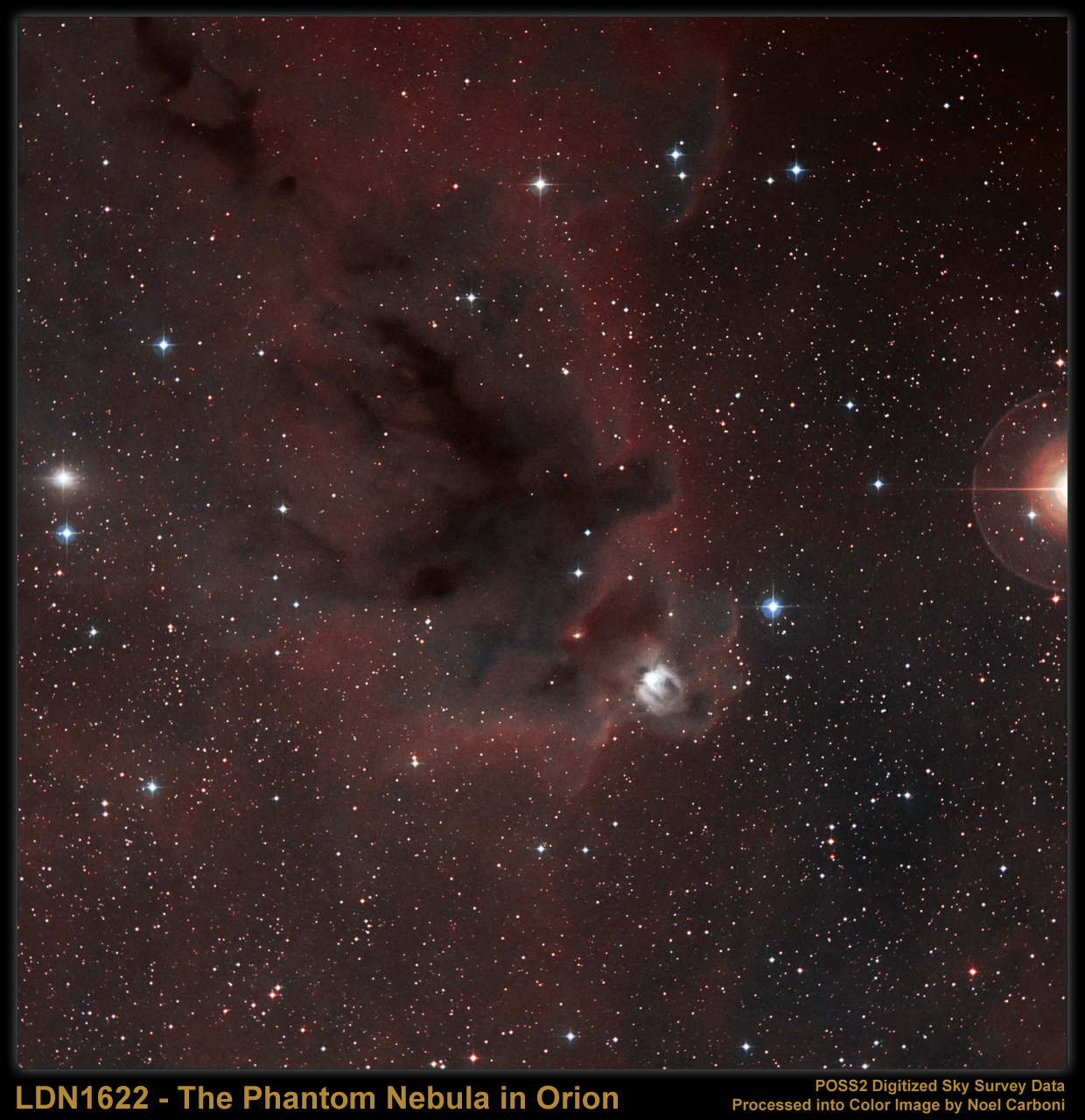
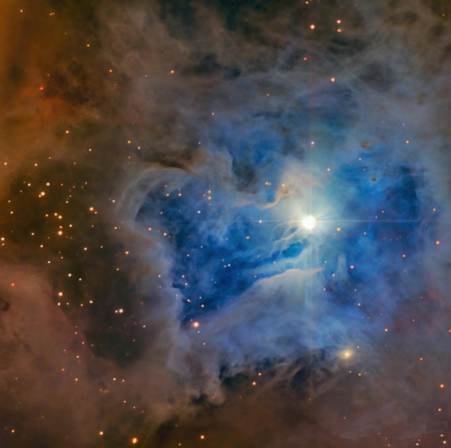
[2] LDN 1622: Dark Nebula in
Orion Data: Digitized Sky Survey
(POSS-II), Color Composite: Noel
Carboni Explanation: The silhouette
of an intriguing dark nebula inhabits
this cosmic scene, based on images from
the Palomar Observatory Sky Survey.
Lynds' Dark Nebula (LDN) 1622 appears
against a faint background of glowing
hydrogen gas only easily seen in long
telescopic exposures of the region. LDN
1622 lies near the plane of our Milky
Way Galaxy, close on the sky to
Barnard's Loop - a large cloud
surrounding the rich complex of
emission nebulae found in the Belt and
Sword of Orion. But the obscuring dust
of LDN 1622 is thought to be much
closer than Orion's more famous
nebulae, perhaps only 500 light-years
away. At that distance, this 1 degree
wide field of view would span less than
10 light-years. PD
source: http://apod.nasa.gov/apod/image/
0705/ldn1622_carboni.jpg
LIFE
form.
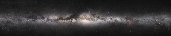
[1] Description This image is
mosaic of multiple shots on
large-format film. It comprises all 360
degrees of the galaxy from our vantage.
Photography was done in Ft. Davis,
Texas for the Northern hemisphere shots
and from Broken Hill, New South Wales,
Australia, for the southern portions.
Note the dust lanes, which obscure our
view of some features beyond them.
Infrared imaging reaches into these
regions, and radio astronomy can look
all the way through with less detail.
The very center, however, shows a
window to the farther side. In the
center, stars are mostly very old and
this causes the more yellow color. The
final file is 1.5GB, and resolves
details of less than one arcminute.
Faintest stars are magnitude 11. There
are 21 pixels of horizontal overlap at
the ends, with the right end slightly
brighter than the corresponding pixels
on the left. Date Source
http://www.digitalskyllc.com (The
image was uploaded to en.wiki at 17:16,
21 September 2006 by Twtunes. Author
Digital Sky LLC CC
source: http://upload.wikimedia.org/wiki
pedia/commons/0/0a/Milkyway_pan1.jpg
[2] note
Hubble_ultra_deep_field_high_rez_edit1
is much larger [2] Hubble ultra deep
field high rez
edit1_small.jpg Deutsch: Das Hubble
Ultra Deep Field ist ein Bild einer
kleinen Himmelsregion aufgenommen vom
Hubble-Weltraumteleskop über einen
Zeitraum vom 3. September 2003 bis 16.
Januar 2004. Dabei wurde eine
Himmelsregion ausgewählt, die kaum
störende helle Sterne im Vordergrund
enthält. Man entschied sich für ein
Zielgebiet südwestlich von Orion im
Sternbild Chemischer Ofen. English:
The Hubble Ultra Deep Field, is an
image of a small region of space in the
constellation Fornax, composited from
Hubble Space Telescope data accumulated
over a period from September 3, 2003
through January 16, 2004. The patch of
sky in which the galaxies reside was
chosen because it had a low density of
bright stars in the
near-field. Español: El Campo Ultra
Profundo del Hubble, es una imagen de
una pequeña región del espacio en la
constelación Fornax, compuesta de
datos obtenidos por el telescopio
espacial Hubble durante el período
entre el 3 de Septiembre de 2003 y el
16 de Enero de 2004. Esta parte del
cielo fue escogida por su baja densidad
de estrellas brillantes en sus
proximidades. Français : Le champ
ultra profond de Hubble, une image
d'une petite portion du ciel dans la
constellation du Fourneau, prise par le
télescope spatial Hubble du 3
septembre 2003 au 16 juillet 2004. La
portion de ciel a été choisie car
elle possède peu d'étoiles brillantes
proches. Date 2003-09-03 -
2004-01-16 Source
http://hubblesite.org/newscenter/ar
chive/releases/2004/07/image/a/warn/ Au
thor NASA and the European Space
Agency. Edited by Noodle snacks PD
source: http://upload.wikimedia.org/wiki
pedia/commons/0/0d/Hubble_ultra_deep_fie
ld_high_rez_edit1.jpg
Galaxy forms.

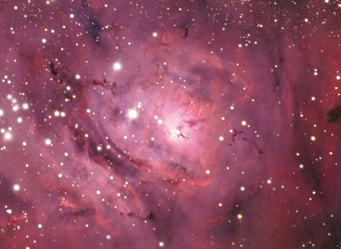
[1] Description English: M8 Lagoon
Nebula in Sagittarius Date 26 June
2009 Source Own
work Author Hewholooks CC
source: http://upload.wikimedia.org/wiki
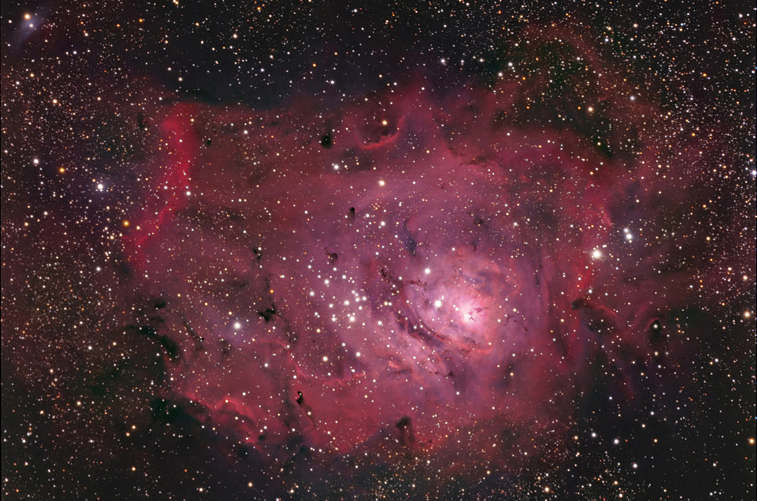
pedia/commons/2/2f/M8HunterWilson.jpg
[2] NGC 7023: The Iris Nebula Credit
& Copyright: Daniel López,
IAC Explanation: Like delicate cosmic
petals, these clouds of interstellar
dust and gas have blossomed 1,300
light-years away in the fertile star
fields of the constellation Cepheus.
Sometimes called the Iris Nebula and
dutifully cataloged as NGC 7023, this
is not the only nebula in the sky to
evoke the imagery of flowers. Still,
this beautiful digital image shows off
the Iris Nebula's range of colors and
symmetries in impressive detail. Within
the Iris, dusty nebular material
surrounds a hot, young star. The
dominant color of the brighter
reflection nebula is blue,
characteristic of dust grains
reflecting starlight. Central filaments
of the dusty clouds glow with a faint
reddish photoluminesence as some dust
grains effectively convert the star's
invisible ultraviolet radiation to
visible red light. Infrared
observations indicate that this nebula
may contain complex carbon molecules
known as PAHs. As shown here, the
bright blue portion of the Iris Nebula
is about six light-years across. PD
source: http://apod.nasa.gov/apod/image/
1011/IRIS_IAC80_DLopez900c.jpg
Galaxy reach another star using a ship.
[1] close up
of: Description English: M8 Lagoon
Nebula in Sagittarius Date 26 June
2009 Source Own
work Author Hewholooks CC
source: http://upload.wikimedia.org/wiki
pedia/commons/2/2f/M8HunterWilson.jpg
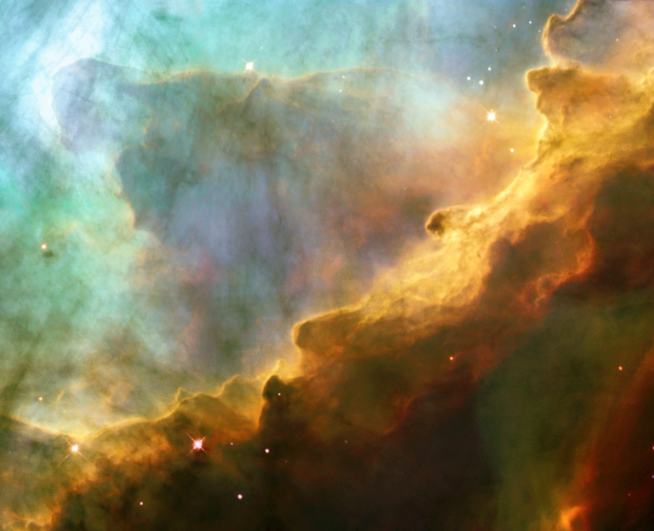
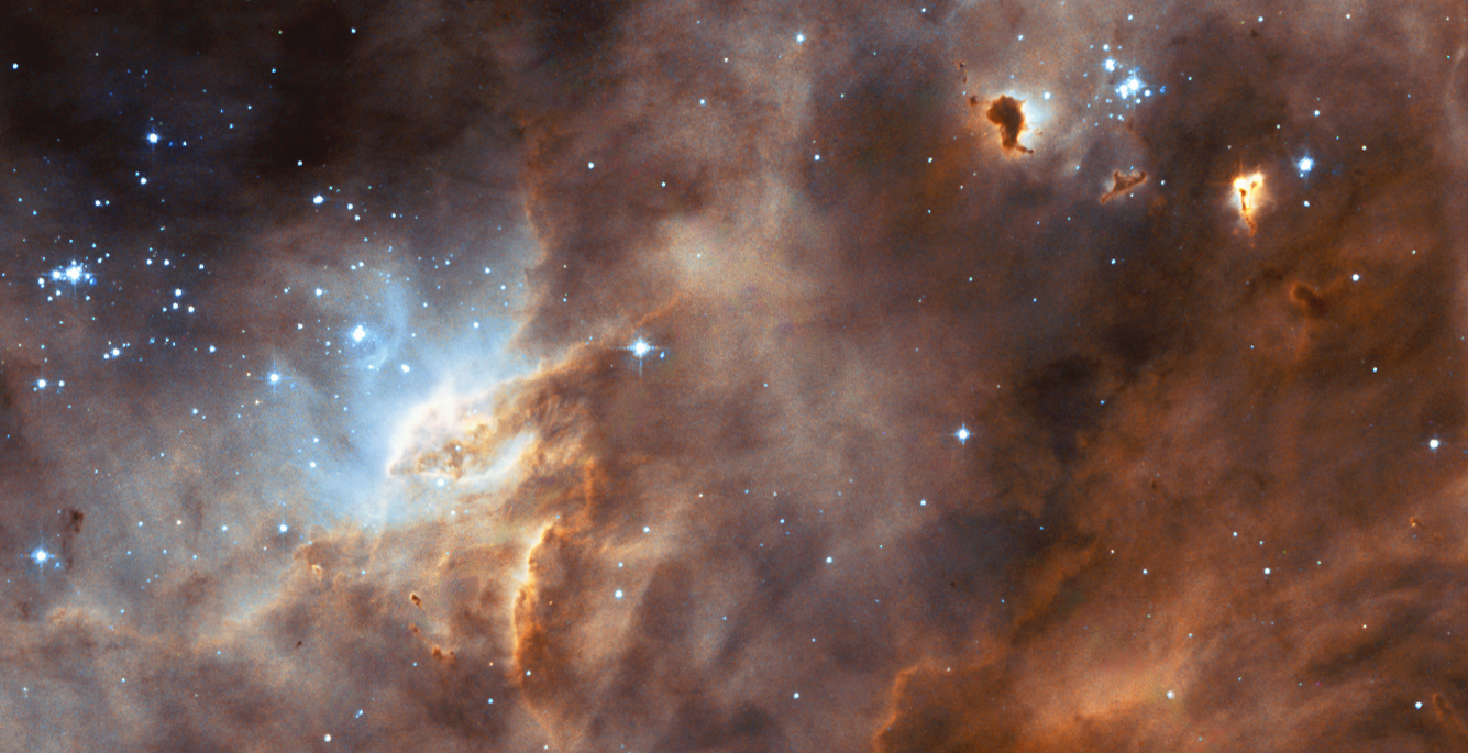
[2] Description The photograph,
taken by NASA's Hubble Space Telescope,
captures a small region within M17, a
hotbed of star formation. M17, also
known as the Omega or Swan Nebula, is
located about 5500 light-years away in
the constellation Sagittarius. The
wave-like patterns of gas have been
sculpted and illuminated by a torrent
of ultraviolet radiation from young,
massive stars, which lie outside the
picture to the upper left. The glow of
these patterns accentuates the
three-dimensional structure of the
gases. The ultraviolet radiation is
carving and heating the surfaces of
cold hydrogen gas clouds. The warmed
surfaces glow orange and red in this
photograph. The intense heat and
pressure cause some material to stream
away from those surfaces, creating the
glowing veil of even hotter greenish
gas that masks background structures.
The pressure on the tips of the waves
may trigger new star formation within
them. The image, roughly 3
light-years across, was taken May
29-30, 1999, with the Wide Field
Planetary Camera 2. The colors in the
image represent various gases. Red
represents sulfur; green, hydrogen; and
blue, oxygen. Date 24 April
2003 Source
http://spacetelescope.org/images/html/he
ic0305a.html (direct link)
http://hubblesite.org/newscenter/archive
/releases/2003/13/image/a/ Author
NASA, ESA and J. Hester (ASU) PD
source: http://upload.wikimedia.org/wiki
pedia/commons/7/72/Omega_Nebula.jpg
100,000 stars in the Milky Way Galaxy.

[1] Description The globular
cluster Omega Centauri — with as many
as ten million stars — is seen in all
its splendour in this image captured
with the WFI camera from ESO's La Silla
Observatory. The image shows only the
central part of the cluster — about
the size of the full moon on the sky
(half a degree). North is up, East is
to the left. This colour image is a
composite of B, V and I filtered
images. Note that because WFI is
equipped with a mosaic detector, there
are two small gaps in the image which
were filled with lower quality data
from the Digitized Sky Survey. Date
2008 Source
http://www.eso.org/public/outreach/
press-rel/pr-2008/phot-44-08.html Autho
r ESO CC
source: http://upload.wikimedia.org/wiki
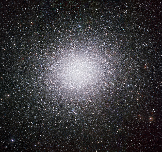
pedia/commons/thumb/e/e6/Omega_Centauri_
by_ESO.jpg/638px-Omega_Centauri_by_ESO.j
pg
[2] Description This image is
mosaic of multiple shots on
large-format film. It comprises all 360
degrees of the galaxy from our vantage.
Photography was done in Ft. Davis,
Texas for the Northern hemisphere shots
and from Broken Hill, New South Wales,
Australia, for the southern portions.
Note the dust lanes, which obscure our
view of some features beyond them.
Infrared imaging reaches into these
regions, and radio astronomy can look
all the way through with less detail.
The very center, however, shows a
window to the farther side. In the
center, stars are mostly very old and
this causes the more yellow color. The
final file is 1.5GB, and resolves
details of less than one arcminute.
Faintest stars are magnitude 11. There
are 21 pixels of horizontal overlap at
the ends, with the right end slightly
brighter than the corresponding pixels
on the left. Date Source
http://www.digitalskyllc.com (The
image was uploaded to en.wiki at 17:16,
21 September 2006 by Twtunes. Author
Digital Sky LLC CC
source: http://upload.wikimedia.org/wiki
pedia/commons/0/0a/Milkyway_pan1.jpg

[1] Description English: The Sun
photographed by the Atmospheric Imaging
Assembly (AIA 304) of NASA's Solar
Dynamics Observatory (SDO). This is
a false color image of the sun observed
in the extreme ultraviolet region of
the spectrum. For example,similar
image Français : Le soleil,
photographié depuis le Solar Dynamics
Observatory de la NASA. Date
2010-08-19T00:32:21Z (ISO
8601) Source NASA/SDO
(AIA). Author NASA/SDO (AIA). PD
source: http://upload.wikimedia.org/wiki
pedia/commons/thumb/b/b4/The_Sun_by_the_
Atmospheric_Imaging_Assembly_of_NASAs_So
lar_Dynamics_Observatory_-_20100819.jpg/
628px-The_Sun_by_the_Atmospheric_Imaging
_Assembly_of_NASAs_Solar_Dynamics_Observ
atory_-_20100819.jpg

[2] Summary Description The star
formation region N11B in the LMC taken
by WFPC2 on the NASA/ESA Hubble Space
Telescope. Date Source
http://www.spacetelescope.org/image
s/html/heic0411a.html Author
NASA/ESA and the Hubble Heritage
Team
(AURA/STScI)/HEIC Permission (Reusing
this file) ESA Public Domain, as
per
http://www.spacetelescope.org/copyright.
html PD
source: http://upload.wikimedia.org/wiki
pedia/commons/6/6c/Heic0411a.jpg
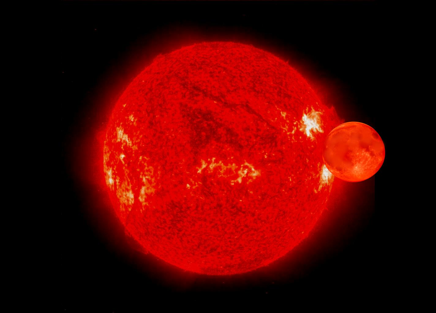
the star, they are red hot with liquid
rock and metals on the surface. Lighter
atoms move to the surface of the
planets. Larger planets are surrounded
by gas.

[1] an 19, 2005 � For the past five
days, forecasters at the NOAA Space
Environment Center in Boulder, Colo.,
have observed all types of space
weather: radio blackouts, solar
radiation storms and geomagnetic
storms. Currently, space weather
forecasters are observing a moderate
geomagnetic storm (G-2 on the NOAA
Space Weather Scales) and a minor (S-1)
solar radiation storm. Earlier
Wednesday an X-class flare produced a
strong (R-3) radio blackout. (Click
image for larger view of the sun taken
on Jan. 19, 2005, at 2:19 p.m. EST.
Click here for high resolution version,
which is a large file. Please credit
European Space Agency-NASA.) PD
source: http://www.noaanews.noaa.gov/sto
ries2005/images/sun-soho011905-1919z.jpg

[2] This artist’s impression shows
the disk of gas and cosmic dust around
the young star HD 142527. Astronomers
using the Atacama Large
Millimeter/submillimeter Array (ALMA)
telescope have seen vast streams of gas
flowing across the gap in the disc
UNKNOWN
source: http://l2.yimg.com/bt/api/res/1.
2/kB0xEBWbOe3fUGcRF7Y3RA--/YXBwaWQ9eW5ld
3M7Zmk9aW5zZXQ7aD00MDg7cT03OTt3PTU3NQ--/
http://media.zenfs.com/en_US/News/SPACE.
com/Never-Before-Seen_Stage_of_Planet_Bi
rth-893372caafae611ec5e71458c2f79fb8
[1] Image of moon superimposed on
Venus PD
source: http://upload.wikimedia.org/wiki
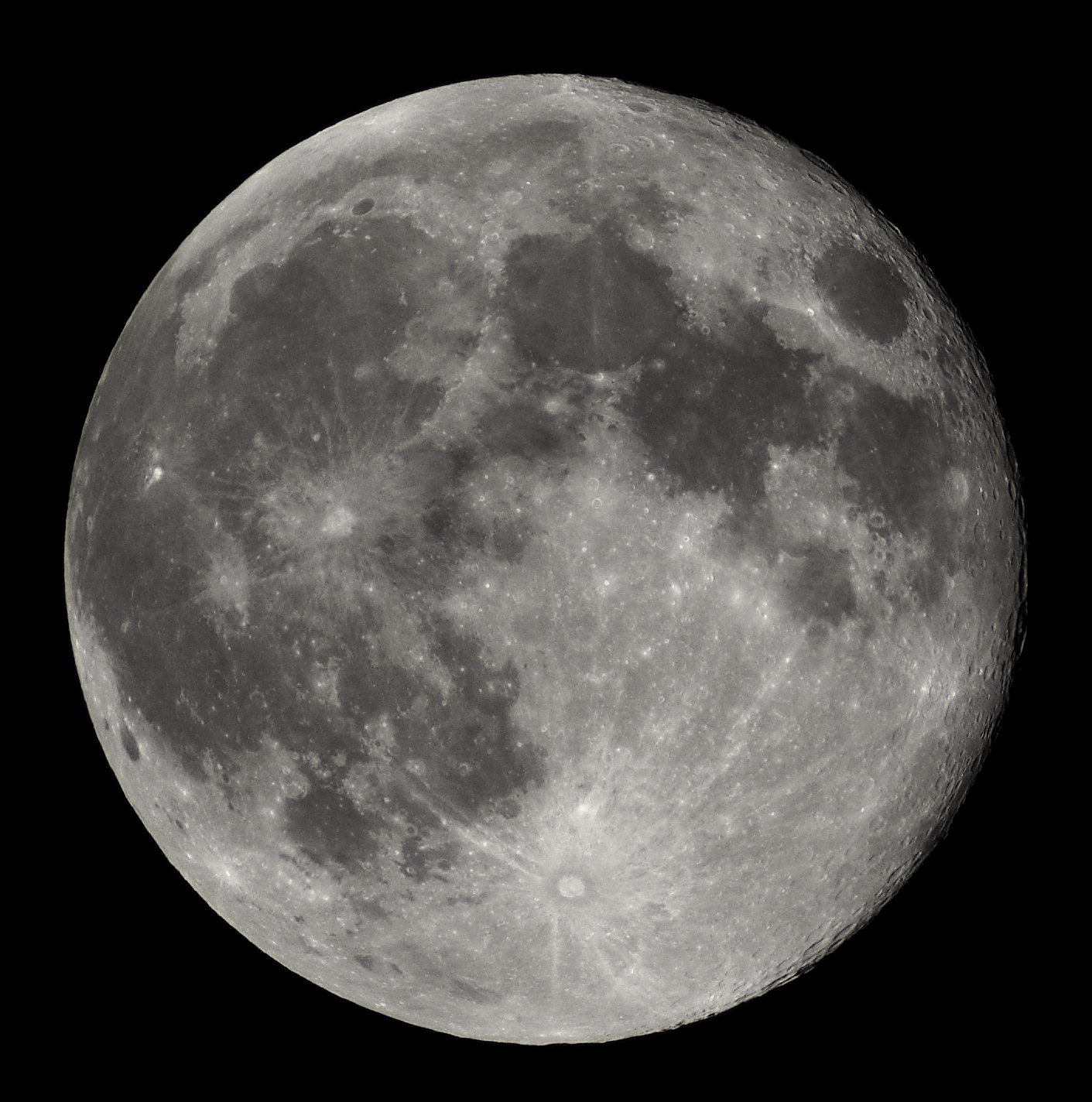
pedia/commons/d/dd/Full_Moon_Luc_Viatour
.jpg

[2] an 19, 2005 � For the past five
days, forecasters at the NOAA Space
Environment Center in Boulder, Colo.,
have observed all types of space
weather: radio blackouts, solar
radiation storms and geomagnetic
storms. Currently, space weather
forecasters are observing a moderate
geomagnetic storm (G-2 on the NOAA
Space Weather Scales) and a minor (S-1)
solar radiation storm. Earlier
Wednesday an X-class flare produced a
strong (R-3) radio blackout. (Click
image for larger view of the sun taken
on Jan. 19, 2005, at 2:19 p.m. EST.
Click here for high resolution version,
which is a large file. Please credit
European Space Agency-NASA.) PD
source: http://www.noaanews.noaa.gov/sto
ries2005/images/sun-soho011905-1919z.jpg
rock turns into a solid thin crust.
Water condenses and falls to the
surface, filling the lowest parts of
the land to make the first Earth
oceans, lakes, and rivers.
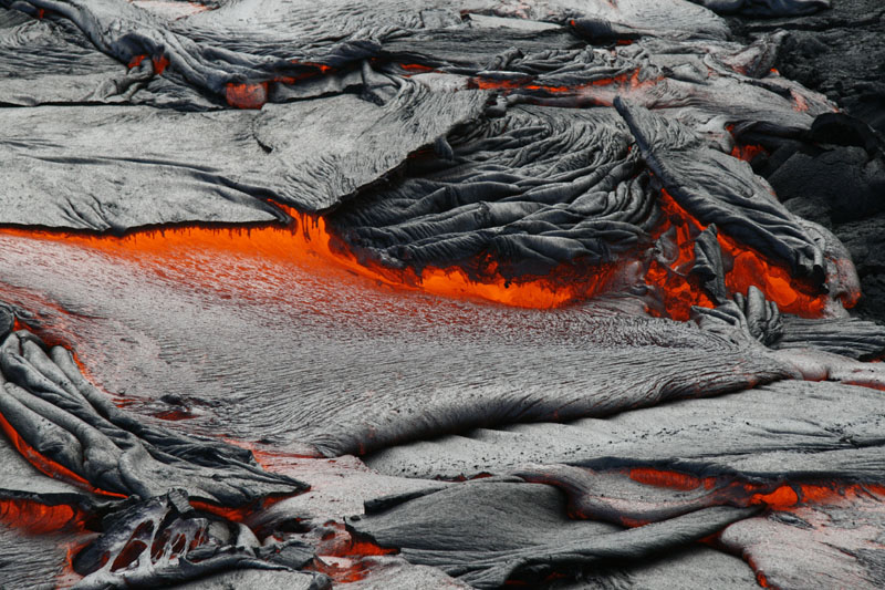
[1] USGS Photo by Tim Orr Pahoehoe
lava breaks out of the crust along a
flow margin PD
source: http://www.nps.gov/havo/parkmgmt
/upload/havo_manage_usgs_20080304_tro381
7_x800.jpg

[2] English: Ultraviolet image of
Venus' clouds as seen by the Pioneer
Venus Orbiter (February 26, 1979). The
immense C- or Y-shaped features which
are visible only in these wavelengths
are individually short lived, but
reform often enough to be considered a
permanent feature of Venus' clouds. The
mechanism by which Venus' clouds absorb
ultraviolet is not well understood. PD
source: http://upload.wikimedia.org/wiki
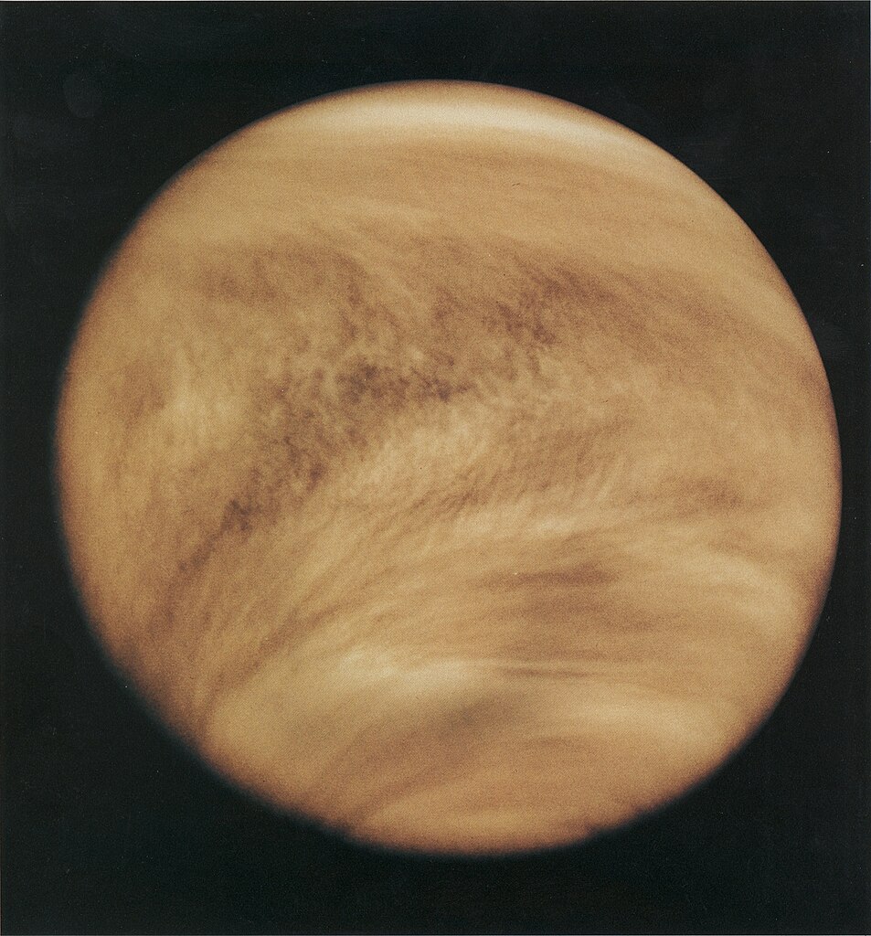
pedia/commons/thumb/b/bc/Venuspioneeruv.
jpg/953px-Venuspioneeruv.jpg
like amino acids, phosphates, and
sugars, the components of living
objects.

[1] The two optical isomers of alanine,
D-Alanine and
L-Alanine D-glucose BOTH PD
source: http://upload.wikimedia.org/wiki
pedia/commons/6/65/D%2BL-Alanine.gif
and http://upload.wikimedia.org/wikiped
ia/commons/thumb/5/5a/D-glucose-chain-3D
-balls.png/640px-D-glucose-chain-3D-ball
s.png

[1] Ribonucleic acid (English
pronunciation:
/raɪbɵ.njuːˌkleɪ.ɨk ˈæsɪd/),
or RNA, is one of the three major
macromolecules (along with DNA and
proteins) that are essential for all
known forms of life. UNKNOWN
source: http://dna-rna.net/wp-content/up
loads/2011/07/rna.jpg
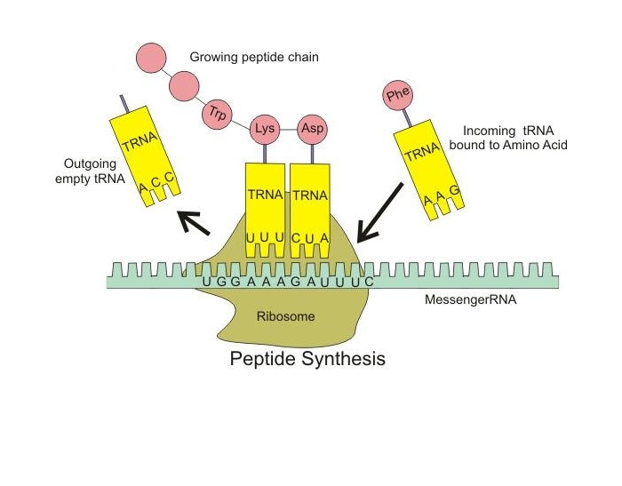
Transfer RNA molecules evolve (tRNA),
and link amino acids into proteins
using other RNA molecules as a
template.
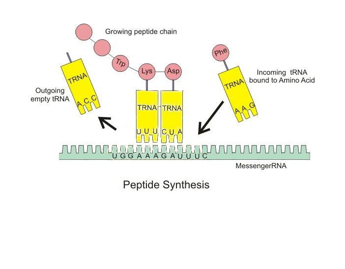
[1] Description English:
Illustration of tRNA building peptide
chain Date 1 March 2009 Source
Own work Author
Boumphreyfr CC
source: http://upload.wikimedia.org/wiki
pedia/commons/0/0f/Peptide_syn.png

[2] Source : ''Role of the
Ribosome'' University of Texas Medical
Branch UNKNOWN
source: http://ead.univ-angers.fr/~jaspa
rd/Page2/COURS/7RelStructFonction/2Bioch
imie/1SyntheseProteines/3Figures/4Organi
tes/2Ribosomes/6Polysome.gif
[1] RNA is a versatile molecule. In its
most familiar role, RNA acts as an
intermediary, carrying genetic
information from the DNA to the
machinery of protein synthesis. RNA
also plays more active roles,
performing many of the catalytic and
recognition functions normally reserved
for proteins. In fact, most of the RNA
in cells is found in ribosomes--our
protein-synthesizing machines--and the
transfer RNA molecules used to add each
new amino acid to growing proteins. In
addition, countless small RNA molecules
are involved in regulating, processing
and disposing of the constant traffic
of messenger RNA. The enzyme RNA
polymerase carries the weighty
responsibility of creating all of these
different RNA molecules. The RNA
Factory RNA polymerase is a huge
factory with many moving parts. The one
shown here, from PDB entry 1i6h, is
from yeast cells. It is composed of a
dozen different proteins. Together,
they form a machine that surrounds DNA
strands, unwinds them, and builds an
RNA strand based on the information
held inside the DNA. Once the enzyme
gets started, RNA polymerase marches
confidently along the DNA copying RNA
strands thousands of nucleotides
long. Accuracy As you might expect,
RNA polymerase needs to be accurate in
its copying of genetic information. To
improve its accuracy, it performs a
simple proofreading step as it builds
an RNA strand. The active site is
designed to be able to remove
nucleotides as well as add them to the
growing strand. The enzyme tends to
hover around mismatched nucleotides
longer than properly added ones, giving
the enzyme time to remove them. This
process is somewhat wasteful, since
proper nucleotides are also
occasionally removed, but this is a
small price to pay for creating better
RNA transcripts. Overall, RNA
polymerase makes an error about once in
10,000 nucleotides added, or about once
per RNA strand created. Poisoning
Polymerase Since RNA polymerase is
absolutely essential for the life of
the cell, it is a sensitive target for
poisons and toxins. The most powerful
of these poisons is alpha-amanitin, a
small circular peptide created by the
death cap mushroom. Eating even one of
these mushrooms will lead to coma and
death in a manner of days, as the
poison attacks RNA polymerase
throughout the body. Surprisingly, it
binds on the back side of RNA
polymerase, away from the active site
and away from the binding site for the
DNA and RNA. It does not physically
block the active site, like most
inhibitors, but instead jams the
mechanism of the enzyme. RNA polymerase
is a highly mobile enzyme, that flexes
and changes shape as it performs the
sequential steps of binding to DNA,
unwinding it, and then building the RNA
strand. As seen in PDB entry 1k83, the
poison binds between two subunits of
the protein, gluing them together and
blocking these essential motions. PD
source: http://www.pdb.org/pdb/education
_discussion/molecule_of_the_month/images
/1i6h-composite.gif
[2] [t Notice that many RNA molecules
are being produced all in sequence,
with each RNA molecule getting longer
as each protein reaches the end of the
DNA molecule.] Micrograph of gene
transcription of ribosomal RNA
illustrating the growing primary
transcripts. ''Begin'' indicates the 5'
end of the coding strand of DNA, where
new RNA synthesis begins; ''end''
indicates the 3' end, where the primary
transcripts are almost
complete. This is an alternate
version of
Image:RibosomaleTranskriptionsEinheit.jp
g, original author identified as Dr.
Hans-Heinrich Trepte, labeled in
German. This version with English
labels is from en:Image:Transcription
label fromcommons.jpg, by
en:UserOpabinia regalis, licensed under
GFDL. GNU
source: http://upload.wikimedia.org/wiki
pedia/commons/4/43/Transcription_label_e
n.jpg
may function as a protocell, providing
a platform for more efficient protein
production. A single RNA may contain
all the instructions needed to make
more ribosomes.
[1] Description English:
Illustration of tRNA building peptide
chain Date 1 March 2009 Source
Own work Author
Boumphreyfr CC
source: http://upload.wikimedia.org/wiki
pedia/commons/0/0f/Peptide_syn.png
[2] Source : ''Role of the
Ribosome'' University of Texas Medical
Branch UNKNOWN
source: http://ead.univ-angers.fr/~jaspa
rd/Page2/COURS/7RelStructFonction/2Bioch
imie/1SyntheseProteines/3Figures/4Organi
tes/2Ribosomes/6Polysome.gif
be made from RNA.
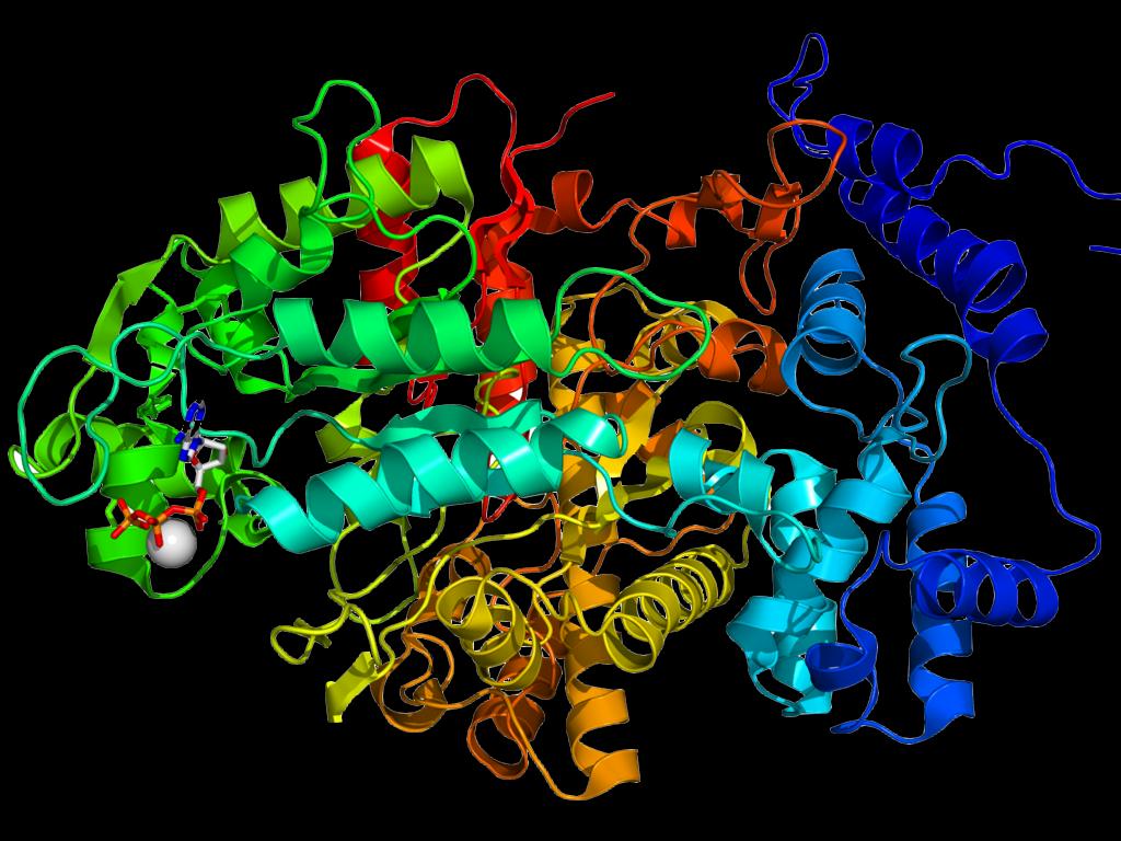
[1] Description Crystallographic
structure of the ribonucleotide
reductase protein R1E from Salmonella
typhimurium. The protein is rainbow
colored (N-terminus = blue, C-terminus
= red) while deoxyadenosine
triphosphate is show as sticks and a
complexed magnesium ion as a grey
sphere.[1] ↑ PDB 1PEU; Uppsten M,
Färnegårdh M, Jordan A, Eliasson R,
Eklund H, Uhlin U (June 2003).
''Structure of the large subunit of
class Ib ribonucleotide reductase from
Salmonella typhimurium and its
complexes with allosteric effectors''.
J. Mol. Biol. 330 (1): 87–97. PMID
12818204. Date 28 February
2008 Source Own
work Author Boghog2 PD
source: http://upload.wikimedia.org/wiki
pedia/commons/thumb/e/e3/1PEU_R1E.png/10
24px-1PEU_R1E.png

[2] Description English: The
reaction mechanism of ribonucleotide
reductase Date 14 January 2006
(original upload
date) Source Transferred from
en.wikipedia; transferred to Commons by
User:Michał Sobkowski using
CommonsHelper. Author Original
uploader was BorisTM at
en.wikipedia PD
source: http://upload.wikimedia.org/wiki
pedia/commons/2/2c/RNR_reaction.png
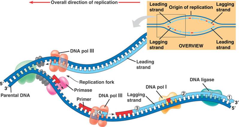
[1] A look at DNA replication, with the
inset showing a larger and general
view. ''Pol'' stands for polymerase, a
key enzyme. Note how each enzyme works
in a 'biochemical team' to complete the
process efficiently COPYRIGHTED
source: http://genmed.yolasite.com/resou
rces/DNA20replication.jpg
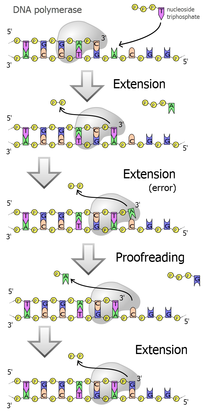
[2] Description Diagram of DNA
polymerase extending a DNA strand and
proof-reading. Date Source Own
work Author Madprime GNU
source: http://upload.wikimedia.org/wiki
pedia/commons/6/6f/DNA_polymerase.svg
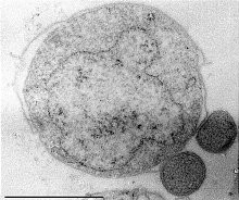
bacterium). DNA is surrounded by a
membrane made of proteins.
This cell may form near the sunlit
water surface or near underwater
volcanoes.

[1] Deutsch: Bild über den Reitenden
Urzwerg English: Image of Nanoarchaeum
equitans Date 2005-09-10 (original
upload date) Source Originally
from de.wikipedia; description page
is/was here. Author Original
uploader was Eber-Jimmy at
de.wikipedia Permission (Reusing
this file) This image is in the
public domain due to its
age. Licensing According to this
article, ''Es wurde von dem
Mikrobiologen Karl O. Stetter entdeckt.
Bildrechte: Public domain.'' PD
source: http://upload.wikimedia.org/wiki
pedia/commons/d/dc/Urzwerg.jpg
[2] Hydrogenobacter thermophilus
(strain TK-6) is an obligately
chemolithoautotrophic, extremely (and
strictly) thermophilic
hydrogen-oxidizing bacterium whose
optimal growth temperature is around 70
to 75°C and was isolated from hot
springs. UNKNOWN
source: http://standardsingenomics.org/i
ndex.php/sigen/article/viewFile/146/534/
4368
Earth; (fats, oils, waxes).

[1] Figure1: Lipid accumulation in
differentiating 3T3-L1 pre-adipocyte
cell line (days in culture) UNKNOWN
source: http://www.emsdiasum.com/microsc
opy/products/sem/wet/images/lipid_accumu
lation.jpg
[2] Lipid Structures under the
microscope. Image by Alison North, The
Rockefeller University. UNKNOWN
source: http://selections.rockefeller.ed
u/cms/images/stories/2010/may/lipid.gif
assembly.

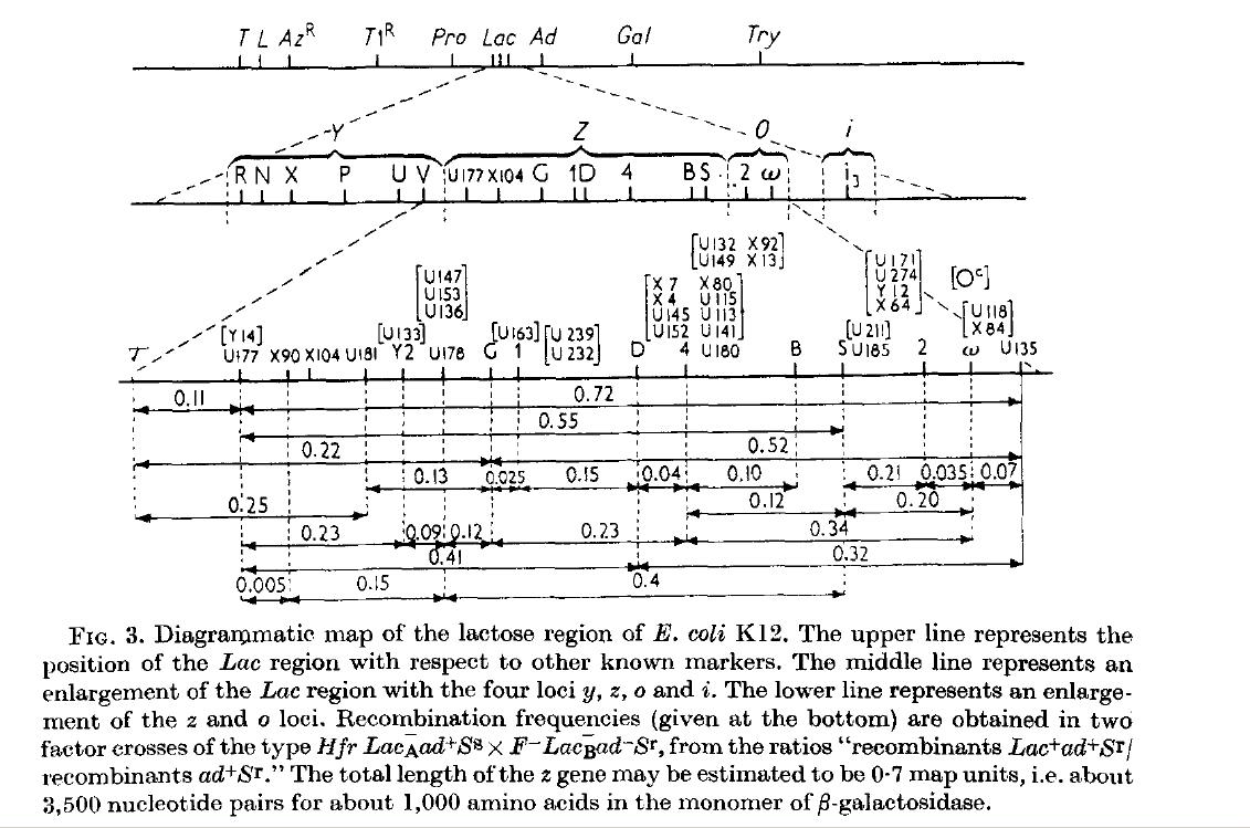
[1] Figure 6 from: Jacob, F. & Monod,
J. Genetic regulatory mechanisms in the
synthesis of proteins. J. Mol. Biol. 3,
318–356 (1961)
http://www.sciencedirect.com/science?_
ob=ArticleURL&_udi=B6WK7-4Y39HH7-B&_user
=4422&_coverDate=06%2F30%2F1961&_alid=17
23143833&_rdoc=1&_fmt=high&_orig=search&
_origin=search&_zone=rslt_list_item&_cdi
=6899&_sort=r&_st=13&_docanchor=&view=c&
_ct=5&_acct=C000059600&_version=1&_urlVe
rsion=0&_userid=4422&md5=c2699b72c7c5bee
4e2c31224c6261556&searchtype=a {Jacob_F
rancois_19601228.pdf} COPYRIGHTED
source: http://www.sciencedirect.com/sci
ence?_ob=ArticleURL&_udi=B6WK7-4Y39HH7-B
&_user=4422&_coverDate=06%2F30%2F1961&_a
lid=1723143833&_rdoc=1&_fmt=high&_orig=s
earch&_origin=search&_zone=rslt_list_ite
m&_cdi=6899&_sort=r&_st=13&_docanchor=&v
iew=c&_ct=5&_acct=C000059600&_version=1&
_urlVersion=0&_userid=4422&md5=c2699b72c
7c5bee4e2c31224c6261556&searchtype=a
[2] Figure 3 from: Jacob, F. & Monod,
J. Genetic regulatory mechanisms in the
synthesis of proteins. J. Mol. Biol. 3,
318–356 (1961)
http://www.sciencedirect.com/science?_
ob=ArticleURL&_udi=B6WK7-4Y39HH7-B&_user
=4422&_coverDate=06%2F30%2F1961&_alid=17
23143833&_rdoc=1&_fmt=high&_orig=search&
_origin=search&_zone=rslt_list_item&_cdi
=6899&_sort=r&_st=13&_docanchor=&view=c&
_ct=5&_acct=C000059600&_version=1&_urlVe
rsion=0&_userid=4422&md5=c2699b72c7c5bee
4e2c31224c6261556&searchtype=a {Jacob_F
rancois_19601228.pdf} COPYRIGHTED
source: http://www.sciencedirect.com/sci
ence?_ob=ArticleURL&_udi=B6WK7-4Y39HH7-B
&_user=4422&_coverDate=06%2F30%2F1961&_a
lid=1723143833&_rdoc=1&_fmt=high&_orig=s
earch&_origin=search&_zone=rslt_list_ite
m&_cdi=6899&_sort=r&_st=13&_docanchor=&v
iew=c&_ct=5&_acct=C000059600&_version=1&
_urlVersion=0&_userid=4422&md5=c2699b72c
7c5bee4e2c31224c6261556&searchtype=a
evolves. Cells can make ATP from
glucose.
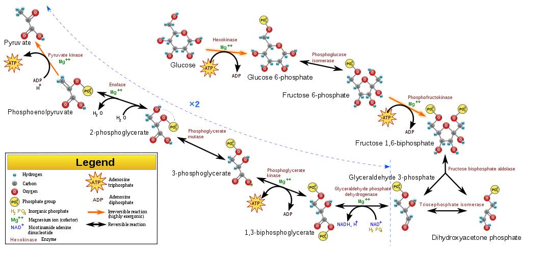
[1] Description English: Glycolysis
pathway overview. Date 3
September 2009 Source Own
work Author
WYassineMrabetTalk✉ Inkscape
Logo.svg This vector image was
created with
Inkscape. Permission (Reusing this
file) GFDL license (see below). GFDL
source: http://upload.wikimedia.org/wiki
pedia/commons/thumb/a/a0/Glycolysis.svg/
1024px-Glycolysis.svg.png

[2] Figure 9.6 from: Campbell, Reece,
et al, ''Biology'', 8th edition, 2008,
p166. COPYRIGHTED
source: Campbell, Reece, et al,
"Biology", 8th edition, 2008, p166.
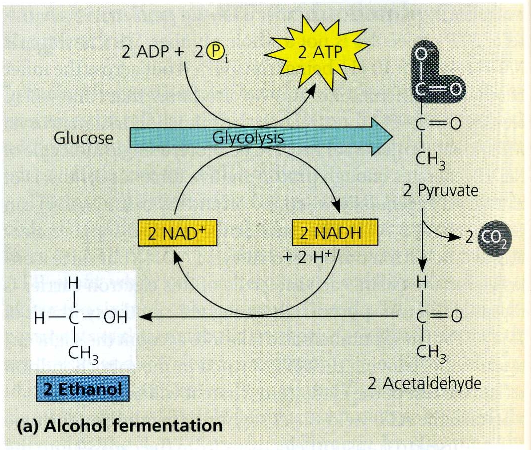
make lactic acid.
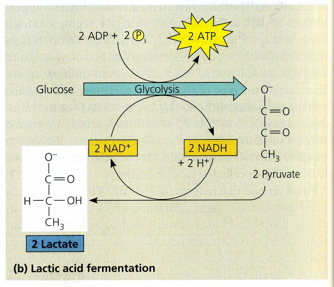
[1] Campbell, Reece, et al,
''Biology'', 8th edition, 2008,
p178. COPYRIGHTED
source: Campbell, Reece, et al,
"Biology", 8th edition, 2008, p178.

[2] IUPAC
name[hide] 2-Hydroxypropanoic
acid Other names[hide] Milk
acid Description de: Struktur
von Milchsäure; en: Structure of
lactic acid Date 12 February
2007 Source Own work Author
NEUROtiker Permission (Reusing
this file) Own work, all rights
released (Public domain) PD
source: http://upload.wikimedia.org/wiki
pedia/commons/5/59/Lactic-acid-3D-balls.
pnghttp://upload.wikimedia.org/wikipedia
/commons/thumb/d/d3/Lactic-acid-skeletal
.svg/1000px-Lactic-acid-skeletal.svg.png
[1] Campbell, Reece, et al,
''Biology'', 8th edition, 2008,
p178. COPYRIGHTED
source: Campbell, Reece, et al,
"Biology", 8th edition, 2008, p178.

[2] Ethanol Full structural
formula, Ball and Stick Model, and
Space-Filling Model of Ethanol PD
source: http://upload.wikimedia.org/wiki
pedia/commons/3/37/Ethanol-2D-flat.pnght
tp://upload.wikimedia.org/wikipedia/comm
ons/b/b0/Ethanol-3D-balls.pnghttp://uplo
ad.wikimedia.org/wikipedia/commons/0/00/
Ethanol-3D-vdW.png
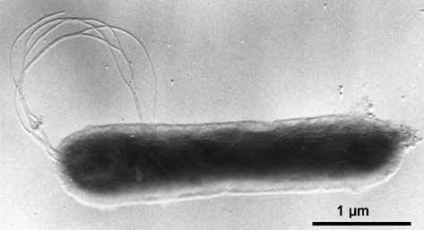
[1] Aquifex pyrophilus (platinum
shadowed). © K.O. Stetter & Reinhard
Rachel, University of Regensburg.
COPYRIGHTED
source: http://biology.kenyon.edu/Microb
ial_Biorealm/bacteria/aquifex/aquifex.ht
m
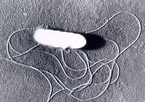
[2] Description English: A
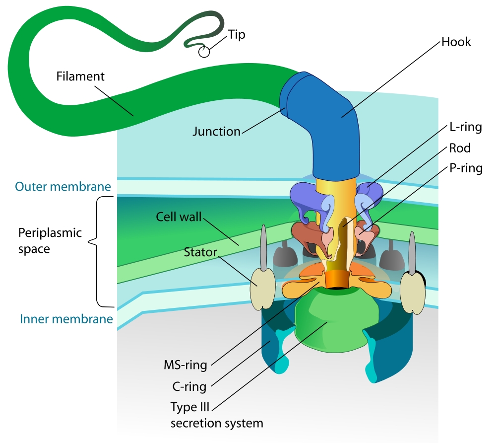
Gram-negative bacterial flagellum. A
flagellum (plural: flagella) is a long,
slender projection from the cell body,
whose function is to propel a
unicellular or small multicellular
organism. The depicted type of
flagellum is found in bacteria such as
E. coli and Salmonella, and rotates
like a propeller when the bacterium
swims. The bacterial movement can be
divided in 2 kinds: run, resulting from
a counterclockwise rotation of the
flagellum, and tumbling, from a
clockwise rotation of the
flagellum. Français : Flagelle de
bactérie Gram-négative. Le flagelle
est une projection longue et fine hors
du corps cellulaire, dont la fonction
est de propulser l'organisme. Ce type
de flagelle est présent dans des
bactéries comme Escherichia coli et
Salmonella, et tourne comme une hélice
quand la bactérie se déplace. Le
flagelle peut provoquer deux types de
déplacement selon son sens de
rotation. Date November 2007 Source
self-made References: [1],[2], [3]
(main 3), [4], [5] (propeller
rotation), PMID 17142059
(bend). Author LadyofHats PD
source: http://upload.wikimedia.org/wiki
pedia/commons/thumb/1/15/Flagellum_base_
diagram_en.svg/1000px-Flagellum_base_dia
gram_en.svg.png
archaebacteria) evolve.
[1] Deutsch: Bild über den Reitenden
Urzwerg English: Image of Nanoarchaeum
equitans Date 2005-09-10 (original
upload date) Source Originally
from de.wikipedia; description page
is/was here. Author Original
uploader was Eber-Jimmy at
de.wikipedia Permission (Reusing
this file) This image is in the
public domain due to its
age. Licensing According to this
article, ''Es wurde von dem
Mikrobiologen Karl O. Stetter entdeckt.
Bildrechte: Public domain.'' PD
source: http://upload.wikimedia.org/wiki
pedia/commons/d/dc/Urzwerg.jpg
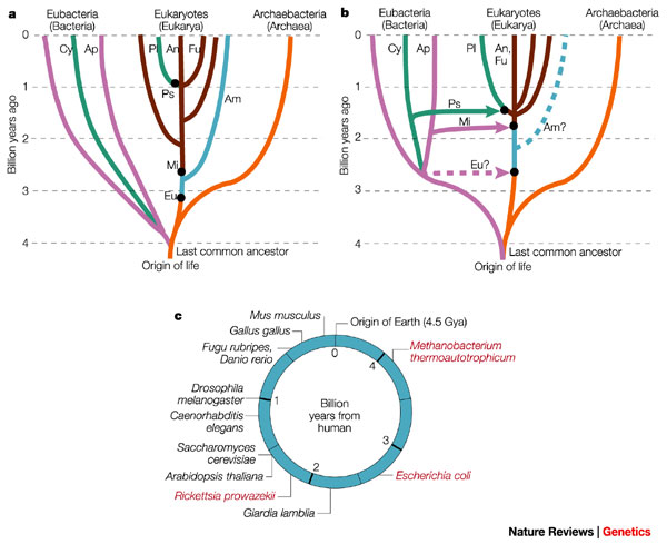
[2] Figure 1) Changing views of the
tree and timescale of life. a) An
early-1990s view, with the tree
determined mostly from ribosomal RNA
(rRNA) sequence analysis. This tree
emphasizes vertical (as opposed to
horizontal) evolution and the close
relationship between eukaryotes and the
Archaebacteria. The deep branching
(>3.5 Giga (109) years ago, Gya) of
CYANOBACTERIA (Cy) and other Eubacteria
(purple), the shallow branching
(approx1 Gya) of plants (Pl), animals
(An) and fungi (Fu), and the early
origin of mitochondria (Mi), were based
on interpretations of the geochemical
and fossil record7, 8. Some deeply
branching amitochondriate (Am) species
were believed to have arisen before the
origin of mitochondria44. Major
symbiotic events (black dots) were
introduced to explain the origin of
eukaryotic organelles42, but were not
assumed to be associated with large
transfers of genes to the host nucleus.
They were: Eu, joining of an
archaebacterium host with a eubacterium
(presumably a SPIROCHAETE) to produce
an amitochondriate eukaryote; Mi,
joining of a eukaryote host with an
alpha-proteobacterium (Ap) symbiont,
leading to the origin of mitochondria,
and plastids (Ps), joining of a
eukaryote host with a cyanobacterium
symbiont, forming the origin of
plastids on the plant lineage and
possibly on other lineages. b) The
present view, based on extensive
genomic analysis. Eukaryotes are no
longer considered to be close relatives
of Archaebacteria, but are genomic
hybrids of Archaebacteria and
Eubacteria, owing to the transfer of
large numbers of genes from the
symbiont genome to the nucleus of the
host (indicated by coloured arrows).
Other new features, largely derived
from molecular-clock studies16, 39 (Box
1), include a relatively recent origin
of Cyanobacteria (approx2.6 Gya) and
mitochondria (approx1.8 Gya), an early
origin (approx1.5 Gya) of plants,
animals and fungi, and a close
relationship between animals and fungi.
Coloured dashed lines indicate
controversial aspects of the present
view: the existence of a
premitochondrial symbiotic event and of
living amitochondriate eukaryotes,
ancestors of which never had
mitochondria. c) The times of
divergence of selected model organisms
from humans, based on molecular clocks.
For the prokaryotes (red), because of
different possible origins through
symbiotic events, divergence times
depend on the gene of interest.
source: http://www.nature.com/nrg/journa
l/v3/n11/full/nrg929_fs.html
evolve (Aquifex, Thermotoga).

[1] A timescale of prokaryote
evolution. Letters indicate nodes
discussed in the text. The last common
ancestor was arbitrarily placed at 4.25
Ga in the tree, although this placement
was not part of the analyses. The grey
rectangle shows the time prior to the
initial rise in oxygen (presumably
anaerobic conditions). Mtb:
Methanothermobacter, Tab:
Thermoanaerobacter, Tsc:
Thermosynechococcus. Battistuzzi et
al. BMC Evolutionary Biology 2004 4:44
doi:10.1186/1471-2148-4-44 Table
1 Time estimates for selected nodes
in the tree of eubacteria (A-K) and
archaebacteria (L-P). Letters refer to
Fig. 3. Time (Ma)a CIb Node
A 102 57–176 Node
B 2508 2154–2928 Node
C 2800 2452–3223 Node
D 1039 702–1408 Node
E 2558 2310–2969 Node
F 2784 2490–3203 Node
G 2923 2587–3352 Node
H 3054 2697–3490 Node
I 3186 2801–3634 Node
J 3644 3172–4130 Node
K 3977 3434–4464 Node
L 233 118–386 Node
M 3085 2469–3514 Node
N 3566 2876–3948 Node
O 3781 3047–4163 Node
P 4112 3314–4486 a Averages of
the divergence times estimated using
the 2.3 Ga minimum constraint and the
five ingroup root constraints (nodes
A-K) and using the 1.198 ± 0.022 Ga
constraint and the five ingroup root
constraints (nodes L-P). b
Credibility interval (minimum and
maximum averages of the analyses under
the five ingroup root
constraints) Battistuzzi et al. BMC
Evolutionary Biology 2004 4:44
doi:10.1186/1471-2148-4-44 COPYRIGHTED
source: http://www.biomedcentral.com/con
tent/figures/1471-2148-4-44-3-l.jpg
[2] Aquifex pyrophilus (platinum
shadowed). © K.O. Stetter & Reinhard
Rachel, University of Regensburg.
source: http://biology.kenyon.edu/Microb
ial_Biorealm/bacteria/aquifex/aquifex.ht
m
(Sulfolobus).
[1] A timescale of prokaryote
evolution. Letters indicate nodes
discussed in the text. The last common
ancestor was arbitrarily placed at 4.25
Ga in the tree, although this placement
was not part of the analyses. The grey
rectangle shows the time prior to the
initial rise in oxygen (presumably
anaerobic conditions). Mtb:
Methanothermobacter, Tab:
Thermoanaerobacter, Tsc:
Thermosynechococcus. Battistuzzi et
al. BMC Evolutionary Biology 2004 4:44
doi:10.1186/1471-2148-4-44 Table
1 Time estimates for selected nodes
in the tree of eubacteria (A-K) and
archaebacteria (L-P). Letters refer to
Fig. 3. Time (Ma)a CIb Node
A 102 57–176 Node
B 2508 2154–2928 Node
C 2800 2452–3223 Node
D 1039 702–1408 Node
E 2558 2310–2969 Node
F 2784 2490–3203 Node
G 2923 2587–3352 Node
H 3054 2697–3490 Node
I 3186 2801–3634 Node
J 3644 3172–4130 Node
K 3977 3434–4464 Node
L 233 118–386 Node
M 3085 2469–3514 Node
N 3566 2876–3948 Node
O 3781 3047–4163 Node
P 4112 3314–4486 a Averages of
the divergence times estimated using
the 2.3 Ga minimum constraint and the
five ingroup root constraints (nodes
A-K) and using the 1.198 ± 0.022 Ga
constraint and the five ingroup root
constraints (nodes L-P). b
Credibility interval (minimum and
maximum averages of the analyses under
the five ingroup root
constraints) Battistuzzi et al. BMC
Evolutionary Biology 2004 4:44
doi:10.1186/1471-2148-4-44 COPYRIGHTED
source: http://www.biomedcentral.com/con
tent/figures/1471-2148-4-44-3-l.jpg

[2] tree of archaea ?
source: http://www.uni-giessen.de/~gf126
5/GROUPS/KLUG/Stammbaum.html
{YRE-oR-KE-O-Tu} (methanogens,
halobacteria).
Earliest cell response to light.
[1] A timescale of prokaryote
evolution. Letters indicate nodes
discussed in the text. The last common
ancestor was arbitrarily placed at 4.25
Ga in the tree, although this placement
was not part of the analyses. The grey
rectangle shows the time prior to the
initial rise in oxygen (presumably
anaerobic conditions). Mtb:
Methanothermobacter, Tab:
Thermoanaerobacter, Tsc:
Thermosynechococcus. Battistuzzi et
al. BMC Evolutionary Biology 2004 4:44
doi:10.1186/1471-2148-4-44 Table
1 Time estimates for selected nodes
in the tree of eubacteria (A-K) and
archaebacteria (L-P). Letters refer to
Fig. 3. Time (Ma)a CIb Node
A 102 57–176 Node
B 2508 2154–2928 Node
C 2800 2452–3223 Node
D 1039 702–1408 Node
E 2558 2310–2969 Node
F 2784 2490–3203 Node
G 2923 2587–3352 Node
H 3054 2697–3490 Node
I 3186 2801–3634 Node
J 3644 3172–4130 Node
K 3977 3434–4464 Node
L 233 118–386 Node
M 3085 2469–3514 Node
N 3566 2876–3948 Node
O 3781 3047–4163 Node
P 4112 3314–4486 a Averages of
the divergence times estimated using
the 2.3 Ga minimum constraint and the
five ingroup root constraints (nodes
A-K) and using the 1.198 ± 0.022 Ga
constraint and the five ingroup root
constraints (nodes L-P). b
Credibility interval (minimum and
maximum averages of the analyses under
the five ingroup root
constraints) Battistuzzi et al. BMC
Evolutionary Biology 2004 4:44
doi:10.1186/1471-2148-4-44 COPYRIGHTED
source: http://www.biomedcentral.com/con
tent/figures/1471-2148-4-44-3-l.jpg
[2] tree of archaebacteria (archaea)
COPYRIGHTED
source: http://www.uni-giessen.de/~gf126
5/GROUPS/KLUG/Stammbaum.html
(autotrophy).
[1] Description Methanopyrus
kandleri Date July
2006 Source ms:Imej:Arkea.jpg Auth
or ms:User:PM Poon GNU
source: http://upload.wikimedia.org/wiki
pedia/commons/a/aa/Arkea.jpg
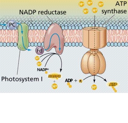
Cells emit free Oxygen.
This is the main system responsible for
producing the Oxygen now in the air of
earth.
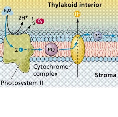
[1] Chemiosmosis as it operates in
photophosphorylation within a
chloroplast. Images from Purves et al.,
Life: The Science of Biology, 4th
Edition, by Sinauer Associates
(www.sinauer.com) and WH Freeman
(www.whfreeman.com) COPYRIGHTED
source: http://www.emc.maricopa.edu/facu
lty/farabee/biobk/0817_1.gif
[2] Chemiosmosis as it operates in
photophosphorylation within a
chloroplast. Images from Purves et al.,
Life: The Science of Biology, 4th
Edition, by Sinauer Associates
(www.sinauer.com) and WH Freeman
(www.whfreeman.com) COPYRIGHTED
source: http://www.emc.maricopa.edu/facu
lty/farabee/biobk/0817_2.gif
evolves in prokaryotes.
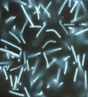
[1] Microgram of filamentous bacteria
from flexible setae. (Courtesy
Zoosystema © 2005) COPYRIGHTED
source: http://bioweb.uwlax.edu/bio203/s
2009/decker_rour/images/yeti-crab-filame
ntous-bacteria.JPG
[2] Filamentous Bacteria Microthrix
Parvicella UNKNOWN
source: http://ebsbiowizard.com/wp-conte
nt/gallery/filamentous-bacteria-microthr
ix-parvicella/filamentous-bacteria-micro
thrix-parvicella.jpg
filamentous prokaryotes, creating
organisms with different kinds of
cells.
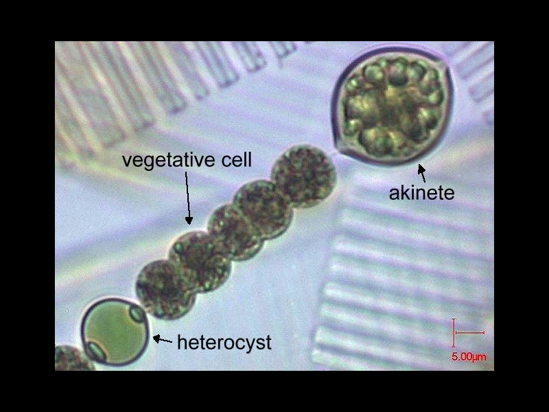
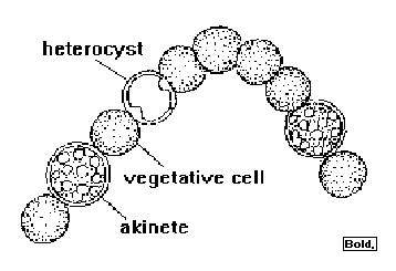
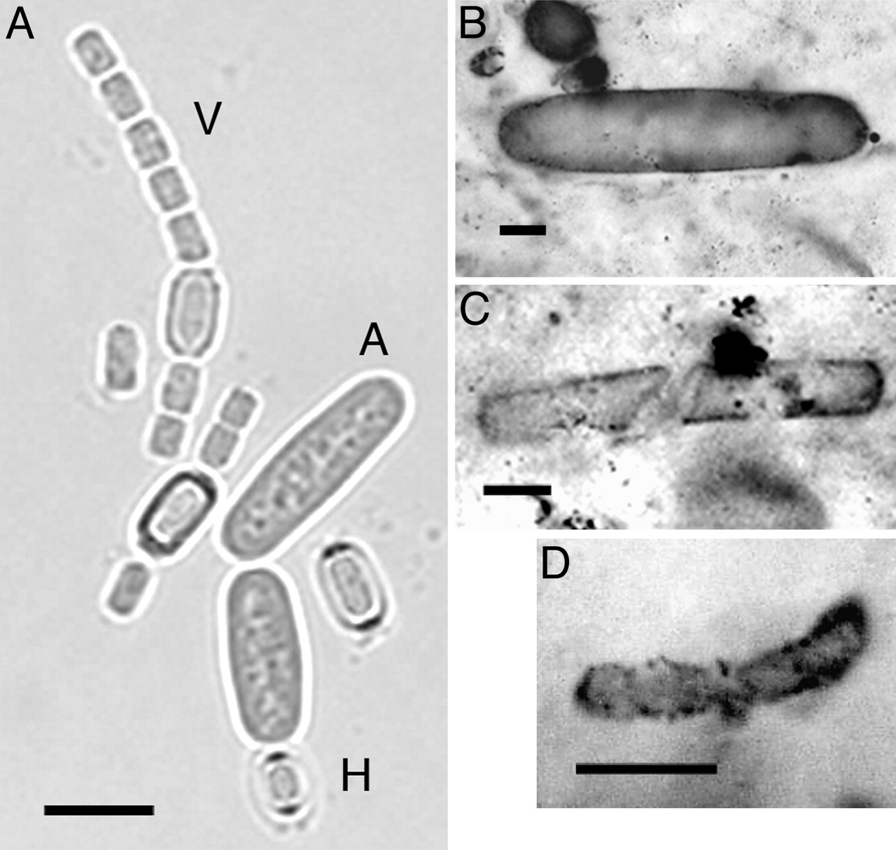
[1] Adapted from: Anabaena smitthi
COPYRIGHTED FRANCE
source: http://www.ac-rennes.fr/pedagogi
e/svt/photo/microalg/anabaena.jpg
[2] Anabaena COPYRIGHTED EDU
source: http://home.manhattan.edu/~franc
es.cardillo/plants/monera/anabaena.gif
nitrogen compounds like ammonia from
Nitrogen gas in the air.

[1] Fig. 2. Modern cyanobacterial
akinetes and Archaeoellipsoides
fossils. (A) Three-month-old culture of
living A. cylindrica grown in a medium
without combined nitrogen. A, akinete;
H, heterocyst; V, vegetative cells.
(B–D) Shown are Archaeoellipsoides
fossils from 1,500-Ma Billyakh Group,
northern Siberia (B); 1,650-Ma McArthur
Group, northern Australia (C); and
2,100-Ma Franceville Group, Gabon (D).
(Scale bars, 10 μm.) COPYRIGHTED
source: http://www.pnas.org/content/103/
14/5442/F2.large.jpg
[2] Fig. 2. Modern cyanobacterial
akinetes and Archaeoellipsoides
fossils. (A) Three-month-old culture of
living A. cylindrica grown in a medium
without combined nitrogen. A, akinete;
H, heterocyst; V, vegetative cells.
(B–D) Shown are Archaeoellipsoides
fossils from 1,500-Ma Billyakh Group,
northern Siberia (B); 1,650-Ma McArthur
Group, northern Australia (C); and
2,100-Ma Franceville Group, Gabon (D).
(Scale bars, 10 μm.) COPYRIGHTED
source: http://www.pnas.org/content/103/
14/5442/F2.large.jpg
aerobic cell. These cells use oxygen to
convert glucose into carbon dioxide,
water, and ATP.
[1] purple aerobic bacteria UNKNOWN
source: http://endosymbiotichypothesis.f
iles.wordpress.com/2010/09/rain-bacteria
.jpg
[2] Organisms of Rickettsia conorii
(r), a close relative of R. rickettsii,
in a cultured human endothelial cell
are located free in the cytosol. One
rickettsia is dividing by binary
fission (arrowhead). (B) These
rickettsiae can move inside the
cytoplasm of the host cell because of
the propulsive force created by the
''tail'' of host cell actin filaments
(arrow). Bars = 0.5 µm. Photo and
text courtesy of David H. Walker -
http://gsbs.utmb.edu/microbook/ch038.htm
UNKNOWN AND Rickettsia prowazekii
(image with Rickettsia outside of
cell) COPYRIGHTED [1] Rickettsia
prowazekii COPYRIGHTED FAIR USE
source: http://www.bio.davidson.edu/peop
le/sosarafova/Assets/Bio307/liwoeste/Pic
tures/Walker%203%5B1%5D.jpghttp://web.ms
t.edu/~microbio/bio221_2001/Image9.jpg
ratio of carbon-13 to carbon-12.
[1] Figure 1 from: Mojzsis, S. J. et
al. ''Evidence for Life on Earth Before
3,800 Million Years Ago.'' Nature
384.6604 (1996):
55–59. http://www.nature.com/nature/j
ournal/v384/n6604/abs/384055a0.html COP
YRIGHTED
source: http://www.nature.com/nature/jou
rnal/v384/n6604/pdf/384055a0.pdf

[2] Figure 1 from: Mojzsis, S. J. et
al. ''Evidence for Life on Earth Before
3,800 Million Years Ago.'' Nature
384.6604 (1996):
55–59. http://www.nature.com/nature/j
ournal/v384/n6604/abs/384055a0.html COP
YRIGHTED
source: http://www.nature.com/nature/jou
rnal/v384/n6604/pdf/384055a0.pdf
Formation" begins.
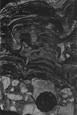
Banded Iron Formation is
sedimentary rock that spans 2 billion
years, made of silica with alternating
layers of black-colored reduced iron
and red-colored oxidized iron, and
represents a seasonal rise and fall of
free oxygen in the ocean, possibly
linked to photosynthetic organisms.
[1] image of BIF from Akilia from
Nature COPYRIGHTED
source: nature 11/7/96
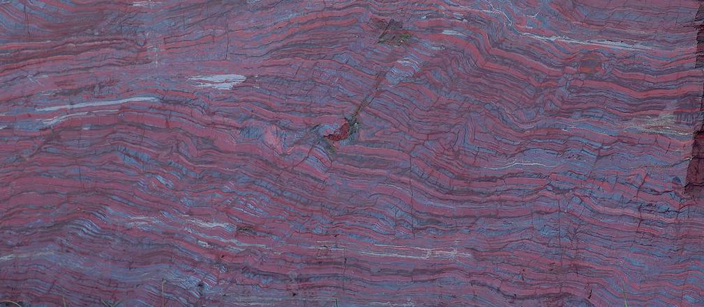
[2] portion taken
from: Description English: This
image shows a 2.1 billion years old
rock containing black-banded ironstone,
which has a weight of about 8.5 tons.
The approximately two meter high, three
meter wide, and one meter thick block
of stone was found in North America and
belongs to the National Museum of
Mineralogy and Geology in Dresden,
Germany. The rock is located at
+51°2'34.84''
+13°45'26.67''. Deutsch: Dieses Bild
zeigt einen etwa 8,5 Tonnen schweren
und 2,1 Milliarden Jahre alten Block
mit Bändereisenerzen. Der etwa zwei
Meter hohe, drei Meter breite und einen
Meter tiefe Gesteinsblock wurde in
Nordamerika gefunden und gehört dem
Staatlichen Museum für Mineralogie und
Geologie Dresden. Der Block befindet
sich bei den Koordinaten +51°2'34.84''
+13°45'26.67''. Camera
data Camera Nikon D70 Lens Tamron
SP AF 90mm/2.8 Di Macro 1:1 Focal
length 90 mm Aperture f/2.8 Exposure
time 1/250 s Sensivity ISO 200 Please
help translating the description into
more languages. Thanks a lot! If
you want a license with the conditions
of your choice, please email me to
negotiate terms. best new
image Date 26 August
2005 Source Own
work Author André Karwath aka
Aka CC
source: http://upload.wikimedia.org/wiki
pedia/commons/thumb/5/5f/Black-band_iron
stone_%28aka%29.jpg/1280px-Black-band_ir
onstone_%28aka%29.jpg
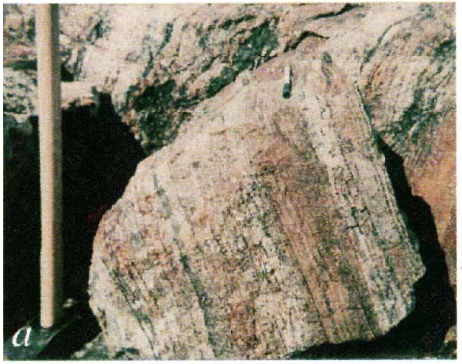
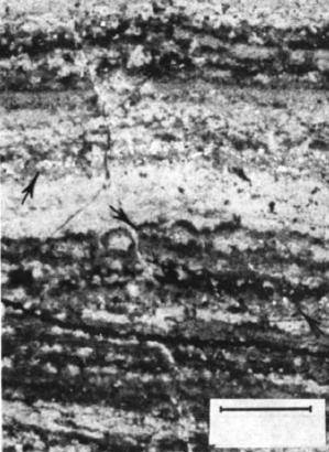
stromatolites.
Tree Group, South Africa
[1] image on left is from swaziland
source: nature feb 6
[2]
source: 1986
2.8 billion years will pass before the
first animal evolves.
Australia and Onverwacht Group,
Barberton Mountain Land, South
Africa
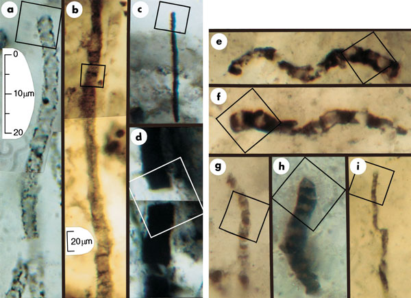
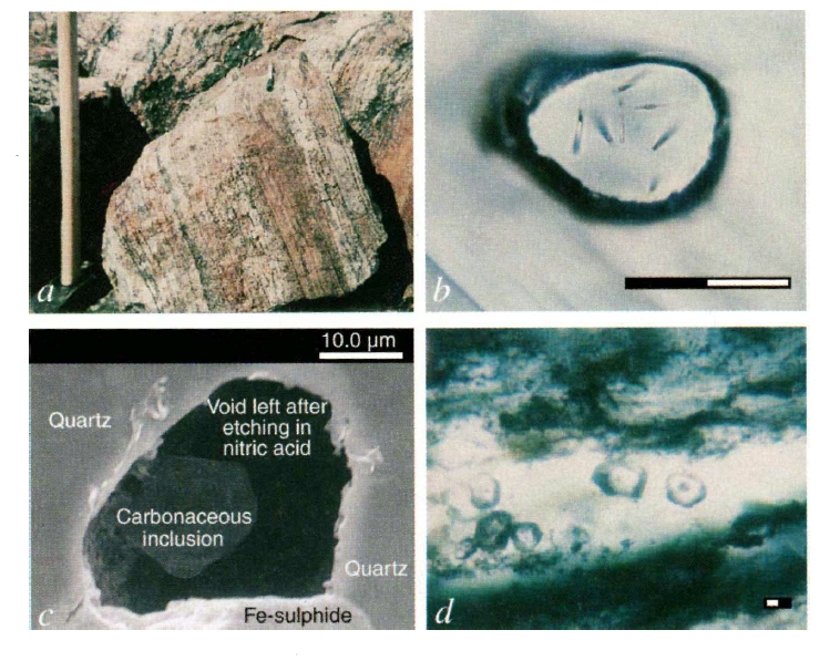
[1] Figure 1 Optical photomicrographs
showing carbonaceous (kerogenous)
filamentous microbial fossils in
petrographic thin sections of
Precambrian cherts. Scale in a
represents images in a and c-i; scale
in b represents image in b. All parts
show photomontages, which is
necessitated by the three-dimensional
preservation of the cylindrical sinuous
permineralized microbes. Squares in
each part indicate the areas for which
chemical data are presented in Figs 2
and 3. a, An unnamed cylindrical
prokaryotic filament, probably the
degraded cellular trichome or tubular
sheath of an oscillatoriacean
cyanobacterium, from the 770-Myr
Skillogalee Dolomite of South
Australia12. b, Gunflintia grandis, a
cellular probably oscillatoriacean
trichome, from the 2,100-Myr Gunflint
Formation of Ontario, Canada13. c, d,
Unnamed highly carbonized filamentous
prokaryotes from the 3,375-Myr Kromberg
Formation of South Africa14: the poorly
preserved cylindrical trichome of a
noncyanobacterial or oscillatoriacean
prokaryote (c); the disrupted,
originally cellular trichomic remnants
possibly of an Oscillatoria- or
Lyngbya-like cyanobacterium (d). e-i,
Cellular microbial filaments from the
3,465-Myr Apex chert of northwestern
Western Australia: Primaevifilum
amoenum4,5, from the collections of The
Natural History Museum (TNHM), London,
specimen V.63164[6] (e); P. amoenum4
(f); the holotype of P.
delicatulum4,5,15, TNHM V.63165[2] (g);
P. conicoterminatum5, TNHM V63164[9]
(h); the holotype of Eoleptonema apex5,
TNHM V.63729[1] (i).
source: Nature416
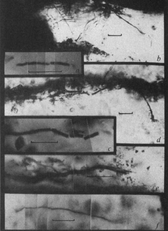
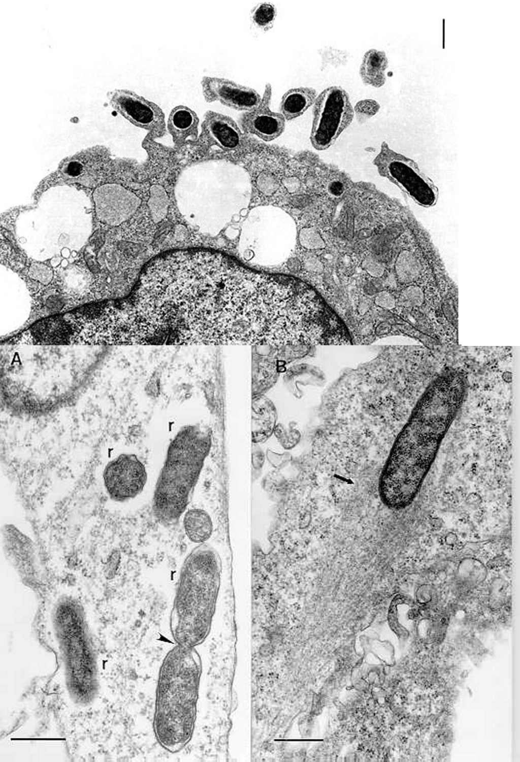
[2] Fig. 3 Filamentous microfossils:
a, cylindrical microfossil from
Hooggenoeg sample; b, threadlike and
tubular filaments extending between
laminae, Kromberg sample; c,d,e,
tubular filamnets oriented subparallel
to bedding, Kromberg sample; f,
threadlike filament flattened parallel
to bedding, Kromberg sample.
source: 73 - 76 (07 Mar 2002) Letters
to Nature
http://www.nature.com/nature/journal/v41
6/n6876/fig_tab/416073a_F1.html

[1] Evolutionary relationships of model
organisms and bacteria that show
unusual reproductive strategies. This
phylogenetic tree (a) illustrates the
diversity of organisms that use the
alternative reproductive strategies
shown in (b). Bold type indicates
complete or ongoing genome projects.
Intracellular offspring are produced by
several low-GC Gram-positive bacteria
such as Metabacterium polyspora,
Epulopiscium spp. and the segmented
filamentous bacteria (SFB). Budding and
multiple fission are found in the
proteobacterial genera Hyphomonas and
Bdellovibrio, respectively. In the case
of the Cyanobacteria, Stanieria
produces baeocytes and Chamaesiphon
produces offspring by budding.
Actinoplanes produce dispersible
offspring by multiple fission of
filaments within the sporangium.
source: http://www.nature.com/nrmicro/jo
urnal/v3/n3/full/nrmicro1096_fs.html
(Nature Reviews Microbiology 3
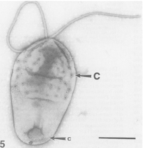
[2] Electron micrograph of a Pirellula
bacterium from giant tiger prawn tissue
(Penaeus monodon). Notice the large
crateriform structures (C) on the cell
surface and flagella. From Fuerst et
al.
source: 214-224 (2005);
doi:10.1038/nrmicro1096)
(unicellular microfossils with
uncertain affinity).

[1] Figure from: Javaux, Emmanuelle
J., Craig P. Marshall, and Andrey
Bekker. “Organic-walled microfossils
in 3.2-billion-year-old shallow-marine
siliciclastic deposits.” Nature
463.7283 (2010):
934-938. http://www.nature.com/nature/j
ournal/v463/n7283/full/nature08793.html
COPYRIGHTED
source: http://www.nature.com/nature/jou
rnal/v463/n7283/full/nature08793.html
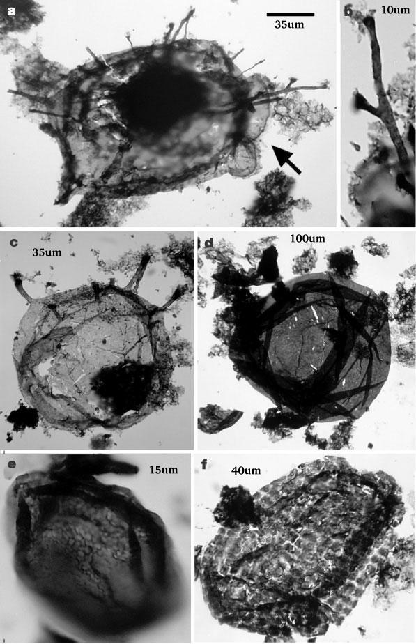
[2] Figure from: Javaux, Emmanuelle
J., Andrew H. Knoll, and Malcolm R.
Walter. “Morphological and ecological
complexity in early eukaryotic
ecosystems.” Nature 412.6842 (2001):
66-69. http://www.nature.com/nature/jou
rnal/v412/n6842/abs/412066a0.html Figur
e 1 Protistan microfossils from the
Roper Group. a, c, Tappania plana,
showing asymmetrically distributed
processes and bulbous protrusions
(arrow in a). b, detail of a, showing
dichotomously branching process. d,
Valeria lophostriata. e, Dictyosphaera
sp. f, Satka favosa. The scale bar in a
is 35 µm for a and c; 10 µm for b;
100 µm for d; 15 µm for e; and 40 µm
for f.
source: http://www.nature.com/nature/jou
rnal/v412/n6842/abs/412066a0.html
evolve (Gram positive bacteria: cause
of botulism, tetanus, anthrax). First
endospores.
[1] Listeria monocytogenes is a
Gram-positive bacterium, in the
division Firmicutes, named for Joseph
Lister. It is motile by means of
flagella. Some studies suggest that 1
to 10% of humans may carry L.
monocytogenes in their
intestines. Researchers have found L.
monocytogenes in at least 37 mammalian
species, both domesticated and feral,
as well as in at least 17 species of
birds and possibly in some species of
fish and shellfish. Laboratories can
isolate L. monocytogenes from soil,
silage, and other environmental
sources. L. monocytogenes is quite
hardy and resists the deleterious
effects of freezing, drying, and heat
remarkably well for a bacterium that
does not form spores. Most L.
monocytogenes are pathogenic to some
degree.
source: http://en.wikipedia.org/wiki/Ima
ge:Listeria.jpg

[2] These are bacteria (about 0.3 µm
in diameter) that do not have outer
walls, only cytoplasmic membranes.
However, they do have cytoskeletal
elements that give them a distinct
non-spherical shape. They look like
schmoos that are pulled along by their
heads. How they are able to glide is a
mystery.
source: http://webmac.rowland.org/labs/b
acteria/projects_glide.html
{ancestor of all mitochondria},
gonorrhea, Salmonella, E. coli).

[1] Figure 1. Transmission electron
micrograph of the ELB agent in XTC-2
cells. The rickettsia are free in the
cytoplasm and surrounded by an electron
transparent halo. Original
magnification X 30,000. CDC PD
source: www.cdc.gov/ncidod/
eid/vol7no1/raoultG1.htm
[2] Caulobacter crescentus. From
http://sunflower.bio.indiana.edu/~ybrun/
L305.html COPYRIGHTED EDU was in wiki
but appears to be removed
source: http://upload.wikimedia.org/wiki
pedia/en/4/42/Caulobacter.jpg
evolve in bacteria.

[1] the fertility factor or F factor is
a very large (94,500 bp) circular dsDNA
plasmid; it is generally independent of
the host chromosome. COPYRIGHTED
source: http://www.mun.ca/biochem/course
s/3107/images/Fplasmidmap.gif
[2] conjugation (via pilus)
COPYRIGHTED EDU
source: http://www.bio.miami.edu/dana/16
0/conjugation.jpg

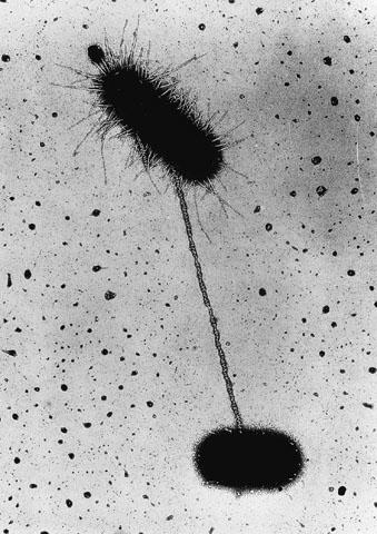
[1] Description Electron
micrograph of Bacteriophages Date
Source
en:Image:Phage.jpg Author
en:User:GrahamColm PD
source: http://upload.wikimedia.org/wiki
pedia/commons/5/52/Phage.jpg
{PlaNK-TO-mI-SETS}.

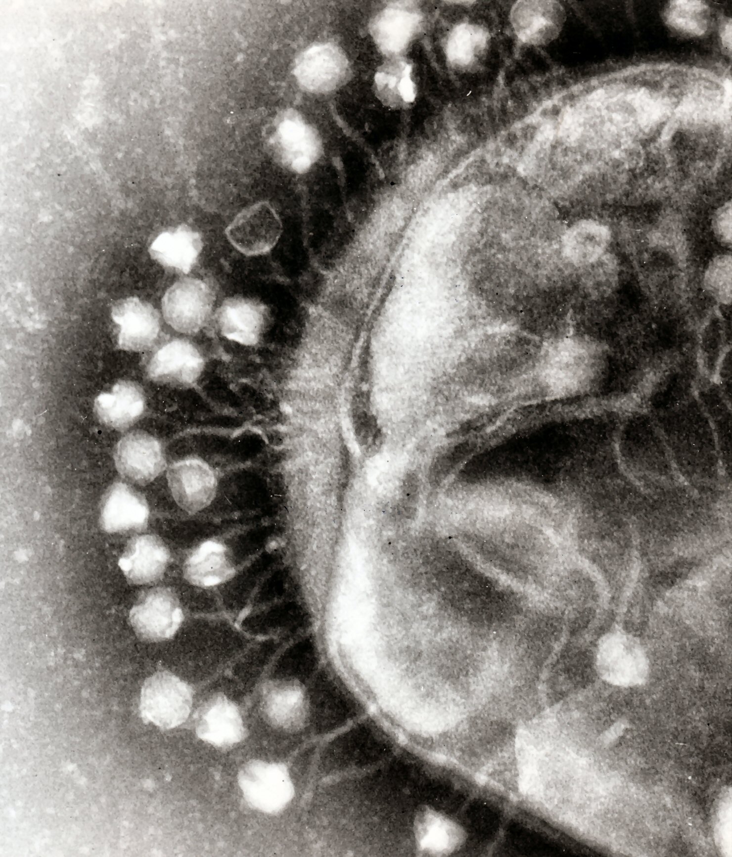
[1] Electron micrographs of cells of
new Gemmata-like and Isosphaera-like
isolates. (A) Negatively stained cell
of the Gemmata-like strain JW11-2f5
showing crateriform structures
(arrowhead) and coccoid cell
morphology. Bar marker, 200 nm. (B)
Negatively stained budding cell of
Isosphaera-like strain CJuql1 showing
uniform crateriform structures
(arrowhead) on the mother cell and
coccoid cell morphology. Bar marker,
200 nm. (C) Thin section of
Gemmata-like cryosubstituted cell of
strain JW3-8s0 showing the
double-membrane-bounded nuclear body
(NB) and nucleoid (N) enclosed within
it. Bar marker, 200 nm. (D) Thin
section of Isosphaera-like strain C2-3
possessing a fibrillar nucleoid (N)
within a cytoplasmic compartment
bounded by a single membrane (M) only.
Bar marker, 200 nm. Appl Environ
Microbiol. 2002 January; 68(1):
417-422. doi:
10.1128/AEM.68.1.417-422.2002.
source: http://www.pubmedcentral.gov/art
iclerender.fcgi?tool=pubmed&pubmedid=117
72655
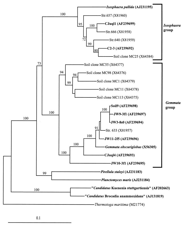
[2] Evolutionary distance tree derived
from comparative analysis of 16S rDNAs
from freshwater and soil isolates and
reference strains of the order
Planctomycetales. Database accession
numbers are shown in parentheses after
species, strain, or clone names.
Bootstrap values of greater than 70%
from 100 bootstrap resamplings from the
distance analysis are presented at
nodes. Thermotoga maritima was used as
an outgroup. Isolates from this study
and representative named species of the
planctomycetes are indicated in bold.
The scale bar represents 0.1 nucleotide
substitution per nucleotide
position. Appl Environ Microbiol.
2002 January; 68(1): 417-422. doi:
10.1128/AEM.68.1.417-422.2002.
source: http://florey.biosci.uq.edu.au/m
ypa/images/fuerst2.gif
{aKTinO-BaK-TER-Eu} (source of
streptomycin).

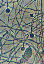
[1] Aerial mycelium and spore of
Streptomyces coelicolor. The mycelium
and the oval spores are about 1µm
wide, typical for bacteria and much
smaller than fungal hyphae and spores.
(Scanning electron micrograph, Mark
Buttner, Kim Findlay, John Innes
Centre). COPYRIGHT UK
source: http://www.sanger.ac.uk/Projects
/S_coelicolor/micro_image4.shtml
[2] Frankia is a genus of
nitrogen-fixing soil bacteria, which
possesses a set of features that are
unique amongst symbiotic
nitrogen-fixing microorganisms,
including rhizobia, making it an
attractive taxon to study. These
heterotrophic Gram-positive bacteria
which are able to induce symbiotic
nitrogen-fixing root nodules
(actinorhizas) in a wide range of
dicotyledonous species (actinorhizal
plants), have also the capacity to fix
atmospheric nitrogen in culture and
under aerobic conditions.
source: http://www.ibmc.up.pt/webpagesgr
upos/cam/Frankia.htm
(Syphilis, Lyme disease).
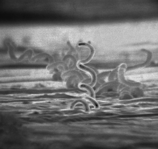
[1] Syphilis is a complex, sexually
transmitted disease (STD) with a highly
variable clinical course. The disease
is caused by the bacterium, Treponema
pallidum. In the United States, 32,871
cases of syphilis, including 432 cases
of congenital syphilis, were detected
by public health officials in 2002.
Eight of the ten states with the
highest rates of syphilis are located
in the southern region of the United
States.
source: http://www.cdc.gov/nchstp/od/tus
kegee/syphilis.htm
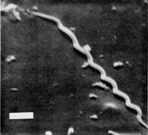
[2] unknown
source: http://uhavax.hartford.edu/bugl/
images/Treponema%20pallidum.jpg
{BaKTRrOEDiTEZ}.

[1] Description Bacteroides
biacutis—one of many en:commensal
anaerobic en:Bacteroides spp. in the
en:gastrointestinal tract—cultured in
blood agar medium for 48
hours. Obtained from the CDC Public
Health Image Library. Image credit:
CDC/Dr. V.R. Dowell, Jr. (PHIL #3087),
1972. Date 2006-03-11 (original
upload date) Source Originally from
en.wikipedia; description page is/was
here. Author Original uploader was
MarcoTolo at
en.wikipedia Permission (Reusing this
file) PD-USGOV-HHS-CDC. PD
source: http://upload.wikimedia.org/wiki
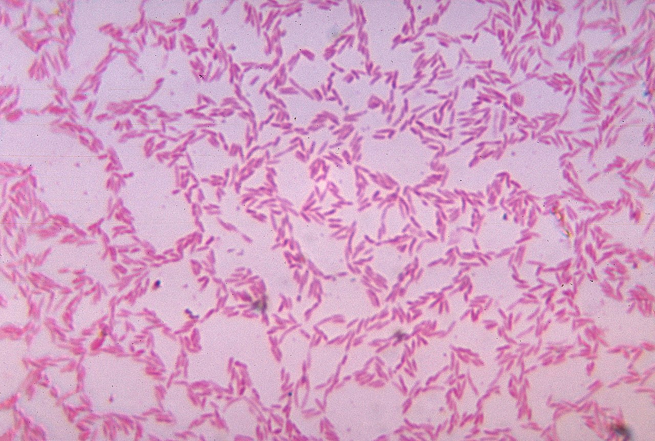
pedia/commons/thumb/6/6c/Bacteroides_bia
cutis_01.jpg/1280px-Bacteroides_biacutis
_01.jpg
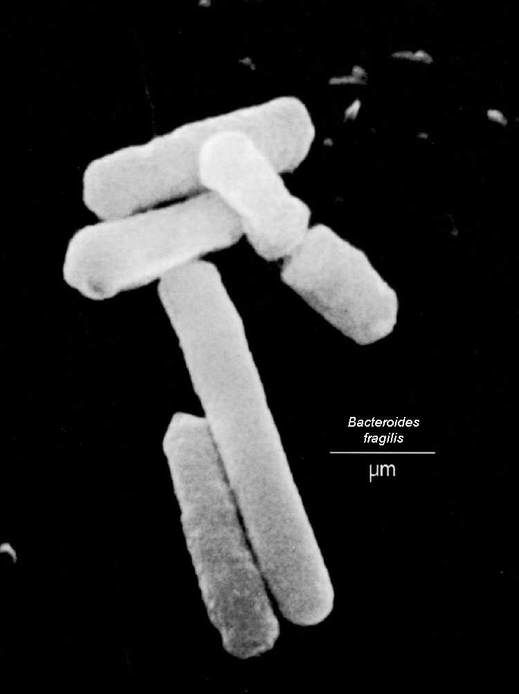
[2] Bacteroides fragilis . From the
Zdravotni University
source: http://biology.kenyon.edu/Microb
ial_Biorealm/bacteria/bacteroidete_chlor
ob_group/bacteroides/bacteroides.htm
or Klo-mi-DE-E}.

[1] cell infected with Chlamydia The
Bavoil laboratory studies the
pathogenesis of the obligate
intracellular pathogen, Chlamydia, and
its bacteriophages. Specific research
areas include the role of Chlamydia
type III secretion in pathogenesis and
development, the impact of Chlamydia
phage infection on disease, the role of
the polymorphic membrane protein family
of C. trachomatis in infection and
disease and comparative genomics within
the Chlamydiaceae. [1] Chlamydia
trachomatis wiki, is copyrighted
source: http://www.dental.umaryland.edu/
sebin/p/o/chlamydia_infected_cell2.jpg
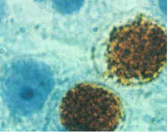
[2] wiki, public domain
source: http://en.wikipedia.org/wiki/Chl
amydia_trachomatis
bacteria).
[1] Description Deutsch: Grüne
Schwefelbakterien (Chlorobiaceae) im
unteren Bereich einer
Winogradsky-Säule Date
20.03.2007 (20 March 2007
(original upload date)) Source
Transferred from de.wikipedia;
transfer was stated to be made by
User:Jacopo Werther. (Original text :
Mikrobiologie Praktikum Universität
Kassel März 2007) Author
kOchstudiO. Original uploader was
KOchstudiO at
de.wikipedia Permission (Reusing this
file) Released into the public
domain (by the author). (Original text
: uneingeschränkte Nutzung) PD
source: http://upload.wikimedia.org/wiki
pedia/commons/e/e7/Green_d_winogradsky.j
pg
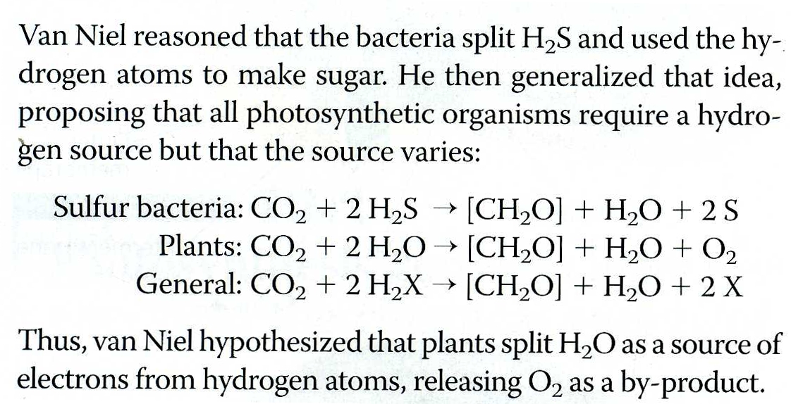
[2] Campbell, N.A., and J.B. Reece.
Biology. Pearson Benjamin Cummings,
2008. Alternative eText Formats Series,
p194. COPYRIGHTED
source: Campbell, N.A., and J.B. Reece.
Biology. Pearson Benjamin Cummings,
2008. Alternative eText Formats Series,
p194.
(VeR-rUKO-mI-KrO-BEo).

[1] Figure 1 Transmission electron
micrographs of high-pressure frozen and
cryosubstituted Verrucomicrobium
spinosum. A. Cell prepared by
high-pressure freezing and
cryosubstitution showing prostheca
(PT), paryphoplasm (P), and an
intracytoplasmic membrane (ICM)
enclosing a pirellulosome region
containing a condensed fibrillar
nucleoid (N). Inset: enlarged view of
area of cell outlined in the white box
showing cytoplasmic membrane (CM),
paryphoplasm and ICM. B.
freeze-fracture replica of cell showing
cross-fractured paryphoplasm (P) and
fracture faces of ICM and CM. Bar –
500 nm Lee et al. BMC Microbiology
2009 9:5
doi:10.1186/1471-2180-9-5 CC
source: http://www.biomedcentral.com/con
tent/figures/1471-2180-9-5-1-l.jpg
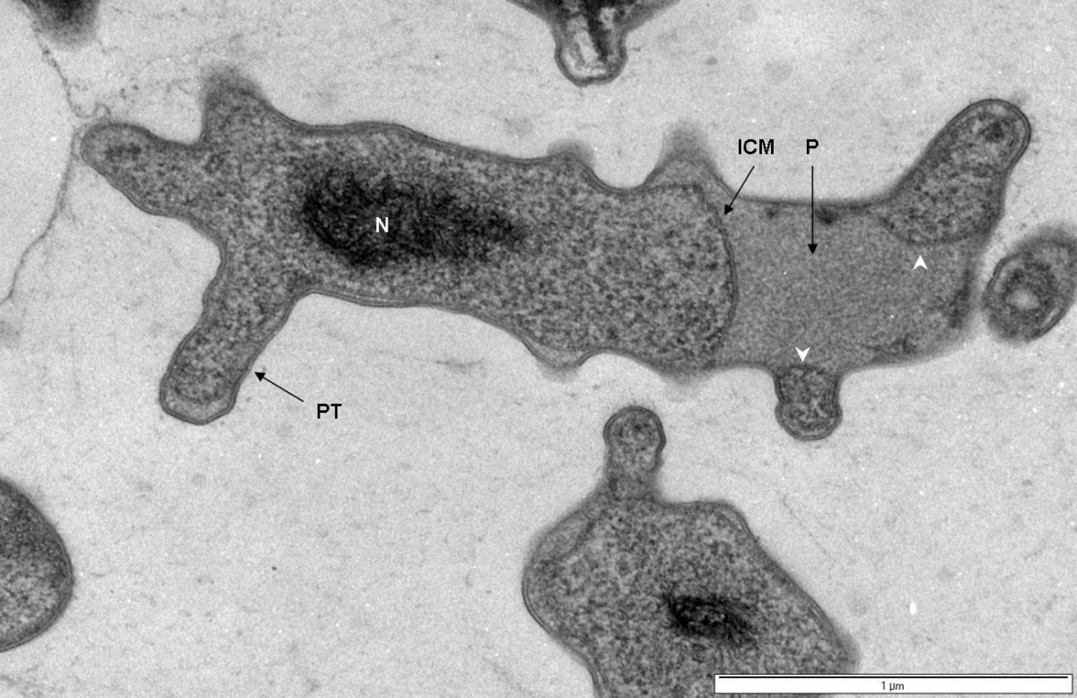
[2] Figure 2 Transmission electron
micrograph of high-pressure frozen and
cryosubstituted Verrucomicrobium
spinosum. Cell prepared by
high-pressure freezing and
cryosubstitution showing prostheca
(PT), ribosome-free paryphoplasm (P),
and an intracytoplasmic membrane (ICM)
enclosing a pirellulosome region
containing a condensed fibrillar
nucleoid (N). Membrane-bounded
vesicle-like compartments within some
prosthecae extensions are also present
(see arrowheads). Bar – 1 μm Lee
et al. BMC Microbiology 2009 9:5
doi:10.1186/1471-2180-9-5 CC
source: http://www.biomedcentral.com/con
tent/figures/1471-2180-9-5-2-l.jpg
eat other cells.
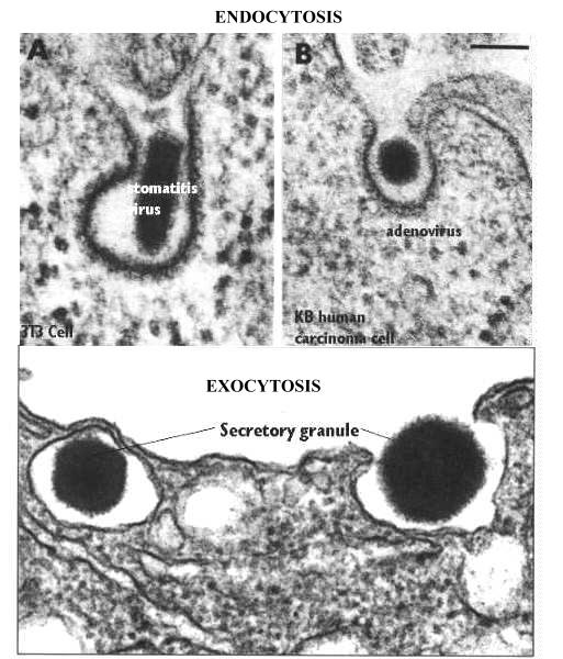
[1] Endocytosis and Exocytosis: For
example, this electron micrograph is
showing the process of exocytosis . The
process begins by fusion of the
membranes at the peripheral pole of the
granule. Then an opening is created
which widens to look like an omicron
figure. This opening allows the
granular material to be released. The
membrane is now part of the plasma
membrane and any proteins carried with
it can be incorporated into the plasma
membrane. Note that there is no coating
on the membrane. This figure was taken
from Alberts et al, Molecular Biology
of the Cell, Garland Publishing Third
Edition, 1994 In contrast, this
micrograph shows a figure which looks
something like an omicron, however,
this view is showing receptor mediated
endocytosis of virus particles. In both
cases, the membrane is coated with
clathrin and these represent classical
receptor mediated endocytosis profiles.
Most ligands cannot be visualized by
themselves, like a virus particle.
Therefore, the cytochemist must attach
label to the ligand. Alternatively, the
cytochemist could immunocytochemically
detect the receptor with antibodies
that recognize the extracellular
domain. This figure was taken from
Endocytosis, Edited by Ira Pastan and
Mark C. Willingham, Plenum Press, N.Y.,
1985 COPYRIGHTED
source: http://www.cytochemistry.net/cel
l-biology/end7.jpg
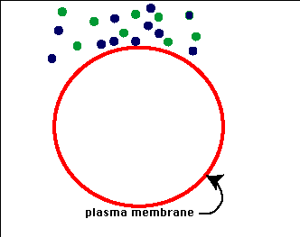
[2] Pinocytosis In the process of
pinocytosis the plasma membrane froms
an invagination. What ever substance
is found within the area of
invagination is brought into the
cell. In general this material will
be dissolved in water and thus this
process is also refered to as
''cellular drinking'' to indicate that
liquids and material dissolved in
liquids are ingested by the
cell. This is opposed to the
ingestion of large particulate material
like bacteria or other cells or cell
debris. UNKNOWN
source: http://academic.brooklyn.cuny.ed
u/biology/bio4fv/page/endocytb.htm
with a nucleus. The first protist. The
nucleus may develop from the infolding
of plasma membrane.

[1] Campbell, Reece, et al,
''Biology'', 2008, p517. COPYRIGHTED
source: Campbell, Reece, et al,
"Biology", 2008, p517.

[2]
http://www.regx.de/m_organisms.php#planc
to
source: http://www.regx.de/m_organisms.p
hp#plancto
of eukaryotes (sterane {STiRAN}
molecules).

[1] Jochen J. Brocks, Graham A. Logan,
Roger Buick, Roger E. Summons,
''Archean Molecular Fossils and the
Early Rise of Eukaryotes'', Science,
Vol 285, Issue 5430, 13 August 1999,
p1033-1036.
http://www.sciencemag.org/content/285/
5430/1033.short
and http://www.jstor.org/stable/2898534
COPYRIGHTED
source: http://www.sciencemag.org/conten
t/285/5430/1033.short
and http://www.jstor.org/stable/2898534
a membrane system that extends from the
nucleus, important in the synthesis of
proteins and lipids.

[1] Figure 1 : Image of n, endoplasmic
reticulum and Golgi apparatus. (1)
Nucleus. (2) Nuclear pore. (3) Rough
endoplasmic reticulum (RER). (4) Smooth
endoplasmic reticulum (SER). (5)
Ribosome on the rough ER. (6) Proteins
that are transported. (7) Transport
vesicle. (8) Golgi apparatus. (9) Cis
face of the Golgi apparatus. (10) Trans
face of the Golgi apparatus. (11)
Cisternae of the Golgi apparatus. I
am the copyright holder of that image
(I might even have the CorelDraw file
around somewhere:-), and I hereby place
the image and all partial images
created from it in the public domain.
So, you are free to use it any way you
like. In fact, I am delighted that one
of my drawings makes it into
print! I can mail you the .cdr file,
if you like (and if I can find it), if
you need a better resolution for
printing. Yours, Magnus
Manske Source: See also User:Magnus
Manske
source: http://en.wikipedia.org/wiki/Ima
ge:Nucleus_ER_golgi.jpg
[2] Description English: The
elongation and membrane targeting
stages of eukaryotic translation. The
ribosome is green and yellow, the tRNAs
are dark blue, and the other proteins
involved are light blue. CC
source: http://upload.wikimedia.org/wiki
pedia/commons/3/3c/Translation.gif
forms in eukaryote cytoplasm.

[1] English: Endothelial cells under
the microscope. Nuclei are stained blue
with DAPI, microtubles are marked green
by an antibody bound to FITC and actin
filaments are labelled red with
phalloidin bound to TRITC. Bovine
pulmonary artery endothelial
cells http://rsb.info.nih.gov/ij/images
/ PD
source: http://upload.wikimedia.org/wiki
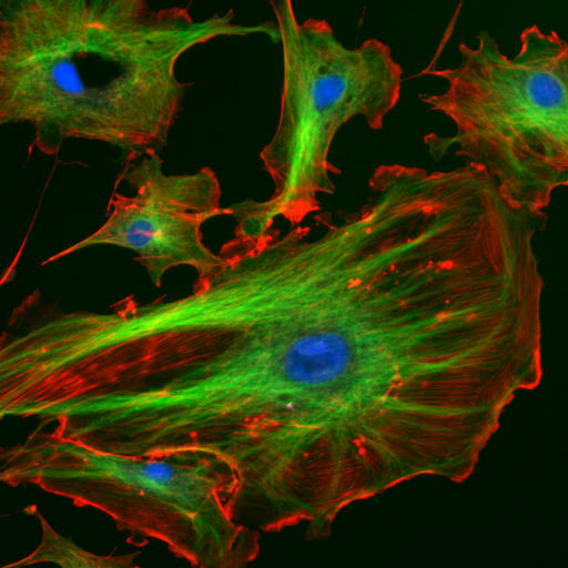
pedia/commons/0/09/FluorescentCells.jpg

[2] FIG. 7. In vitro polymerization
of cytoskeletal proteins of the
MinD/ParA superfamily. (A) Formation of
MinD filament bundles in the presence
of MinE, ATP, and phospholipid
vesicles. One end of the bundle is
markedly frayed because of the presence
of MinE. (Reprinted from reference 198
with permission of the publisher.
Copyright 2003 National Academy of
Sciences, U.S.A.) (B) Formation of a
ParApTP228(ParF) filament bundle in the
presence of ParBpTP228(ParG) and ATP.
ParBpTP228(ParG) stimulates formation
of the frayed end(s) of the
ParApTP228(ParF) bundle. (Reprinted
from reference 11 by permission from
Macmillan Publishers Ltd.) (C)
Formation of Soj filaments in the
presence of DNA and ATP. (Reprinted
from reference 116 by permission from
Macmillan Publishers Ltd.) UNKNOWN
source: http://www.ncbi.nlm.nih.gov/pmc/
articles/PMC1594594/bin/zmr0030621350007
.jpg
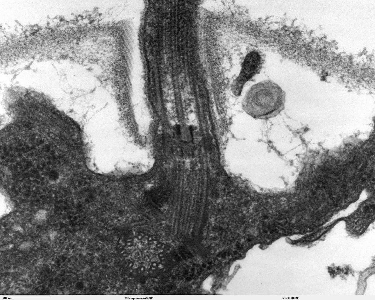
[1] Cilia and flagella are projections
from the cell. They are made up of
microtubules , as shown in this cartoon
and are covered by an extension of the
plasma membrane. They are motile and
designed either to move the cell itself
or to move substances over or around
the cell. The primary purpose of cilia
in mammalian cells is to move fluid,
mucous, or cells over their surface.
Cilia and flagella have the same
internal structure. The major
difference is in their length. This
figure shows a cross section of a
cilium next to a longitudinal section.
Below, we will see how the microtubules
are organized in the core (shown in the
cartoon in this figure). Also shown is
the centriole or basal body that
organizes the formation and direction
of the cilia. COPYRIGHTED
source: Description Transmission
electron microscope image, showing an
example of green algae
(Chlorophyta). Chlamydomanas
reinhardtii is a unicellular flagellate
used as a model system in molecular
genetics work and flagellar motility
studies. This image is a
longitudinal section through the
flagella area. In the cell apex is the
basal body that is the anchoring site
for a flagella. Basal bodies originate
from and have a substructure similar to
that of centrioles, with nine
peripheral microtubule triplets(see
structure at bottom center of image).
The two inner microtubules of each
triplet in a basal body become the two
outer doublets in the flagella. This
image also shows the transition region,
with its fibers of the stellate
structure. The top of the image shows
the flagella passing through the cell
wall. Date 20 September
2007 Source Source and public domain
notice at
http://remf.dartmouth.edu/imagesindex.ht
ml Author Dartmouth Electron
Microscope Facility, Dartmouth
College PD

[2] This figure shows an electron
micrograph of a cross section of a
cilium. Note that you can see the
dynein arms and the nexin links. The
dynein arms have ATPase activity. In
the presence of ATP, they can move from
one tubulin to another. They enable the
tubules to slide along one another so
the cilium can bend. The dynein
bridges are regulated so that sliding
leads to synchronized bending. Because
of the nexin and radial spokes, the
doublets are held in place so sliding
is limited lengthwise. If nexin and the
radial spokes are subjected to enzyme
digestion, and exposed to ATP, the
doublets will continue to slide and
telescope up to 9X their length.
COPYRIGHTED
source: http://upload.wikimedia.org/wiki
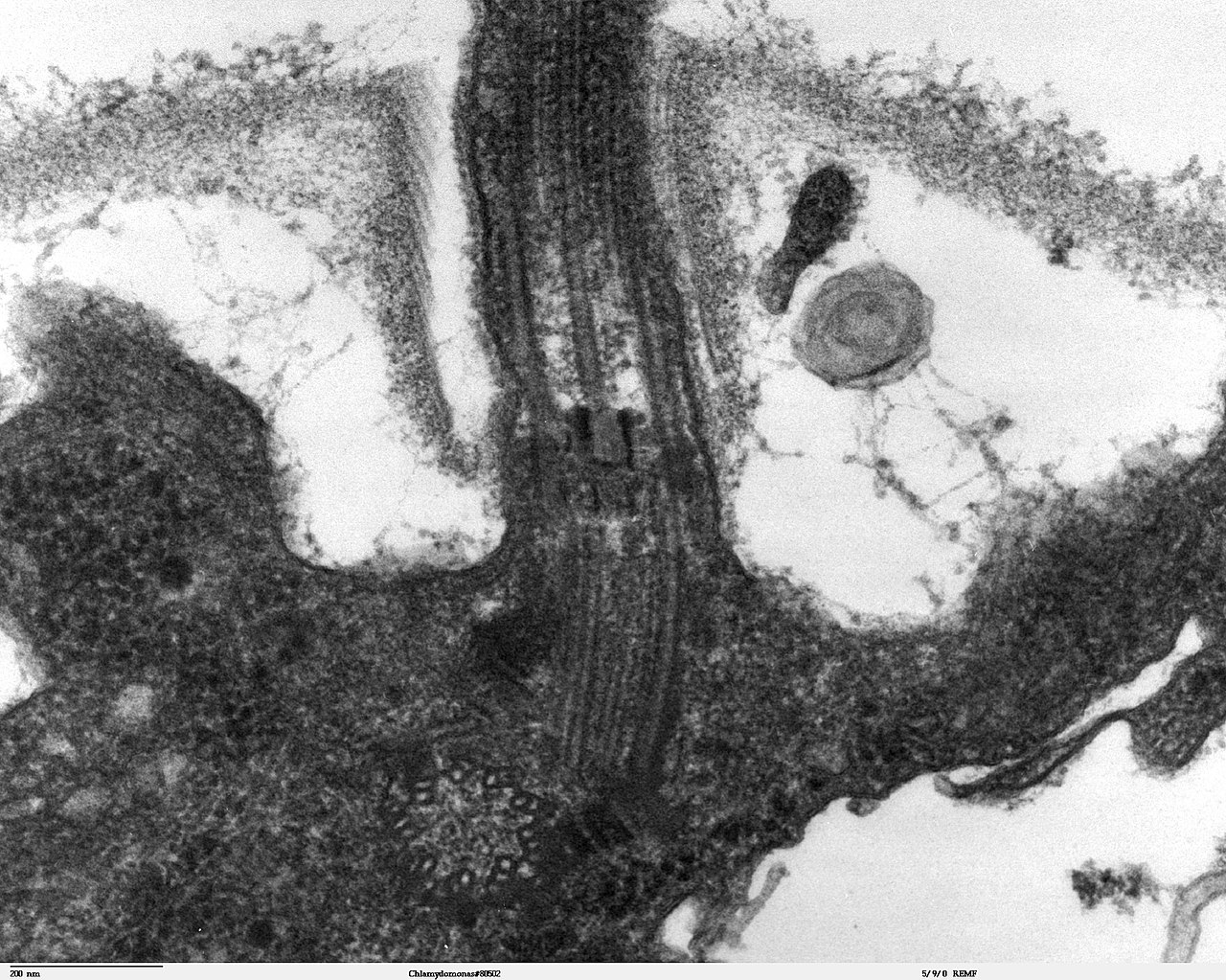
pedia/commons/thumb/9/99/Chlamydomonas_T
EM_09.jpg/1280px-Chlamydomonas_TEM_09.jp
g
nucleus changes into linear
chromosomes.

[1] A DNA molecule is very long (a few
meters) but extremely thin (narrow;
measured in nanometers). Here is an
electron microscope photo of a DNA
strand: PD
source: http://rst.gsfc.nasa.gov/Sect20/
dna1.jpg

[2] [t Is this an accurate image? - Is
a chromosome made of a single wound
strand of DNA? update- no see image
8] Every cell in the human body
(except red blood cells) contains 23
pairs of chromosomes. (a) Each
chromosome is made up of a tightly
coiled strand of DNA. (b) DNA’s
uncoiled state reveals its familiar
double helix shape. If DNA is pictured
as a twisted ladder, its sides, made of
sugar and phosphate molecules, are
connected by (c) rungs made of
chemicals called bases. DNA has four
bases—adenine, thymine, guanine, and
cytosine—that form interlocking
pairs. The order of the bases along the
length of the ladder is the DNA
sequence. PD
source: https://www.llnl.gov/str/June03/
gifs/Stubbs1.gif
(packages proteins and lipids into
vesicles for delivery to targeted
destinations).
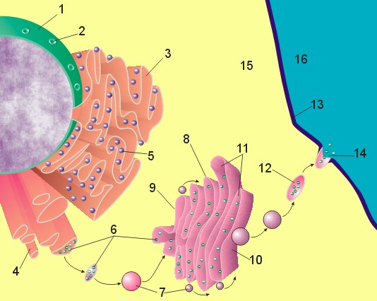
[1] Figure 1: Image of nucleus,
endoplasmic reticulum and Golgi
apparatus: (1) Nucleus, (2) Nuclear
pore, (3) Rough endoplasmic reticulum
(RER), (4) Smooth endoplasmic reticulum
(SER), (5) Ribosome on the rough ER,
(6) Proteins that are transported, (7)
Transport vesicle, (8) Golgi apparatus,
(9) Cis face of the Golgi apparatus,
(10) Trans face of the Golgi apparatus,
(11) Cisternae of the Golgi apparatus,
(12) Secretory vesicle, (13) Plasma
membrane, (14) Exocytosis, (15)
Cytoplasm, (16) Extracellular space.
source: http://en.wikipedia.org/wiki/Ima
ge:Nucleus_ER_golgi_ex.jpg
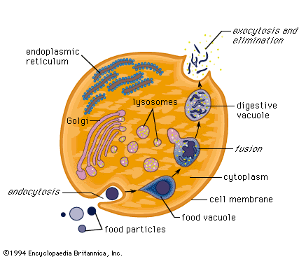
[2] no description UNKNOWN
source: http://sun.menloschool.org/~cwea
ver/cells/e/lysosomes/
nucleus that makes ribosomal RNA).
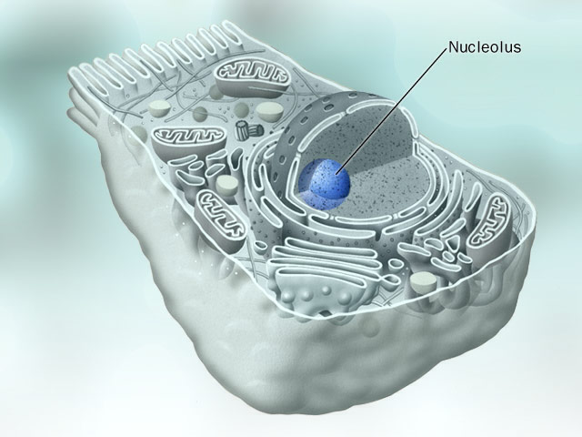
[1] Nucleolus, COPYRIGHTED
source: http://www.eccentrix.com/members
/chempics/Slike/cell/Nucleolus.jpg
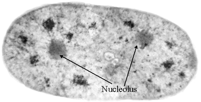
[2] With the combination of x-rays
from the Advanced Light Source and a
new protein-labeling technique,
scientists can see the distribution of
the nucleoli within the nucleus of a
mammary epithelial cell. USG PD
source: http://www.lbl.gov/Science-Artic
les/Archive/xray-inside-cells.html

[1] Mitosis divides genetic information
during cell division Source:
http://www.ncbi.nlm.nih.gov/About/primer
/genetics_cell.html This image is
from the Science Primer, a work of the
National Center for Biotechnology
Information, part of the National
Institutes of Health. As a work of the
U.S. federal government, the image is
in the public domain.
source: http://en.wikipedia.org/wiki/Mit
osis
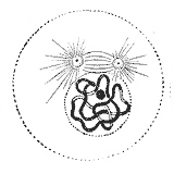
[2] Prophase: The two round objects
above the nucleus are the centrosomes.
Note the condensed chromatin. from
Gray's Anatomy. Unless stated
otherwise, it is from the online
edition of the 20th U.S. edition of
Gray's Anatomy of the Human Body,
originally published in 1918. Online
editions can be found on Bartleby and
also on Yahoo!
source: UNKNOWN
diploid cell (2 sets of chromosomes).
First zygote. Increase in genetic
variety.
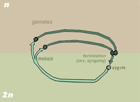
[1] Theoretical first eukaryote
sex adapted from image of gametic
meiosis GNU
source: http://en.wikipedia.org/wiki/Ima
ge:Zygotic_meiosis.jpg
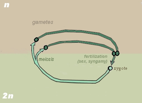
[2] Theoretical first eukaryote
sex adapted from image of gametic
meiosis GNU
source: http://en.wikipedia.org/wiki/Ima
ge:Zygotic_meiosis.jpg
haploid cells).
[1] Theoretical first eukaryote
sex adapted from image of gametic
meiosis GNU
source: http://en.wikipedia.org/wiki/Ima
ge:Zygotic_meiosis.jpg
[2] Theoretical first eukaryote
sex adapted from image of gametic
meiosis GNU
source: http://en.wikipedia.org/wiki/Ima
ge:Zygotic_meiosis.jpg
{aNISoGomE}, sex (cell and nucleus
fusion) between two cells that are
different in size or shape.

[1] Combination of images: Description
English: Different types of
isogamy: A) Isogamy of motile
cells B) Isogamy of non-motile
cells C) Conjugation of
gametangia Date 30 July
2008 Source Vectorised SVG version of
http://en.wikipedia.org/wiki/Image:Isoga
my.png Author Original bitmap version
by Tameeria, SVG version by Qef Other
versions
http://en.wikipedia.org/wiki/Image:
Isogamy.png PD AND Description
Different types of en:anisogamy:
A) Anisogamy of motile gametes B)
Oogamy (non-motile egg cell, motile
sperm cell) C) Anisogamy of
non-motile gametes Date 2008-06-30
02:07 (UTC) Source
Anisogamy.png Author This
SVG version by Qef (talk)
Anisogamy.png: Original uploader was
Tameeria at en.wikipedia Later
versions were uploaded by Helix84 at
en.wikipedia. PD
source: http://upload.wikimedia.org/wiki
pedia/commons/d/d5/Isogamy.svghttp://upl
oad.wikimedia.org/wikipedia/commons/a/a7
/Anisogamy.svg
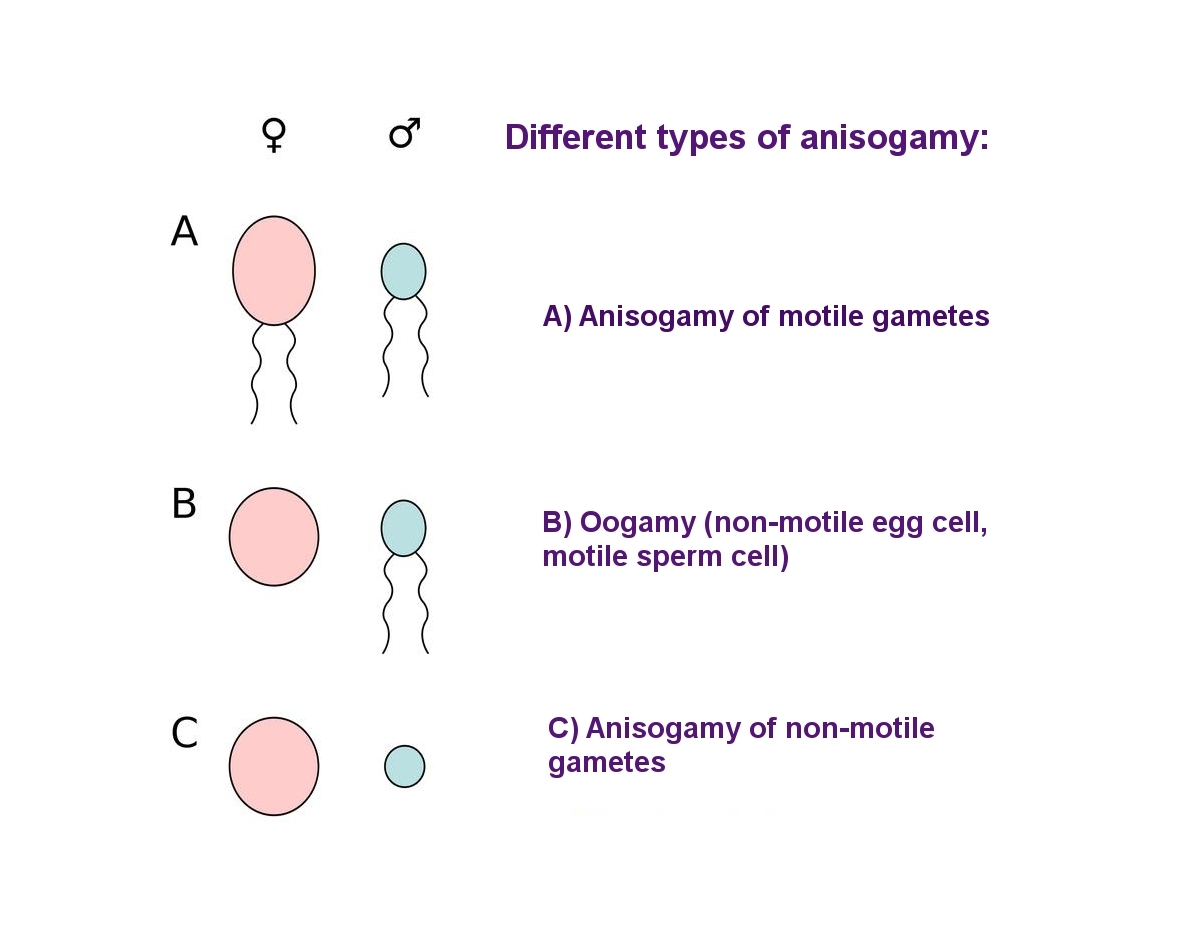
[2] Description Different types of
en:anisogamy: A) Anisogamy of motile
gametes B) Oogamy (non-motile egg
cell, motile sperm cell) C) Anisogamy
of non-motile
gametes Date 2008-06-30 02:07
(UTC) Source Anisogamy.png Author
This SVG version by Qef
(talk) Anisogamy.png: Original
uploader was Tameeria at
en.wikipedia Later versions were
uploaded by Helix84 at
en.wikipedia. PD
source: http://upload.wikimedia.org/wiki
pedia/commons/thumb/a/a7/Anisogamy.svg/1
000px-Anisogamy.svg.png
copies and then the cell divides twice
into four haploid cells).
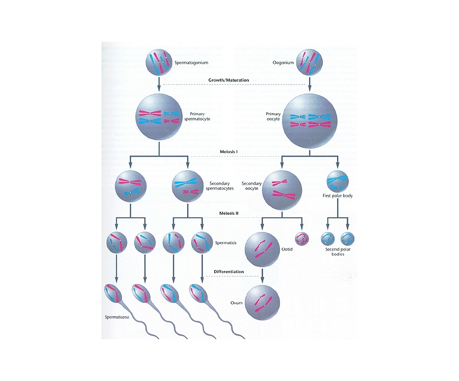
[1] GametoGenesis. COPYRIGHTED EDU
source: http://www.bio.miami.edu/dana/10
4/gametogenesis.jpg

[2] Sexual cycle oxymonas, identical
to saccinobaculus, one step meiosis.
haploid. COPYRIGHTED CANADA
source: http://www.zoology.ubc.ca/~redfi
eld/clevelan/oxymonas.GIF
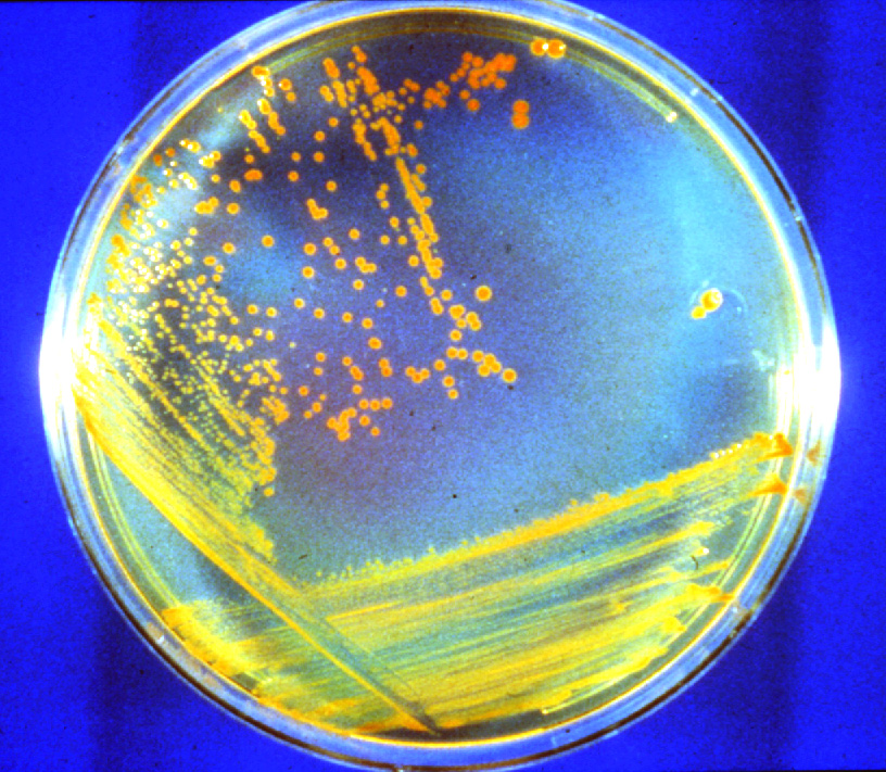
[1] D. radiodurans growing on a
nutrient agar plate. The red color is
due to carotenoid pigment. Links to
816x711-pixel, 351KB JPG. Credit: M.
Daly, Uniformed Services University of
the Health Sciences NASA
source: http://science.nasa.gov/newhome/
headlines/images/conan/D_rad_dish.jpg
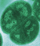
[2] Photomicrograph of Deinococcus
radiodurans, from
www.ornl.gov/ORNLReview/ v34 The Oak
Ridge National Laboratory United
States Federal Government This work
is in the public domain because it is a
work of the United States Federal
Government. This applies worldwide. See
Copyright.
source: http://en.wikipedia.org/wiki/Ima
ge:Deinococcus.jpg
{SIe-NO-BaK-TERE-u} (ancestor of all
plastids).
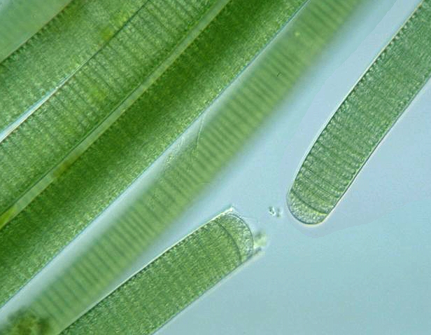
[1] Oscillatoria COPYRIGHTED EDU
source: http://www.stcsc.edu/ecology/alg
ae/oscillatoria.jpg

[2] Lyngbya COPYRIGHTED EDU
source: http://www.stanford.edu/~bohanna
n/Media/LYNGB5.jpg

[1] Chloroflexus photomicrograph from
Doe Joint Genome Institute of US Dept
Energy PD
source: http://en.wikipedia.org/wiki/Ima
ge:Chlorofl.jpg

[1] Acidic waste water from a modern
mining site supports the same oxygen
using bacterial life that appeared on
Earth 2.48 billion years ago. UNKNOWN
source: http://media.news.ualberta.ca/~/
media/University%20of%20Alberta/Administ
ration/External%20Relations/ExpressNews/
Images/2011/10/111020-RocksBanner-cw.jpg

[2] Bacillus specie soil
bacteria. UNKNOWN
source: http://www.scharfphoto.com/fine_
art_prints/archives/199812-054-Soil-Bact
eria.jpg
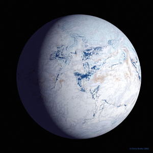
[1] snowball Earth UNKNOWN
source: http://www.cosmosmagazine.com/fi
les/imagecache/feature/files/features/pr
int/20090528_snowball_earth.jpg

[2] Snowball Earth Snowball Earth
describes a theory that for millions of
years the Earth was entirely smothered
in ice, stretching from the poles to
the tropics. This freezing happened
over 650 million years ago in the
Pre-Cambrian, though it's now thought
that there may have been more than one
of these global glaciations. They
varied in duration and extent but
during a full-on snowball event, life
could only cling on in ice-free
refuges, or where sunlight managed to
penetrate through the ice to allow
photosynthesis. UNKNOWN
source: http://www.bbc.co.uk/nature/imag
es/ic/credit/640x395/s/sn/snowball_earth
/snowball_earth_1.jpg
oxygen in the air of Earth.
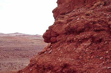
[1]
http://www.kgs.ukans.edu/Extension/redhi
lls/redhills.html
source: http://www.kgs.ukans.edu/Extensi
on/redhills/redhills.html
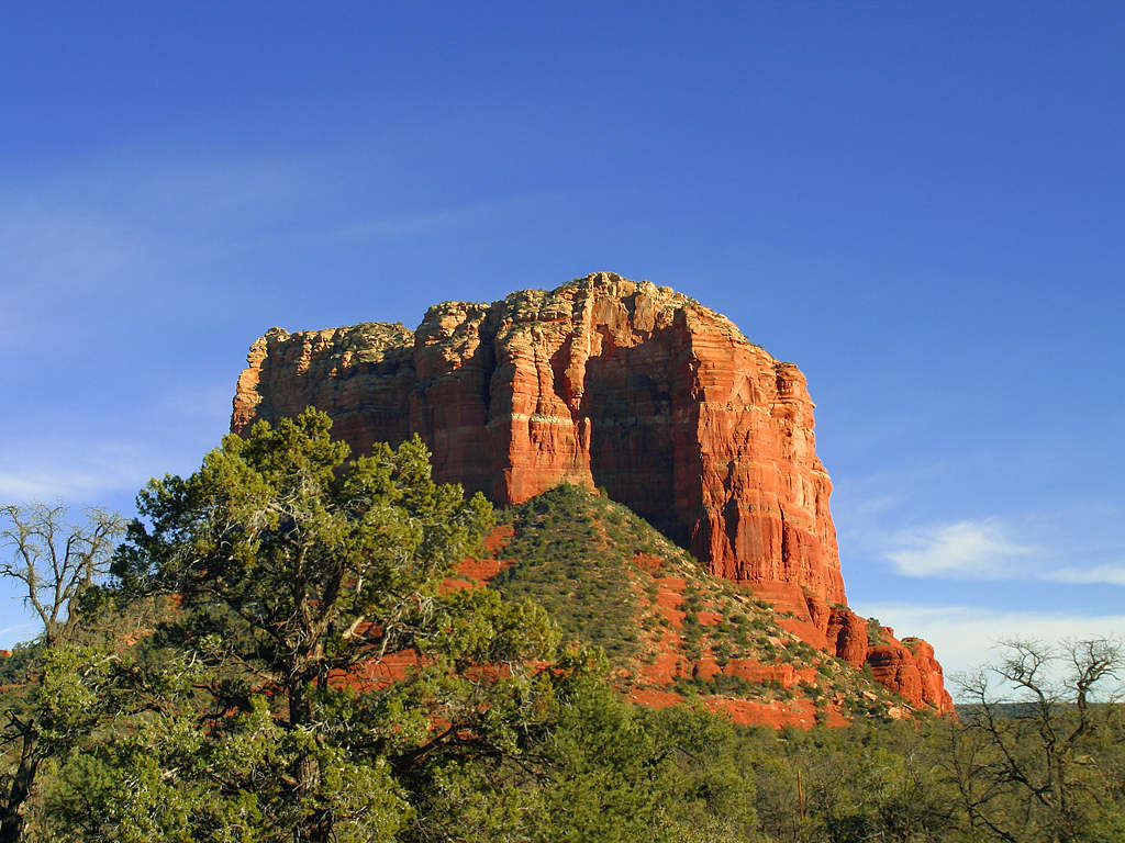
[2] In Archean rocks, metals tend to
occur in low oxidation states (for
example, Fe2+ instead of Fe3+)
indicating a high metal:oxygen ratio in
the oceans and atmosphere. The
sediments are essentially rust-free.
After the late Proterozoic,
sedimentary deposits often have reddish
colors and are called red beds due to
the presence of iron-oxide coatings
between sand grains. From the later
Proterozoic onward, enough free oxygen
has been available to oxidize iron in
sediments. A sandstone butte outside
of Sedona, Arizona. Public domain
image by Jon Sullivan. PD
source: http://upload.wikimedia.org/wiki
pedia/commons/3/38/Butte_pdphoto_roadtri
p_24_bg_021604.jpg
is captured by a eukaryote and through
endosymbiosis, becomes the
mitochondria, organelles of most
eukaryotes.

[1] Figure from: Michael W. Gray, et
al, ''Genome structure and gene content
in protist mitochondrial DNAs'',
Nucl. Acids Res. (1998) 26(4):
865-878 doi:10.1093/nar/26.4.865
http://nar.oxfordjournals.org/content/
26/4/865.full Phylogenetic hypothesis
of the eukaryotic lineage based on
ultrastructural and molecular data.
Organisms are divided into three main
groups distinguished by mitochondrial
cristal shape (either discoidal,
flattened or tubular). Unbroken lines
indicate phylogenetic relationships
that are firmly supported by available
data; broken lines indicate
uncertainties in phylogenetic
placement, resolution of which will
require additional data. Color coding
of organismal genus names indicates
mitochondrial genomes that have been
completely (Table 1), almost completely
(Jakoba, Naegleria and
Thraustochytrium) or partially (*)
sequenced by the OGMP (red), the FMGP
(black) or other groups (green). Names
in blue indicate those species whose
mtDNAs are currently being sequenced by
the OGMP or are future candidates for
complete sequencing. Amitochondriate
retortamonads are positioned at the
base of the tree, with broken arrows
denoting the endosymbiotic origin(s) of
mitochondria from a Rickettsia-like
eubacterium. Macrophar.,
Macropharyngomonas.
source: http://nar.oxfordjournals.org/co
ntent/vol26/issue4/images/gkb18201.gif

[2] Figure 1 Phylogenetic tree of
eukaryotes based on ultrastructural and
molecular data. Organisms are
sub-divided into main groups as
discussed in the text. Only a few
representative species for which
complete (or almost complete) mtDNA
sequences are known are shown in each
lineage. In some cases, line drawings
or actual pictures of the organisms are
provided (Acanthamoeba, M. Nagata; URL:
http://protist.i.hosei.ac.jp/PDB/PCD3379
/htmls/21.html; Allomyces, Tom Volk;
URL:
http://botit.botany.wisc.edu/images/332/
Chytridiomycota/Allomyces_r_So_pa/A._arb
uscula_pit._sporangia_tjv.html;
Amoebidium, URL:
http://cgdc3.igmors.upsud.fr/microbiolog
ie/mesomycetozoaires.htm; Marchantia,
URL:
http://www.science.siu.edu/landplants/He
patophyta/images/March.female.JPEG
Scenedesmus, Entwisle et al.,
http://www.rbgsyd.gov.au/_data/page/1824
/Scenedesmus.gif). The color-coding of
the main groups (alternating between
dark and light blue) on the outer
circle corresponds to the color-coding
of the species names. Unbroken lines
indicate phylogenetic relationships
that are firmly supported by available
molecular data; broken lines indicate
uncertainties in phylogenetic
placement, resolution of which will
require additional sequence data. [t:
why not color code or add which type of
mito?]
source: http://arjournals.annualreviews.
org/doi/full/10.1146/annurev.genet.37.11
0801.142526
(Grypania).
USA
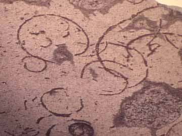
[1]
file:/root/web/Grypania_spiralis_wmel000
0.htm
source: file:/root/web/Grypania_spiralis
_wmel0000.htm
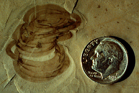
[2]
http://www.peripatus.gen.nz/paleontology
/lrgGrypaniaspiralis.jpg
source: http://www.peripatus.gen.nz/pale
ontology/lrgGrypaniaspiralis.jpg
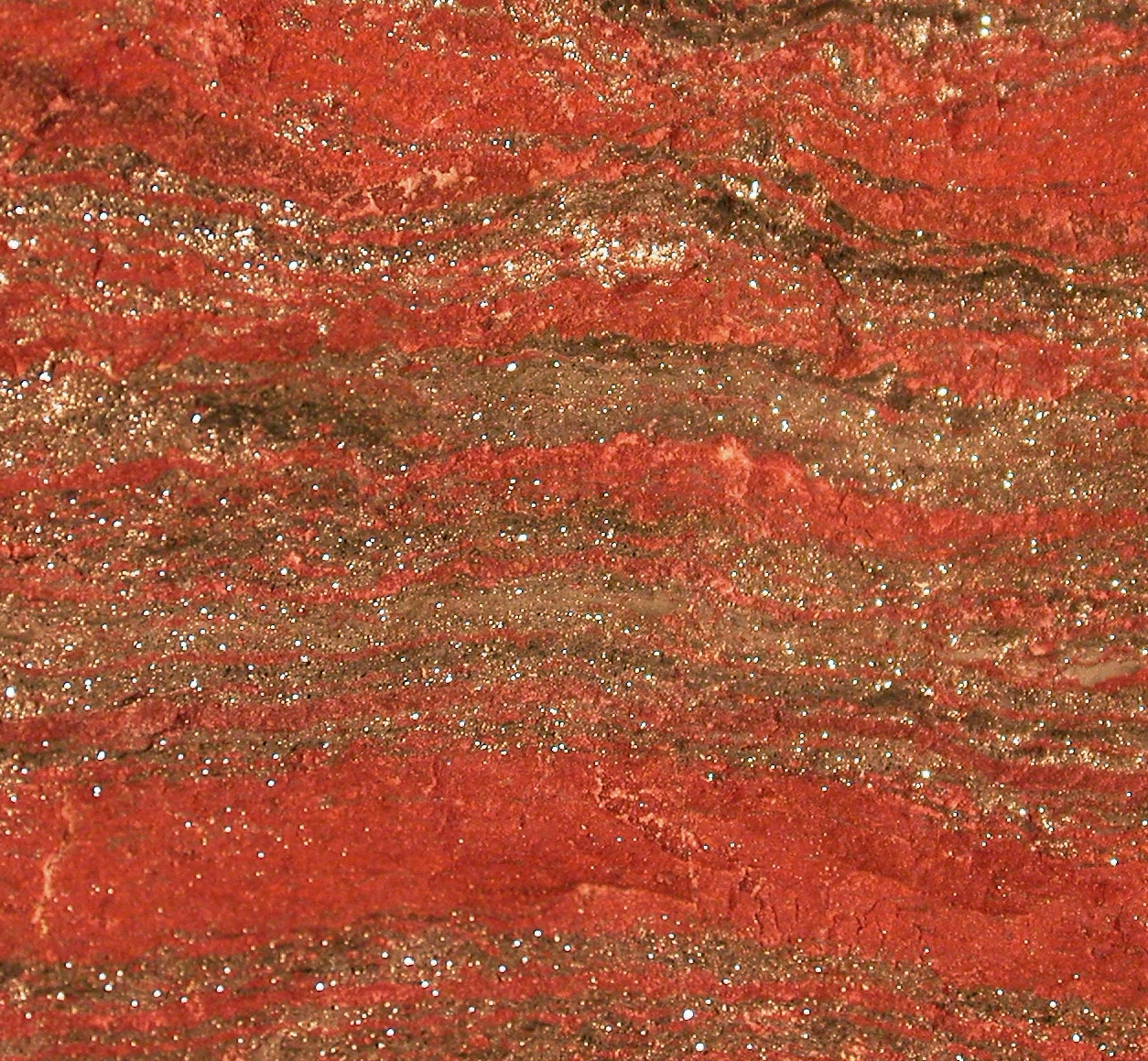
[1] Ted Huntington PD
source: http://upload.wikimedia.org/wiki
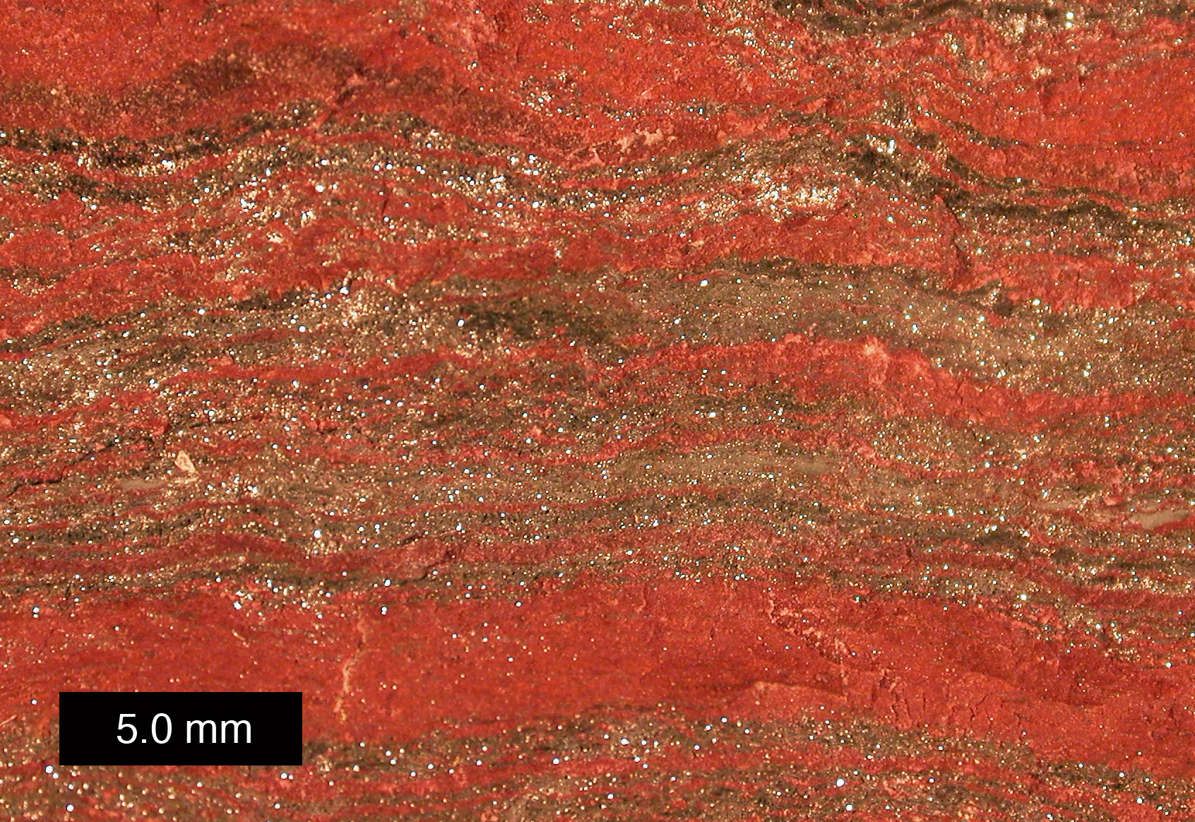
pedia/commons/6/62/MichiganBIF.jpg
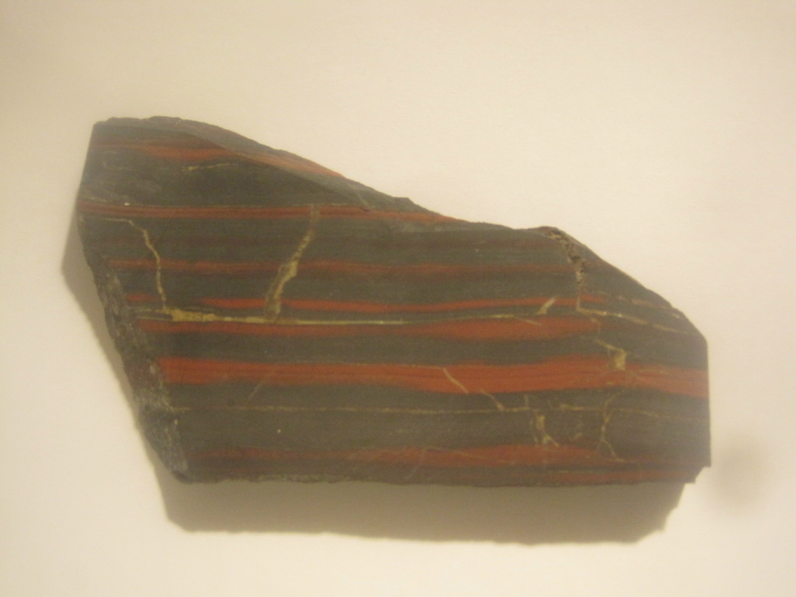
[2] Ted Huntington PD
source: Ted Huntington
genes regulate the building of major
body parts.
[1] {ULSF: Homeobox genes} Desajustes
en el modelo UNKNOWN
source: http://cnho.files.wordpress.com/
2010/07/hox_genes_illus.png
[2] {ULSF: Homeobox genes} UNKNOWN
source: http://cnho.files.wordpress.com/
2010/07/homeobox1.jpg
eukaryotes divides into bikont and
unikont descendants. Bikonts lead to
all Chromalveolates, Excavates,
Rhizaria, and Plants. Unikonts lead to
all Amoebozoa, Animals and Fungi.

[1] Figure 1: Figure 1. Eukaryote
phylogeny integrating ultrastructure,
sequence trees, gene fusions and
molecular cladistic markers. The
unikont topology is established, but
the branching order of the six bikont
groups remains uncertain. The single
enslavement [12] of a red alga (R) to
create chromalveolates is supported by
a plastid glyceraldehyde phosphate
dehydrogenase (GAPDH) replacement [13].
Whether there was a single enslavement
of a green alga (G) to form cabozoa or
two separate enslavements (asterisks)
to form Cercozoa and Excavata is
uncertain [12], as is the position of
Heliozoa [14]. Polyubiquitin [15] and
EF-1α[16] insertions strongly support
the clades core Rhizaria and
opisthokonts. The inset shows the BamHI
restriction fragment from H.
cantabrigiensis that was sequenced and
analysed in this study, spanning the
DHFR and the amino terminus of the TS
gene (red, introns are green). The
length of the noncoding regions
upstream and downstream of the DHFR
gene from one of the clones is
indicated. Figure 1 from: Stechmann
A, Cavalier-Smith T, ''The root of the
eukaryote tree pinpointed.'', 2003,
Curr. Biol. 13, R665–R666.
doi:10.1016/S0960-9822(03)00602-X. http
://www.sciencedirect.com/science/article
/pii/S096098220300602X COPYRIGHTED
source: http://www.sciencedirect.com/sci
ence?_ob=MiamiCaptionURL&_method=retriev
e&_eid=1-s2.0-S096098220300602X&_image=1
-s2.0-S096098220300602X-gr1_lrg.jpg&_ba=
&_fmt=full&_orig=na&_issn=09609822&_pii=
S096098220300602X&_isHiQual=Y&_acct=C000
059600&_version=1&_urlVersion=0&_userid=
4422&md5=cec46b2161caca87740f4ff34545ab6
9

[2] cavalier-smith diagram COPYRIGHTED
source: cavalier_jmolevol_2003_56_540-56
3.pdf
slime molds). Feeding using pseudopods.
[1] SUBPHYLUM Lobosa CLASS Amoebaea
Chaos diffluens, an amoeba. Photo
released by Dr. Ralf Wagner.
source: http://en.wikipedia.org/wiki/Ima
ge:Chaos_diffluens.jpg
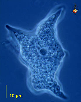
[2] CLASS Amoebaea Mayorella (may-or
-ell-a) a medium sized free-living
naked amoeba with conical pseudopodia.
Central body is the nucleus. Phase
contrast. This picture was taken by
David Patterson of material from
Limulus-ridden sediments at Plum Island
(Massachusetts USA) in spring and
summer, 2001. NONCOMMERCIAL USE
source: http://microscope.mbl.edu/script
s/microscope.php?func=imgDetail&imageID=
515

[1] [t Note that this Chrysophytes
{golden algae} do not evolve
genetically until much later - but I
can't find colonial euglinas or
kinetoplasts- dinobryon look very
similar to euglenas however, even with
a red eyespot- which implies a close
relation.] [1] Dinobryon, a colony of
Chrysophytes showing flagella and red
eyespots UNKNOWN
source: http://www.microscopy-uk.org.uk/
mag//imagsmall/Dinobryonb.jpg

[2] [t Note that this CHrysophytes
{golden algae} do not evolve
genetically until much later - but I
can't find colonial euglinas or
kinetoplasts] [2] golden algae colony
(synura) Scanning EM showing the
colony of cells covered with scales By
Joel Mancuso UNKNOWN
source: http://farm1.staticflickr.com/38
/110623789_7d189c795b_b.jpg
form plastids through symbiosis, within
a eukaryote cell. Like mitochondria,
these organelles copy themselves and
are not made by the cell DNA.

[1] Description Plagiomnium
affine, Laminazellen, Rostock Date
created 01.11.2006 Source
photographed by myself Author
Kristian Peters --
Fabelfroh Permission (Reusing this
file) GFDL
source: http://upload.wikimedia.org/wiki
pedia/commons/4/49/Plagiomnium_affine_la
minazellen.jpeg
and red algae and land plants).
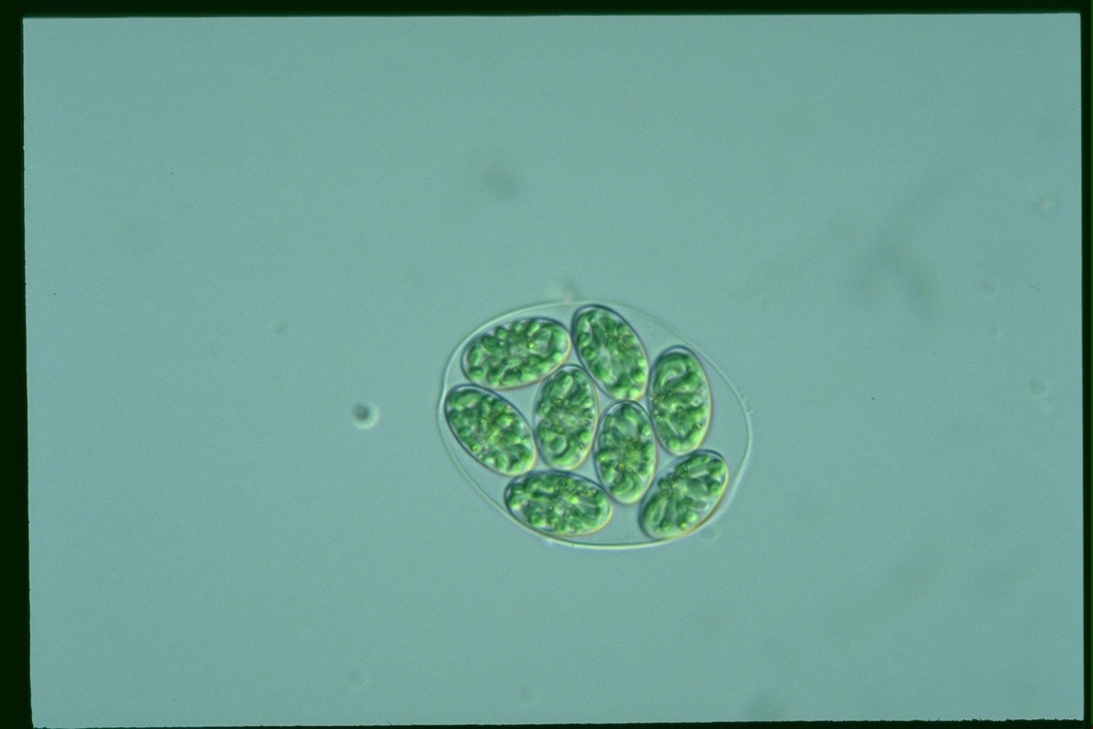
[1] ? COPYRIGHTED
source: http://protist.i.hosei.ac.jp/PDB
3/PCD3711/htmls/86.html

[2] (See Image) COPYRIGHTED
source: Richard Dawkins, "The
Ancestor's Tale", (Boston, MA: Houghton
Mifflin Company, 2004). (c1500)
Fungi, Choanoflagellates and Animals).

[1] Parasite spore, SEM Z115/0073
Rights Managed Credit: EYE OF
SCIENCE/SCIENCE PHOTO
LIBRARY Caption: Parasite spore.
Coloured scanning electron micrograph
(SEM) of a microsporidian (Tubulinosema
ratisbonensis) spore cultured on human
lung fibroblast cells (brown).
Microsporidia are single-celled
parasites. T. ratisbonenesis is a
parasite of the fruit fly (Drosophila
melanogaster), but may also be able to
infect humans with weakened immune
systems. The spore is the infective
phase of the life cycle. It is excreted
by the old host and enters the gut of a
new host. The contents of the spore,
the sporoplasm, is injected into the
host's cell via the polar tubule. Once
in the cell the organism divides many
times with the resultant organisms
producing more spores. Magnification:
x10,000 at 10 centimetres
wide. Release details: Model and
property releases are not available
UNKNOWN
source: http://www.sciencephoto.com/imag
e/365473/large/Z1150073-Parasite_spore,_
SEM-SPL.jpg
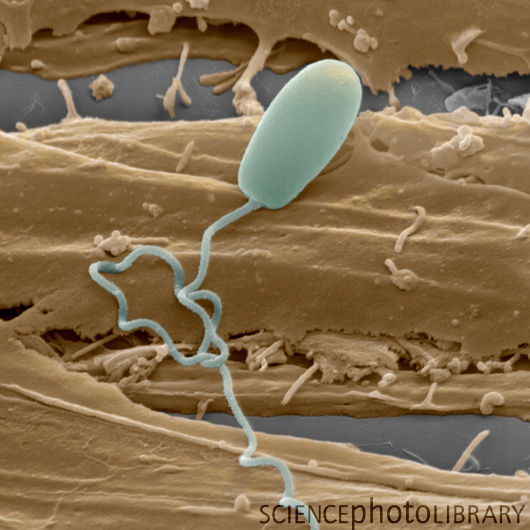
[2] Parasite spore, SEM Z115/0073
Rights Managed Credit: EYE OF
SCIENCE/SCIENCE PHOTO
LIBRARY Caption: Parasite spore.
Coloured scanning electron micrograph
(SEM) of a microsporidian (Tubulinosema
ratisbonensis) spore cultured on human
lung fibroblast cells (brown).
Microsporidia are single-celled
parasites. T. ratisbonenesis is a
parasite of the fruit fly (Drosophila
melanogaster), but may also be able to
infect humans with weakened immune
systems. The spore is the infective
phase of the life cycle. It is excreted
by the old host and enters the gut of a
new host. The contents of the spore,
the sporoplasm, is injected into the
host's cell via the polar tubule. Once
in the cell the organism divides many
times with the resultant organisms
producing more spores. Magnification:
x10,000 at 10 centimetres
wide. Release details: Model and
property releases are not available
UNKNOWN
source: http://www.sciencephoto.com/imag
e/365473/large/Z1150073-Parasite_spore,_
SEM-SPL.jpg
[1] ? COPYRIGHTED
source: http://protist.i.hosei.ac.jp/PDB
3/PCD3711/htmls/86.html

[2] ? COPYRIGHTED
source: http://protist.i.hosei.ac.jp/PDB
/Images/Others/Glaucocystis/
lettuce, Spirogyra).
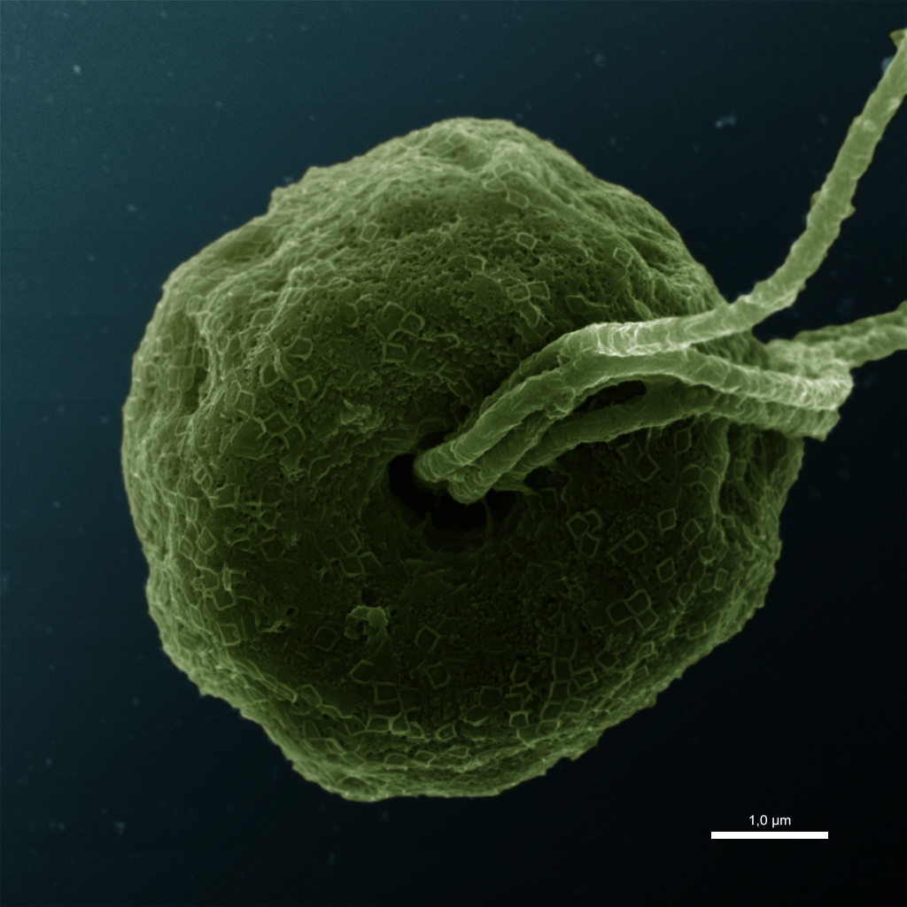
[1] Description Flagellar pit of
Pyramimonas sp. / from Nigaku-Ike of
University of Tsukuba, Tsukuba, Ibaraki
Pref., Japan / SEM:JEOL JSM-6330F /
scale bar = 1.0μm Date 2009-05-04
18:30 (UTC) Source
Pyramimonas_sp.jpg Author
Pyramimonas_sp.jpg: ja:User:NEON /
User:NEON_ja derivative work:
Addicted04 (talk) CC
source: http://upload.wikimedia.org/wiki
pedia/commons/c/cb/Pyramimonas_sp_color.
jpg
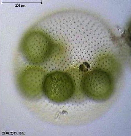
[2] Micrograph of Volvox aureus.
Copyright held by Dr. Ralf Wagner,
uploaded to German Wikipedia under
GFDL. Permission is granted to copy,
distribute and/or modify this document
under the terms of the GNU Free
Documentation License, Version 1.2 or
any later version published by the Free
Software Foundation; with no Invariant
Sections, no Front-Cover Texts, and no
Back-Cover Texts. Subject to
disclaimers.
source: http://en.wikipedia.org/wiki/Vol
vox
(Rhodophyta {rODOFITu}).

[1] Close-up of a red alga (Genus?
Laurencia), Class Florideophyceae,
Order=? a marine seaweed from Hawaii.
GNU
source: http://en.wikipedia.org/wiki/Ima
ge:Laurencia.jpg
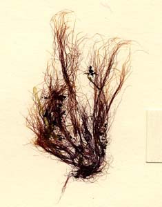
[2] Bangia atropurpurea Profile:
unbranched filaments in tufts. Often
forming dense fringes in the spalsh
zone. Uniseriate at base, multiseriate
above with protoplasts separate in a
firm gelatinous sheath. Stellate
chloroplasts. US NOAA PD
source: http://www.glerl.noaa.gov/seagra
nt/GLWL/Algae/Rhodophyta/Cards/Bangia.ht
ml
{JE-oR-DE-u}).
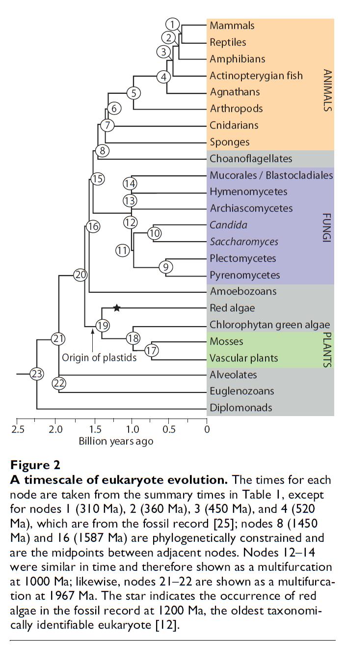
[1] A timescale of eukaryote evolution.
The times for each node are taken from
the summary times in Table 1, except
for nodes 1 (310 Ma), 2 (360 Ma), 3
(450 Ma), and 4 (520 Ma), which are
from the fossil record [25]; nodes 8
(1450 Ma) and 16 (1587 Ma) are
phylogenetically constrained and are
the midpoints between adjacent nodes.
Nodes 12–14 were similar in time and
therefore shown as a multifurcation at
1000 Ma; likewise, nodes 21–22 are
shown as a multifurcation at 1967 Ma.
The star indicates the occurrence of
red algae in the fossil record at 1200
Ma, the oldest taxonomically
identifiable eukaryote [12]. Hedges
et al. BMC Evolutionary Biology 2004
4:2
doi:10.1186/1471-2148-4-2 COPYRIGHTED
source: http://www.biomedcentral.com/con
tent/figures/1471-2148-4-2-2.jpg
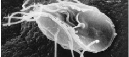
[2] Giardia lamblia, a parasitic
flagellate that causes giardiasis.
Image from public domain source at
http://www.nigms.nih.gov/news/releases/i
mages/para.jpg
source: http://www.nigms.nih.gov/news/re
leases/images/para.jpg
Eukaryotes evolves.
Formation) Somerset Island, arctic
Canada
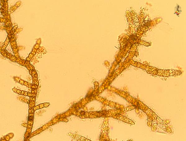
[1] Bodanella (bow-dan-ell-a)
lauterbornii, a branching filamentous
brown alga. Nearly all brown algae are
marine organisms, but this species is
found in the bottoms of freshwater
lakes. Bright field. data on this
strain. This image is of material
from Provasoli-Guillard National Center
for Culture of Marine Phytoplankton,
images taken by David Patterson and Bob
Andersen. Image copyright: Bob Andersen
and D. J. Patterson, image used under
license to MBL
(micro*scope). NONCOMMERCIAL USE ONLY
source: http://starcentral.mbl.edu/msr/r
awdata/files/bodonella_bgz.zip
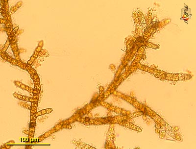
[2] Bodanella (bow-dan-ell-a)
lauterbornii, a branching filamentous
brown alga. Nearly all brown algae are
marine organisms, but this species is
found in the bottoms of freshwater
lakes. Bright field. data on this
strain. This image is of material
from Provasoli-Guillard National Center
for Culture of Marine Phytoplankton,
images taken by David Patterson and Bob
Andersen. Image copyright: Bob Andersen
and D. J. Patterson, image used under
license to MBL
(micro*scope). NONCOMMERCIAL USE ONLY
source: http://starcentral.mbl.edu/msr/r
awdata/viewable/bodonella_bgw.jpg
eukaryote. Gamete (or spore) cells and
somatic cells. Unlike gamete cells,
somatic cells are asexual (non-fusing).
Start of death by aging.

[1] Volvoxcell differentiation. The
pathways leading to germ cells or
somatic cells are controlled by genes
that cause cells to follow one or the
other fate. Mutations can prevent the
formation of one of these lineages.
http://www.devbio.com/chap02/link0204.sh
tml Although all the volvocaceans,
like their unicellular relative
Chlamydomonas, reproduce predominantly
by asexual means, they are also capable
of sexual reproduction, which involves
the production and fusion of haploid
gametes. In many species of
Chlamydomonas, including the one
illustrated in Figure 2.10, sexual
reproduction is isogamous (“the same
gametes”), since the haploid gametes
that meet are similar in size,
structure, and motility. However, in
other species of Chlamydomonas—as
well as many species of colonial
volvocaceans—swimming gametes of very
different sizes are produced by the
different mating types. This pattern is
called heterogamy (“different
gametes”). But the larger
volvocaceans have evolved a specialized
form of heterogamy, called oogamy,
which involves the production of large,
relatively immotile eggs by one mating
type and small, motile sperm by the
other (see Sidelights and
Speculations) UNKNOWN
source: http://www.ncbi.nlm.nih.gov/book
s/NBK10031/bin/ch2f12.jpg

[2] Description English: Four
Different Species of Volvocales Algae.
(A) Gonium pectorale, (B) Eudorina
elegans, (C) Pleodorina californica,
and (D) Volvox carteri. These are
unicellular organisms that live in
colonies and have both large and small
gametes. Date Published: June 15,
2004 Source Whitfield J:
Everything You Always Wanted to Know
about Sexes. PLoS Biol 2/6/2004: e183.
http://dx.doi.org/10.1371/journal.pbio.0
020183 Author Photo courtesy of
Aurora M. Nedelcu, from the Volvocales
Information Project
(http://www.unbf.ca/vip/index.htm). Per
mission (Reusing this file) See
below. CC
source: http://upload.wikimedia.org/wiki
pedia/commons/c/c5/Volvocales.png
occurs in both haploid and diploid life
stages).

[1] Drawn by self for Biological life
cycle Based on Freeman & Worth's
Biology of Plants (p. 171). GNU
source: http://en.wikipedia.org/wiki/Ima
ge:Sporic_meiosis.png
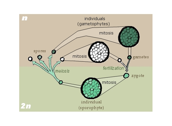
[2] Drawn by self for Biological life
cycle Based on Freeman & Worth's
Biology of Plants (p. 171). GNU
source: http://en.wikipedia.org/wiki/Ima
ge:Sporic_meiosis.png
plastid in the ancestor of all
chromalveolates.

[1] Fig. 2. The tree of life based
on molecular, ultrastructural and
palaeontological evidence. Contrary to
widespread assumptions, the root is
among the eubacteria, probably within
the double-enveloped Negibacteria, not
between eubacteria and archaebacteria
(Cavalier-Smith, 2002b); it may lie
between Eobacteria and other
Negibacteria (Cavalier-Smith, 2002b).
The position of the eukaryotic root has
been nearly as controversial, but is
less hard to establish: it probably
lies between unikonts and bikonts (Lang
et al., 2002; Stechmann and
Cavalier-Smith, 2002, 2003). For
clarity the basal eukaryotic kingdom
Protozoa is not labelled; it comprises
four major groups (alveolates, cabozoa,
Amoebozoa and Choanozoa) plus the small
bikont phylum Apusozoa of unclear
precise position; whether Heliozoa are
protozoa as shown or chromists is
uncertain (Cavalier-Smith, 2003b).
Symbiogenetic cell enslavement occurred
four or five times: in the origin of
mitochondria and chloroplasts from
different negibacteria, of
chromalveolates by the enslaving of a
red alga (Cavalier-Smith, 1999, 2003;
Harper and Keeling, 2003) and in the
origin of the green plastids of
euglenoid (excavate) and chlorarachnean
(cercozoan) algae—a green algal cell
was enslaved either by the ancestral
cabozoan (arrow) or (less likely) twice
independently within excavates and
Cercozoa (asterisks) (Cavalier-Smith,
2003a). The upper thumbnail sketch
shows membrane topology in the
chimaeric cryptophytes (class
Cryptophyceae of the phylum Cryptista);
in the ancestral chromist the former
food vacuole membrane fused with the
rough endoplasmic reticulum placing the
enslaved cell within its lumen (red) to
yield the complex membrane topology
shown. The large host nucleus and the
tiny nucleomorph are shown in blue,
chloroplast green and mitochondrion
purple. In chlorarachneans (class
Chlorarachnea of phylum Cercozoa) the
former food vacuole membrane remained
topologically distinct from the ER to
become an epiplastid membrane and so
did not acquire ribosomes on its
surface, but their membrane topology is
otherwise similar to the cryptophytes.
The other sketches portray the four
major kinds of cell in the living world
and their membrane topology. The upper
ones show the contrasting ancestral
microtubular cytoskeleton (ciliary
roots, in red) of unikonts (a cone of
single microtubules attaching the
single centriole to the nucleus, blue)
and bikonts (two bands of microtubules
attached to the posterior centriole and
an anterior fan of microtubules
attached to the anterior centriole).
The lower ones show the single plasma
membrane of unibacteria (posibacteria
plus archaebacteria), which were
ancestral to eukaryotes and the double
envelope of negibacteria, which were
ancestral to mitochondria and
chloroplasts (which retained the outer
membrane, red). COPYRIGHTED
source: http://aob.oxfordjournals.org/co
ntent/95/1/147/F2.large.jpg

[2] Figure 3: Fig. 3. Schematic
representation of the evolutionary
relationships and divergence times for
the red, green, glaucophyte, and
chromist algae. These photosynthetic
groups are outgroup-rooted with the
Opisthokonta which putatively
ancestrally lacked a plastid. The
branches on which the cyanobacterial
(CB) primary and red algal chromist
secondary endosymbioses occurred are
shown Figure 3 from: Yoon, Hwan Su
et al. “A Molecular Timeline for the
Origin of Photosynthetic Eukaryotes.”
Molecular Biology and Evolution 21.5
(2004): 809 -818.
Print. http://mbe.oxfordjournals.org/co
ntent/21/5/809.abstract COPYRIGHTED
source: http://mbe.oxfordjournals.org/co
ntent/21/5/809/F3.large.jpg
{KrOM-aL-VEO-leTS} (ancestor of
Chromista {Cryptophytes, Haptophytes
and Stramenopiles {STro-meN-o-Pi-lEZ}}
and Alveolates {aL-VEO-leTS}).
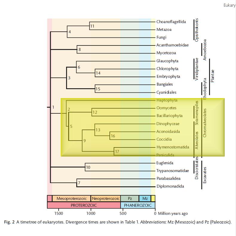
[1] S. Blair Hedges and Sudhir Kumar,
''The TimeTree of Life'', 2009,
p117-118. http://www.timetree.org/book.
php COPYRIGHTED
source: http://www.timetree.org/book.php
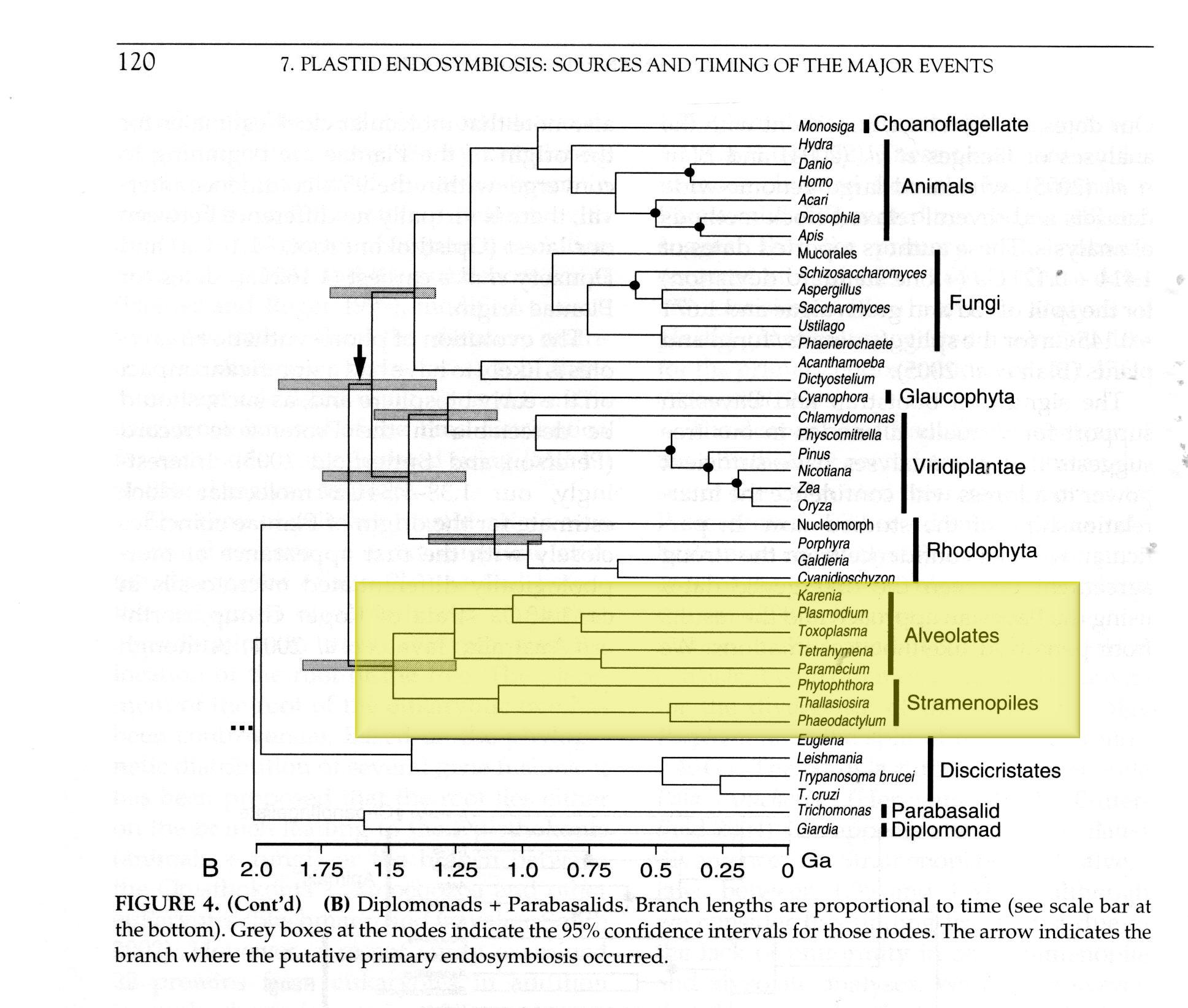
[2] Hackett JD, Yoon HS, Butterfield
NJ, Sanderson MJ, Bhattacharya D,
''Plastid endosymbiosis: Sources and
timing of the major events.'', in:
Falkowski P, Knoll A, editors.
''Evolution of primary producers in the
sea.'', Elsevier; 2007, p120.
COPYRIGHTED
source: Hackett JD, Yoon HS,
Butterfield NJ, Sanderson MJ,
Bhattacharya D, "Plastid endosymbiosis:
Sources and timing of the major
events.", in: Falkowski P, Knoll A,
editors. "Evolution of primary
producers in the sea.", Elsevier; 2007,
p120.
(red algae).
arctic Canada
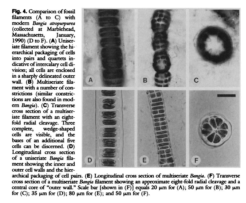
[1] Figure 4 from: Science 1990 vol
250 Butterfield N. J. A. H. Knoll K.
Swett 1990 A bangiophyte red alga from
the Proterozoic of Arctic Canada.
Science 250: 104-107
http://www.jstor.org/stable/2877905
COPYRIGHTED
source: http://www.jstor.org/stable/2877
905

[2] Figure 2 from: Science 1990 vol
250 Butterfield N. J. A. H. Knoll K.
Swett 1990 A bangiophyte red alga from
the Proterozoic of Arctic Canada.
Science 250: 104-107
http://www.jstor.org/stable/2877905
COPYRIGHTED
source: http://www.jstor.org/stable/2877
905

[1] Microsporidia. Image from Sterling
Parasitology Microsporidia
Research. UNKNOWN
source: http://microbewiki.kenyon.edu/im
ages/3/37/Micro2.jpg
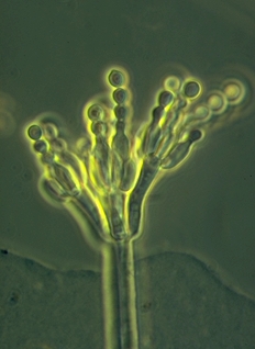
[2] Penicillium [t Note: Penecillium
is a multicellular fungi.] UNKNOWN
source: http://www.mold-help.org/pages/i
mages/Penicillium.jpg
(ancestor of all Ciliates,
Apicomplexans, and Dinoflagellates
{DInOFlaJeleTS}).
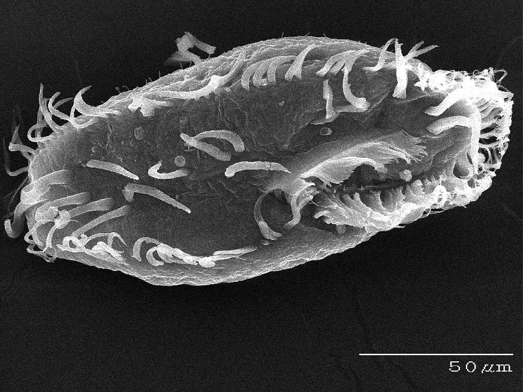
[1]
Unknown http://www.genome.gov/Images/pr
ess_photos/highres/85-300.jpg PD
source: http://upload.wikimedia.org/wiki
pedia/commons/thumb/6/6e/Oxytricha_trifa
llax.jpg/1024px-Oxytricha_trifallax.jpg

[2] Description English: Unknown
species of cilliate in the last stages
of mitosis (cytokinesis), with cleavage
furrow visible. Date Source
Own work Author
TheAlphaWolf CC
source: http://upload.wikimedia.org/wiki
pedia/commons/5/55/Unk.cilliate.jpg
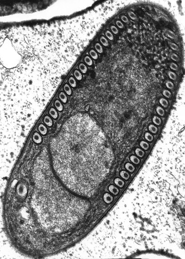
[1] Sporoblast of the Microsporidium
Fibrillanosema crangonycis. Electron
micrograph taken by Leon White. GNU
source: http://en.wikipedia.org/wiki/Ima
ge:Fibrillanosema_spore.jpg
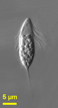
[2] Spironema
multiciliatum Spironema:
Octosporoblastic sporogony producing
horseshoe-shaped monokaryotic spores in
sporophorous vesicles; monomorphic,
diplokaryotic and monokaryotic;
merogony - last generation merozoites
are diplokaryotic; sporogony - initial
division of the sporont nuclei is
meiotic as indicated by the occurrence
of synaptonemal complexes; spores are
horse-shoe-shaped, with swollen ends in
T. variabilis and have one elongate
nucleus; exospore with three layers,
endospore is of medium thickness;
polaroplast composed of two lamellar
parts, an anterior part of closely
packed lamellae and a posterior part of
wider compartments; polar tube is
isofilar and forms, in the posterior
quarter of the spore, 3-4 coils in a
single rank (T. variabilis) or 8-10
coils in a single rank (T. chironomi);
type species Toxoglugea vibrio in
adipose tissue of larvae of Ceratopogon
sp. (Diptera, Ceratopogonidae).
Spironema (spire-oh-knee-ma)
multiciliatum Klebs, 1893. Cells are
lanceolate, relatively flattened and
flexible. The cells have a spiral
groove, long kinetics and a tail, which
tapers posteriorly, and are about 15 -
21 microns without the tail. The
nucleus is located anteriorly or near
the centre of the cell. When the cells
are squashed, the cells are more
flexible. Food materials are seen under
the cell surface. Rarely observed.
This picture was taken by Won Je Lee
using conventional photographic film
using a Zeiss Axiophot microscope of
material collected in marine sediments
of Botany Bay (Sydney, Australia). The
image description refers to material
from Botany Bay. NONCOMMERCIAL USE
source: http://microscope.mbl.edu/script
s/microscope.php?func=imgDetail&imageID=
3928
{DI-nO-Fla-Je-leTS}.

[1] Dinoflagellate Ceratium
sp. Phylum Dinoflagellata Upper
Newport Bay, Orange County, CA.
9/22/12. © Peter J.
Bryant COPYRIGHTED
source: http://nathistoc.bio.uci.edu/Din
oflagellates/DSC_6886b.jpg
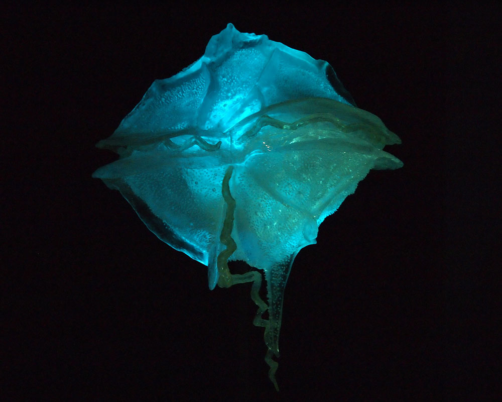
[2] Model of Pyrodinium bahamense, a
dinoflagellate species, in the American
Museum of Natural History Credit:
Life’s Little Mysteries Fire
water Have you ever seen glowing ocean
water, like the bright blue surf
pictured in the intro slide? The neon
water is brimming with dinoflagellates,
single-celled plankton with tails that
slosh around together in vast numbers.
These creatures have been highlighting
Earth’s coastlines for 1.2 billion
years, and for the past few millennia,
they’ve puzzled humans, who used to
attribute the glow of some ocean water
to magic or the gods.Dinoflagellates
still puzzle us; we know how they glow,
but not why. They might have evolved
bioluminescence as a way of frightening
predators, or to reveal those
predators’ locations by flashing when
touched. Alternatively, their
bioluminescence may just be a fancy way
of ridding themselves of oxygen
radicals (because the chemical reaction
requires oxygen). Whatever the answer,
they certainly make for a nice holiday
in the Bahamas. UNKNOWN
source: http://www.lifeslittlemysteries.
com/images/i/1651/original/dinoflagellat
e.jpg
{DiSKIKriSTATS} (includes euglenids).

[1] euglena
source: http://www.fcps.k12.va.us/Stratf
ordLandingES/Ecology/mpages/euglena.htm

[2] euglena
source: http://protist.i.hosei.ac.jp/PDB
/Images/Mastigophora/Euglena/genus1L.jpg
three-dimensional response to light.
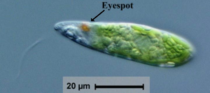
[1] Adapted from: Euglena is a
photosynthetic euglenoid with at least
150 described species. The cells are
cylindrical with a rounded anterior and
tapered posterior. The chloroplasts are
well-developed, bright green, and
sometimes have pyrenoids. ... Euglena
is a photosynthetic euglenoid with at
least 150 described species. The cells
are cylindrical with a rounded anterior
and tapered posterior. The chloroplasts
are well-developed, bright green, and
sometimes have pyrenoids. They are
often discoidal in shape but can also
be ovate, lobate, elongate, U-shaped,
or ribbon-shaped. Some researchers use
the structure and position of the
chloroplasts to divide the group into
three subgenera. Even though they are
able to photosynthesize, Euglena cells
also have a phagotrophic ingestion
apparatus. Euglena has one long,
protruding flagellum and a shorter
flagellum that is not usually
visible. The euglenoids can glide
and swim using their flagella, or can
ooze along a substrate with an
undulating, shape-changing, contraction
motion called metaboly. The cytoplasm
of Euglena and other euglenoids
contains many paramylon starch storage
granules. The euglenoid cells are
covered by a pellicle composed of
ribbonlike, woven strips of
proteinaceous material that cover the
cell in a helical arrangement from apex
to posterior. Freshwater euglenoids
have a contractile vacuole. Euglenoids
sense light using a red pigmented
eyespot or stigma and the paraflagellar
body located at the base of the
emergent flagella. The cytoplasm of
Euglena and other euglenoids contains
many paramylon starch storage granules.
The euglenoid cells are covered by a
pellicle composed of ribbonlike, woven
strips of proteinaceous material that
cover the cell in a helical arrangement
from apex to posterior. Freshwater
euglenoids have a contractile vacuole.
Euglenoids sense light using a red
pigmented eyespot or stigma and the
paraflagellar body located at the base
of the emergent flagella. UNKNOWN
source: http://silicasecchidisk.conncoll
.edu/Pics/Other%20Algae/Other_jpegs/Eugl
ena_Key225.jpg

[2] Figure 1. The distribution of
three-dimensional phototaxis in the
tree of eukaryotes. Red arrows indicate
the likely point of origin of
phototaxis in a given group. Question
marks indicate uncertainties regarding
independent or common origin. Figure
1 from: Jékely, Gáspár. ''Evolution
of phototaxis.'' Philosophical
Transactions of the Royal Society B:
Biological Sciences 364 (October
2009):
2795–2808. http://rstb.royalsocietypu
blishing.org/content/364/1531/2795.short
COPYRIGHTED
source: http://rstb.royalsocietypublishi
ng.org/content/364/1531/2795/F1.large.jp
g
{STro-meN-o-Pi-lEZ} (also called
Heterokonts) (ancestor of all brown and
golden algae, diatoms, and oomycota
{Ou-mI-KO-Tu)).

[1] Phylum Stramenopiles COPYRIGHTED
source: Brusca and Brusca,
"Invertebrates", Second Edition, 2003,
p153-155.

[2] S. Blair Hedges and Sudhir Kumar,
''The TimeTree of Life'', 2009,
p117-118. http://www.timetree.org/book.
php COPYRIGHTED
source: http://www.timetree.org/book.php
{me-ZO-mI-SE-TO-ZO-u} (or DRIPs).
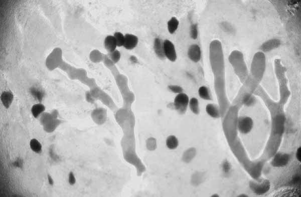
[1] Ichthyophonus, a fungus-like
protistan that occurs in high
prevalence in Pacific Ocean perch
(Sebastes aultus) and yellowtail
rockfish (Sebastes flavedus). Note the
parasite forms branching hyphae-like
structures. Ichthyophonus hoferi has
caused massive mortalities in herring
in the Atlantic ocean, and has recently
been reported to cause disease in wild
Pacific herring from Washington through
Alaska. COPYRIGHTED EDU
source: http://oregonstate.edu/dept/salm
on/projects/images/16Ichthyophonus.jpg
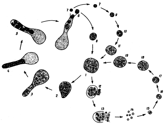
[2] Microscopic appearence of the
organism is dependent on its stage of
development. The stages include (1)
spore at ''resting'' stage, (2)
germinating spore, (3) hyphal
stage. It is believed that there are
two forms of Ichthyophonus, both
belonging to one genus. One of them is
known as the ''salmon'' form, occuring
in freshwater and cold-preferring sea
fishes: this form is characterized by
its ability to produce long tubulose
germ hyphae. The other is called the
''aquarium fish'' form, typical of the
tropical freshwater fishes. This form
is completely devoid of hyphae.
Developmental cycle of Ichthyophonus
hoferi: 1-5 - development of
''daughter'' spores, 7-11 - development
of resting spore from the ''daughter''
spore, 12-19 - development of resting
spore by fragmentation. COPYRIGHTED
source: http://www.fao.org/docrep/field/
003/AC160E/AC160E02.htm
(Water molds).

[1] Figure 2 from: Sandra L. Baldauf,
A. J. Roger, I. Wenk-Siefert, W. F.
Doolittle, ''A Kingdom-Level Phylogeny
of Eukaryotes Based on Combined Protein
Data'', Science, Vol 290, num 5493, p
972, (2000).
http://www.sciencemag.org/content/290/
5493/972.full Figure 2 Single-gene
phylogenies support subsets of the
combined protein tree. (A) A summary of
the tree in Fig. 1is shown with
supergroups indicated beside brackets
to the right. Multi-taxon represented
clusters are given as triangles, with
height proportional to number of taxa
and width proportional to averaged
overall branch length (1) compensated
for missing data (47). (B) Published
support for the numbered nodes in (A)
is shown for commonly used molecular
phylogenetic markers grouped as (a)
ribosomal RNAs, (b) proteins not used
in the current analysis, (c) proteins
used in the current analysis, and (d)
the combined data (Fig. 1). These
markers are, from left to right, SSU
[SSU rRNA (1–4)], LSU [LSU rRNA
(19)], LSU+SSU [combined LSU and SSU
rRNA (48)], EF-2 (10), V/A-ATPases
[vacuolar ATPases (49)], HSP70-cy
[cytosolic 70-kD heat shock protein
(50)], mito [combined mitochondrial
proteins (51)], RPB1 (52), actin (8,
16, 53), α-tubulin (8, 54), β-tubulin
(8, 54), EF-1α (15, 20), and combined
(Fig. 1). Rejected nodes are indicated
in pink and accepted nodes in green,
with checked circles indicating BP < 70% and solid circles indicating BP >
70%. COPYRIGHTED
source: http://www.sciencemag.org/conten
t/290/5493/972/F2.large.jpg
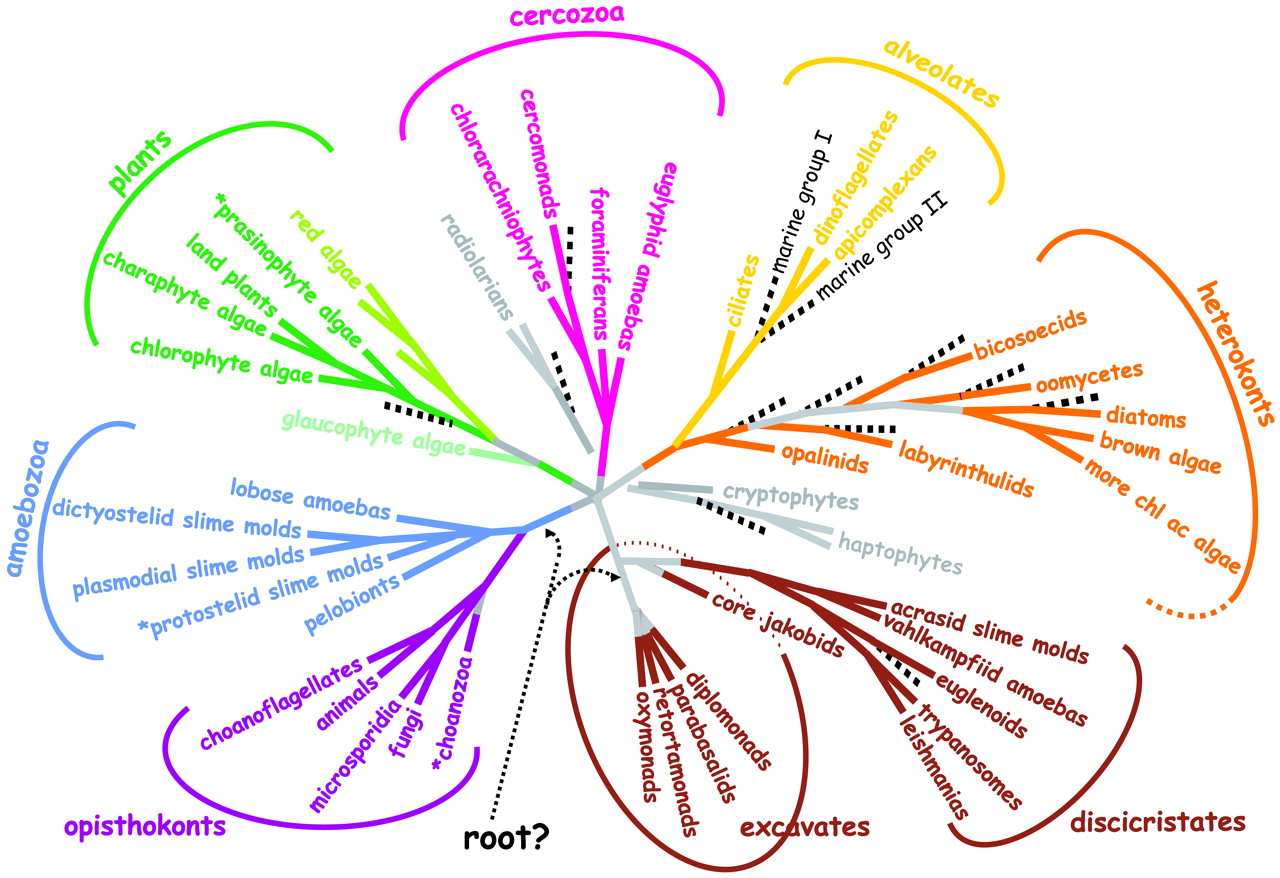
[2] Fig. 1. A consensus phylogeny of
eukaryotes. The vast majority of
characterized eukaryotes, with the
notable exception of major subgroups of
amoebae, can now be assigned to one of
eight major groups. Opisthokonts (basal
flagellum) have a single basal
flagellum on reproductive cells and
flat mitochondrial cristae (most
eukaryotes have tubular ones).
Eukaryotic photosynthesis originated in
Plants; theirs are the only plastids
with just two outer membranes.
Heterokonts (different flagellae) have
a unique flagellum decorated with
hollow tripartite hairs (stramenopiles)
and, usually, a second plain one.
Cercozoans are amoebae with filose
pseudopodia, often living with in tests
(hard outer shells), some very
elaborate (foraminiferans). Amoebozoa
are mostly naked amoebae (lacking
tests), often with lobose pseudopodia
for at least part of their life cycle.
Alveolates have systems of cortical
alveoli directly beneath their plasma
membranes. Discicristates have discoid
mitochondrial cristae and, in some
cases, a deep (excavated) ventral
feeding groove. Amitochondrial
excavates lack substantial molecular
phylogenetic support, but most have an
excavated ventral feeding groove, and
all lack mitochondria. The tree shown
is based on a consensus of molecular
(1-4) and ultrastructural (16, 17) data
and includes a rough indication of new
ciPCR ''taxa'' (broken black lines)
(7-11). An asterisk preceding the taxon
name indicates probable paraphyletic
group COPYRIGHTED
source: http://www.sciencemag.org/cgi/co
ntent/full/300/5626/1703
(ancestor of all Radiolaria,
Foraminifera and Cercozoa).

[1] Figure : Maximum likelihood
phylogeny of Rhizaria inferred from SSU
rRNA gene sequences using the GTR+G+I
model of evolution. UNKNOWN
source: http://www.unige.ch/sciences/bio
logie/biani/msg/Amoeboids/Rhizaria_large
.jpg

[2] Figure 1 from: Keeling, Patrick
J. et al. “The tree of eukaryotes.”
Trends in Ecology & Evolution 20.12
(2005):
670-676. http://www.sciencedirect.com/s
cience/article/pii/S0169534705003046
source: http://www.sciencedirect.com/cac
he/MiamiImageURL/1-s2.0-S016953470500304
6-gr1.jpg/0?wchp=dGLbVBA-zSkWz
[1] Figure 2. Zygomycota A: sporangia
of Mucor sp. B: whorl of sporangia of
Absidia sp. C: zygospore of
Zygorhynchus sp. D: sporangiophore and
sporangiola of Cunninghamella sp.
source: http://www.botany.utoronto.ca/Re
searchLabs/MallochLab/Malloch/Moulds/Cla
ssification.html
[2] Figure 3. Syncephalis, a member of
the Zygomycota parasitic on other
Zygomycota
source: http://www.botany.utoronto.ca/Re
searchLabs/MallochLab/Malloch/Moulds/Cla
ssification.html

[1] Paramecium protozoan,
SEM C001/0068 Rights Managed Credit:
STEVE GSCHMEISSNER/SCIENCE PHOTO
LIBRARY Caption: Paramecium protozoan,
coloured scanning electron micrograph
(SEM). Paramecia are a group of
unicellular ciliate protozoa. They
inhabit fresh water, and feed mainly on
bacteria and smaller protozoa.
Paramecia range from about 50 to 350
micrometres in length, depending on
species. Simple cilia, which cover the
body, are moved in a synchronous motion
to allow the cell to move.
Magnification: x825 when printed at 10
centimetres wide. COPYRIGHTED
source: http://www.nonlocal.com/hbar/par
amecium.gif

[2] Summary Description English:
Scanning electron microscope view of
Oxytricha trifallax Español: Imagen
de microscopía electrónica de barrido
de Oxytricha trifallax Date Unknown
date Source http://www.genome.gov/I
mages/press_photos/highres/85-300.jpg
Author Unknown Permission (Reusin
g this file) See below. PD [1] Fig.
1. A consensus phylogeny of eukaryotes.
The vast majority of characterized
eukaryotes, with the notable exception
of major subgroups of amoebae, can now
be assigned to one of eight major
groups. Opisthokonts (basal flagellum)
have a single basal flagellum on
reproductive cells and flat
mitochondrial cristae (most eukaryotes
have tubular ones). Eukaryotic
photosynthesis originated in Plants;
theirs are the only plastids with just
two outer membranes. Heterokonts
(different flagellae) have a unique
flagellum decorated with hollow
tripartite hairs (stramenopiles) and,
usually, a second plain one. Cercozoans
are amoebae with filose pseudopodia,
often living with in tests (hard outer
shells), some very elaborate
(foraminiferans). Amoebozoa are mostly
naked amoebae (lacking tests), often
with lobose pseudopodia for at least
part of their life cycle. Alveolates
have systems of cortical alveoli
directly beneath their plasma
membranes. Discicristates have discoid
mitochondrial cristae and, in some
cases, a deep (excavated) ventral
feeding groove. Amitochondrial
excavates lack substantial molecular
phylogenetic support, but most have an
excavated ventral feeding groove, and
all lack mitochondria. The tree shown
is based on a consensus of molecular
(1-4) and ultrastructural (16, 17) data
and includes a rough indication of new
ciPCR ''taxa'' (broken black lines)
(7-11). An asterisk preceding the taxon
name indicates probable paraphyletic
group COPYRIGHTED
source: http://upload.wikimedia.org/wiki
pedia/commons/thumb/6/6e/Oxytricha_trifa
llax.jpg/1024px-Oxytricha_trifallax.jpg
{a-PE-KoM-PleK-Su} (Malaria).

[1] Description A thin-film Giemsa
stained micrograph of ring-forms, and
gametocytes of Plasmodium falciparum.
From
http://phil.cdc.gov/phil/home.asp Date
2006-11-16 (original upload
date) Source Originally from
en.wikipedia; description page is/was
here. Author Original uploader was
TimVickers at
en.wikipedia Permission (Reusing this
file) PD
source: http://upload.wikimedia.org/wiki
pedia/commons/3/3c/Plasmodium.jpg
[2] Fig. 1. A consensus phylogeny of
eukaryotes. The vast majority of
characterized eukaryotes, with the
notable exception of major subgroups of
amoebae, can now be assigned to one of
eight major groups. Opisthokonts (basal
flagellum) have a single basal
flagellum on reproductive cells and
flat mitochondrial cristae (most
eukaryotes have tubular ones).
Eukaryotic photosynthesis originated in
Plants; theirs are the only plastids
with just two outer membranes.
Heterokonts (different flagellae) have
a unique flagellum decorated with
hollow tripartite hairs (stramenopiles)
and, usually, a second plain one.
Cercozoans are amoebae with filose
pseudopodia, often living with in tests
(hard outer shells), some very
elaborate (foraminiferans). Amoebozoa
are mostly naked amoebae (lacking
tests), often with lobose pseudopodia
for at least part of their life cycle.
Alveolates have systems of cortical
alveoli directly beneath their plasma
membranes. Discicristates have discoid
mitochondrial cristae and, in some
cases, a deep (excavated) ventral
feeding groove. Amitochondrial
excavates lack substantial molecular
phylogenetic support, but most have an
excavated ventral feeding groove, and
all lack mitochondria. The tree shown
is based on a consensus of molecular
(1-4) and ultrastructural (16, 17) data
and includes a rough indication of new
ciPCR ''taxa'' (broken black lines)
(7-11). An asterisk preceding the taxon
name indicates probable paraphyletic
group COPYRIGHTED
source: http://www.sciencemag.org/cgi/co
ntent/full/300/5626/1703
{KO-e-nO-FlaJ-e-lATS}.

[1] Choanoflagellate single cell
(thecate) UNKNOWN
source: http://behance.vo.llnwd.net/prof
iles22/483113/projects/1558429/6ea555ab5
457e21432def0f2e6b83fe3.jpg

[2] Salpingoeca: Cells solitary or
colonial with a distinct and firm
sheath or theca usually as a cup either
sessile or with a pedicel; theca
colourless or amber; contractile
vacuoles posterior in freshwater
specie; in freshwater, brackish, and
marine habitats. Record information:
Salpingoeca (sal-ping-go-eek-a), a
collar flagellate (choanoflagellate) -
all of which have a single anterior
flagellum surrounded by a collar of
very fine pseudopodia (in cross-section
the collar seems like two arms, one on
either side of the flagellum). The
flagellum beats drawing water through
the collar and bacteria and other small
particles are trapped and then
ingested. Believed to be the source
group of the sponges and the metazoa.
Salpingoeca has an organic lorica.
Phase contrast. This picture was
taken by David Patterson, Linda Amaral
Zettler and Virginia Edgcomb of
material from the salt marsh at Little
Sippewissett (Massachusetts, USA) in
Autumn, 2000 and in Spring and summer,
2001. NONCOMMERCIAL USE
source: http://microscope.mbl.edu/script
s/microscope.php?func=imgDetail&imageID=
746
moving Protist. This allows larger free
moving organisms to evolve.
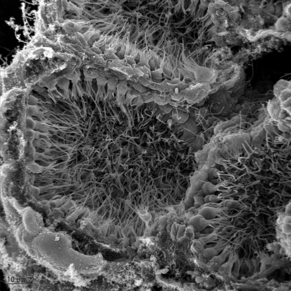
[1] Sponge showing several choanocyte
chambers UNKNOWN
source: http://behance.vo.llnwd.net/prof
iles22/483113/projects/1558429/43a2a4c7e
127f66b7090ed679a8da30a.jpg
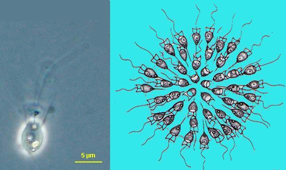
[2] Combination of: Saepicula and
Sphaeroeca NONCOMMERCIAL USE
source: http://microscope.mbl.edu/script
s/microscope.php?func=imgDetail&imageID=
3229
sponges (Porifera). Metazoans are
multicellular and have differentiation
(their cells perform different
functions). There are only three major
kinds of metazoans: sponges,
cnidarians, and bilaterians.

[1] Summary Description English:
Marine sponge. Color adjusted (but not
color accurate) underwater photograph
taken by Dlloyd using a digital camera
at a depth of approximately 100 feet in
Cayman. GNU
source: http://upload.wikimedia.org/wiki
pedia/commons/6/62/SpongeColorCorrect.jp
g
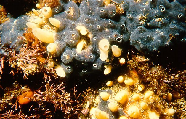
[2]
source: http://www.museums.org.za/bio/me
tazoa.htm
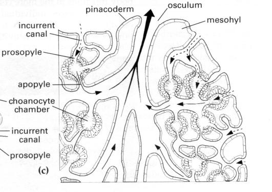
[1] Oocyte (female egg) release from
sponge, sperm release from sponge,
FIgure from: D. T. Anderson,
''Invertebrate Zoology'', Oxford
University Press, Second Edition,
2001. COPYRIGHTED
source: D. T. Anderson, "Invertebrate
Zoology", Oxford University Press,
Second Edition, 2001.

[2] Combination of image from: Brusca
and Brusca, ''Invertebrates'', Second
Edition, 2003,
http://www.oceanicresearch.org/sponges
.html and D. T. Anderson,
''Invertebrate Zoology'', Oxford
University Press, Second Edition,
2001. COPYRIGHTED
source: http://www.museums.org.za/bio/me
tazoa.htm
in metazoans.
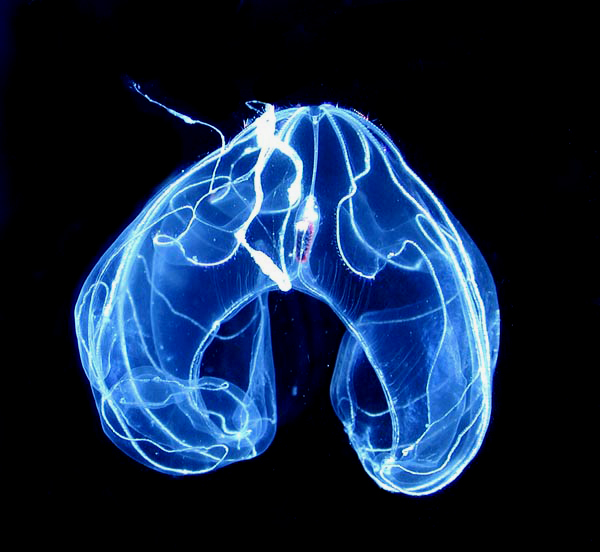
[1] Description This is an example
of a ctenophore, Bathocyroe fosteri,
which is a mesopelagic species. Date
Source Description This is
an example of a ctenophore, Bathocyroe
fosteri, which is a mesopelagic
species. Date Source
[1] Author Photo courtesy of
Marsh Youngbluth Author Photo
courtesy of Marsh Youngbluth PD
source: http://upload.wikimedia.org/wiki
pedia/commons/2/21/Bathocyroe_fosteri.jp
g

[2] Light diffracting along the comb
rows of a Mertensia ovum. The right
lower portion of the body is
regenerating from previous damage.
Source: NOAA Photo Gallery/ Photo by
Kevin Raskoff PD
source: http://upload.wikimedia.org/wiki
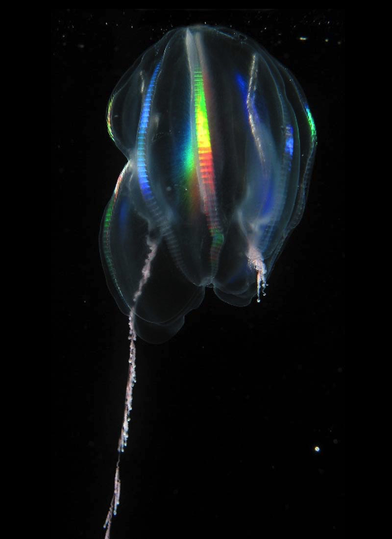
pedia/commons/4/42/LightRefractsOf_comb-
rows_of_ctenophore_Mertensia_ovum.jpg

[1] Description Trichoplax sp.
from Australia in light
microscopy Date February
2006 Source Oliver Voigt Author
Oliver Voigt CC
source: http://upload.wikimedia.org/wiki
pedia/commons/c/c3/Trichoplax_mic.jpg
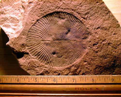
[2] from ediacara of australia
source: http://www.ucmp.berkeley.edu/ven
dian/dickinsonia.html
{KI-TriDEO-mI-KO-Tu).

[1] Chytrids (Chytridiomycota): The
Primitive Fungi These fungi are
mostly aquatic, are notable for having
a flagella on the cells (a flagella is
a tail, somewhat like a tail on a sperm
or a pollywog), and are thought to be
the most primitive type of
fungi. actual photo comes
from: http://www.csupomona.edu/~jcclark
/classes/bot125/resource/graphics/chy_al
l_sph.html
source: http://www.davidlnelson.md/Cazad
ero/Fungi.htm
[2] Chytridiomycota - Blastocladiales
- zoospore of Allomyces (phase contrast
illumination) X 2000
source: http://www.mycolog.com/chapter2b
.htm
nervous system evolve in a metazoan.
Earliest touch and sound detection and
memory.

[1] English: Drawing of Purkinje cells
(A) and granule cells (B) from pigeon
cerebellum by Santiago Ramón y Cajal,
1899; Instituto Santiago Ramón y
Cajal, Madrid, Spain. PD
source: http://upload.wikimedia.org/wiki
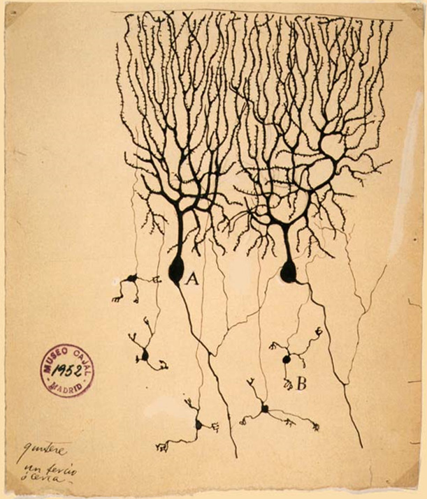
pedia/commons/1/15/PurkinjeCell.jpg

[2] figure from: D. T. Anderson,
''Invertebrate Zoology'', Oxford
University Press, Second Edition, 2001,
p39. COPYRIGHTED
source: D. T. Anderson, "Invertebrate
Zoology", Oxford University Press,
Second Edition, 2001, p39.
[1] Figure from: D. T. Anderson,
''Invertebrate Zoology'', Oxford
University Press, Second Edition, 2001,
p39. COPYRIGHTED
source: D. T. Anderson, "Invertebrate
Zoology", Oxford University Press,
Second Edition, 2001, p39.
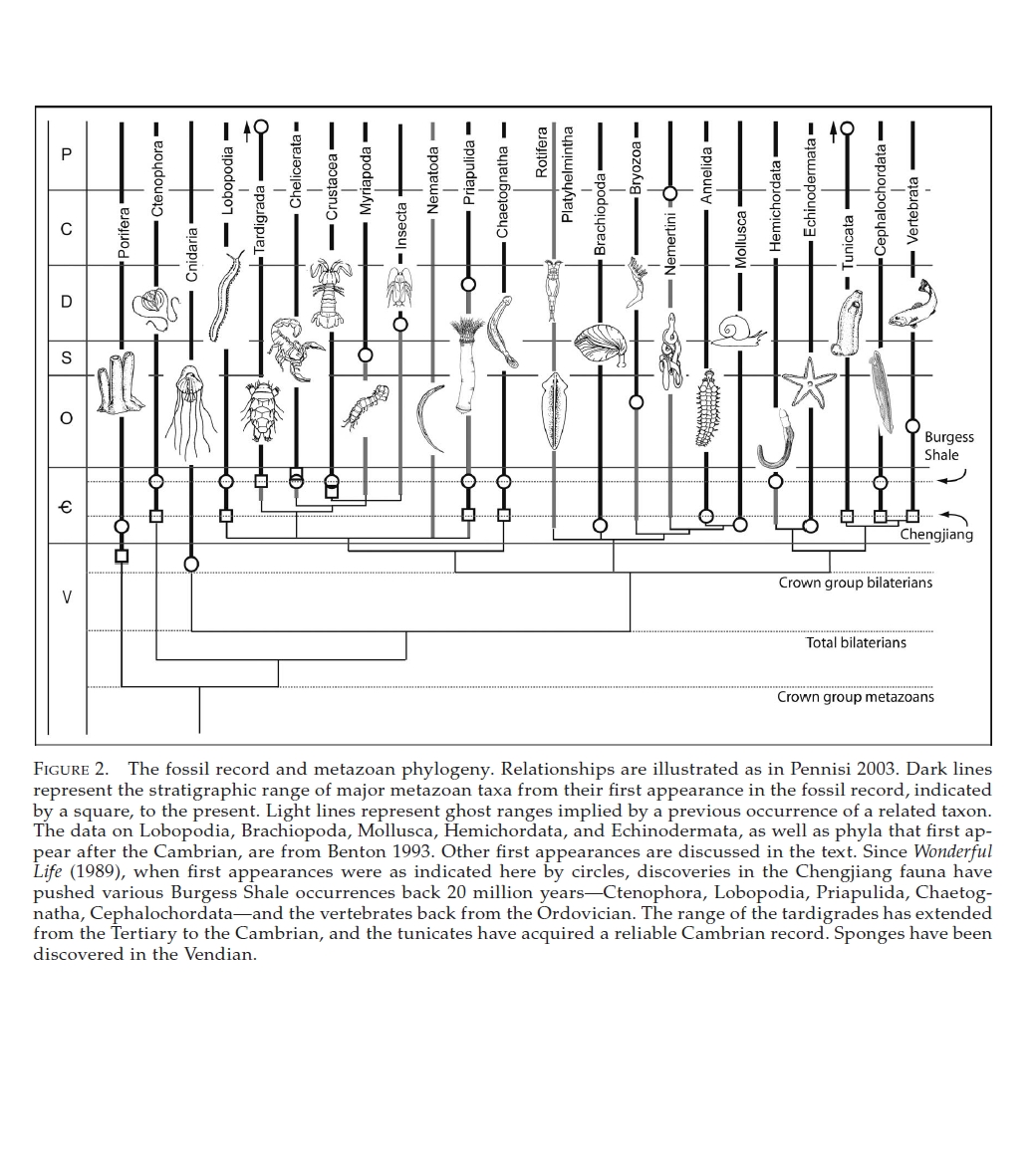
[2] Derek E. G. Briggs and Richard A.
Fortey, ''Wonderful Strife:
Systematics, Stem Groups, and the
Phylogenetic Signal of the Cambrian
Radiation'', Paleobiology , Vol. 31,
No. 2, Supplement. Macroevolution:
Diversity, Disparity, Contingency:
Essays in Honor of Stephen Jay Gould
(Spring, 2005), pp.
94-112 http://www.jstor.org/stable/2548
2671 COPYRIGHTED
source: http://www.jstor.org/stable/2548
2671
metazoans.
[1] Description This is an example
of a ctenophore, Bathocyroe fosteri,
which is a mesopelagic species. Date
Source Description This is
an example of a ctenophore, Bathocyroe
fosteri, which is a mesopelagic
species. Date Source
[1] Author Photo courtesy of
Marsh Youngbluth Author Photo
courtesy of Marsh Youngbluth PD
source: http://upload.wikimedia.org/wiki
pedia/commons/2/21/Bathocyroe_fosteri.jp
g
[2] Light diffracting along the comb
rows of a Mertensia ovum. The right
lower portion of the body is
regenerating from previous damage.
Source: NOAA Photo Gallery/ Photo by
Kevin Raskoff PD
source: http://upload.wikimedia.org/wiki
pedia/commons/4/42/LightRefractsOf_comb-
rows_of_ctenophore_Mertensia_ovum.jpg
metzoans.

[1] From: Brusca and Brusca,
''Invertebrates'', Second Edition,
2003. COPYRIGHTED
source: Brusca and Brusca,
"Invertebrates", Second Edition, 2003

[2] Figure 3.8 Anthozoa. (a) Anemone
(Actiniaria), showing the pharynx,
mesenteries, mesenterial filamnets and
acontia. (b) Structure of a mesenterial
filament in transverse section. (c)
Scleractinian coral, showing calcareous
skeleton and coenenchyme. (d)
Gorgonian, showing skeleton made up of
a horny axial rod and spicules in the
mesogloea (after Pearse et al 1987).
(e) Alcyonarian soft coral, showing
spicular skeleton in the
mesogloea. From: D. T. Anderson,
''Invertebrate Zoology'', Oxford
University Press, Second Edition,
2001. COPYRIGHTED
source: D. T. Anderson, "Invertebrate
Zoology", Oxford University Press,
Second Edition, 2001.
evolve (comb jellies).
[1] Description This is an example
of a ctenophore, Bathocyroe fosteri,
which is a mesopelagic species. Date
Source Description This is
an example of a ctenophore, Bathocyroe
fosteri, which is a mesopelagic
species. Date Source
[1] Author Photo courtesy of
Marsh Youngbluth Author Photo
courtesy of Marsh Youngbluth PD
source: http://upload.wikimedia.org/wiki
pedia/commons/2/21/Bathocyroe_fosteri.jp
g
[2] Light diffracting along the comb
rows of a Mertensia ovum. The right
lower portion of the body is
regenerating from previous damage.
Source: NOAA Photo Gallery/ Photo by
Kevin Raskoff PD
source: http://upload.wikimedia.org/wiki
pedia/commons/4/42/LightRefractsOf_comb-
rows_of_ctenophore_Mertensia_ovum.jpg
evolve (sea anemones, corals,
jellyfish). Earliest animal eye.

[1] Octocorals Stylatula elongata –
White Sea Pen UNKNOWN
source: http://pt-lobos.com/cnidarianimg
/white_sea_pens.jpg
[2] Sea nettles, Chrysaora
quinquecirrha CC
source: http://upload.wikimedia.org/wiki
pedia/commons/3/36/Sea_nettles.jpg
soft-bodied invertebrate fossils.
Lesser Karatau Microcontinent,
Kazakhsta

[1] A general view of the life in the
time frame from about 605 to 542
million years ago (the Vendian), is
found at this New Zealand site which
concentrates on the Ediacaran epoch; it
mentions Australian and other
geographic localities where the
assemblages have been found. The fossil
life is represented entirely by
creatures with soft parts only. It is
suggested that these may be ancestral
to later phylla observed at the
beginning of the Paleozoic. Below is a
chart presenting typical Ediacaran
fauna, followed by an artist's
depiction of life on the sea floor at
that time, and beneath that is a layout
of some actual fossils: PD
source: http://rst.gsfc.nasa.gov/Sect20/
800pxlife_in_the_ediacaran_sea.jpg
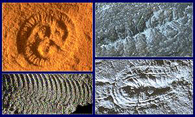
[2] A more general view of the life in
the time frame from about 600+ to 542
million years ago (end of Proterozoic
and Precambrian into the oldest
Cambrian), known as the Ediacaran or
Vendian, is found at this New Zealand
site; it mentions Australian and other
geographic localities where the
assemblages have been found. The fossil
life represents entirely creatures with
soft parts only and suggestions that
these may be ancestral to later phylla
observed at the beginning of the
Paleozoic. Below is an artist's sketch
of some of these creatures: UNKNOWN
source: http://www.fas.org/irp/imint/doc
s/rst/Sect20/vendintro.jpg
sided symmetry).
Earliest animal brain. First
triploblastic species (third embryonic
layer: the mesoderm {meZuDRM}).
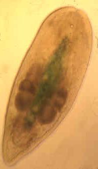
[1] Convoluta pulchra Smith and Bush
1991, a typical mud-inhabiting acoel
that feeds on diatoms
source: ?

[2] Figure from: Giribet, G. (2008).
Assembling the lophotrochozoan
(=spiralian) tree of life.
Philosophical Transactions of the Royal
Society B: Biological Sciences , 363
(1496), 1513-1522. URL
http://dx.doi.org/10.1098/rstb.2007.2241
http://rstb.royalsocietypublishing.org
/content/363/1496/1513 COPYRIGHTED
source: http://rstb.royalsocietypublishi
ng.org/content/363/1496/1513
Acoelomorpha (acoela flat worms and
nemertodermatida).
[1] Convoluta pulchra Smith and Bush
1991, a typical mud-inhabiting acoel
that feeds on diatoms
source: ?
[2] Figure from: Giribet, G. (2008).
Assembling the lophotrochozoan
(=spiralian) tree of life.
Philosophical Transactions of the Royal
Society B: Biological Sciences , 363
(1496), 1513-1522. URL
http://dx.doi.org/10.1098/rstb.2007.2241
http://rstb.royalsocietypublishing.org
/content/363/1496/1513 COPYRIGHTED
source: http://rstb.royalsocietypublishi
ng.org/content/363/1496/1513
bilaterian.
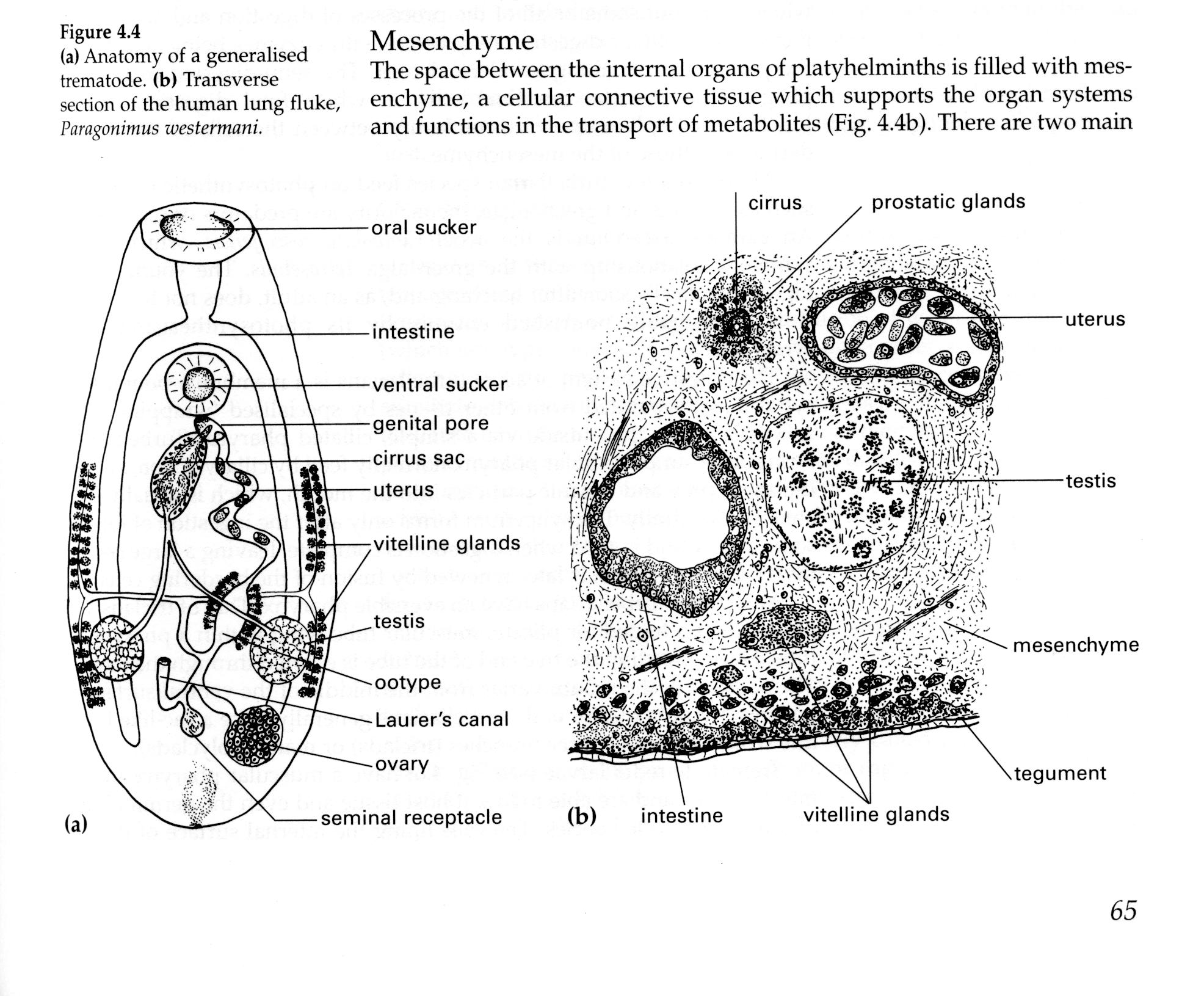
[1] From: D. T. Anderson,
''Invertebrate Zoology'', Oxford
University Press, Second Edition,
2001. COPYRIGHTED
source: D. T. Anderson, "Invertebrate
Zoology", Oxford University Press,
Second Edition, 2001.
[2] Convoluta pulchra Smith and Bush
1991, a typical mud-inhabiting acoel
that feeds on diatoms
source: ?
through-put of food evolves in a
bilaterian.
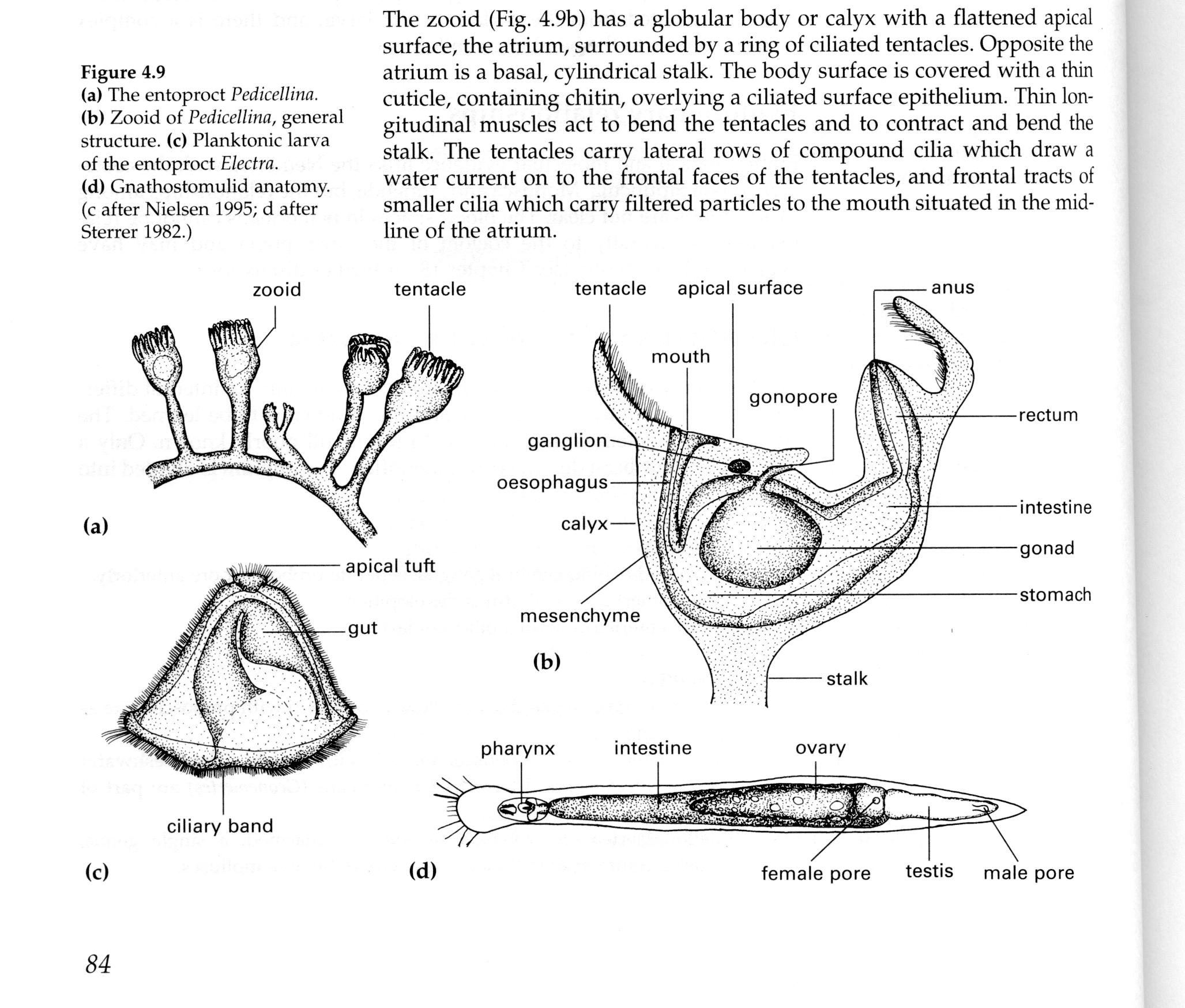
[1] From: D. T. Anderson,
''Invertebrate Zoology'', Oxford
University Press, Second Edition,
2001. COPYRIGHTED
source: D. T. Anderson, "Invertebrate
Zoology", Oxford University Press,
Second Edition, 2001.
[2] Convoluta pulchra Smith and Bush
1991, a typical mud-inhabiting acoel
that feeds on diatoms
source: ?
uterus evolve in a bilaterian.

[1] From: D. T. Anderson,
''Invertebrate Zoology'', Oxford
University Press, Second Edition,
2001. COPYRIGHTED
source: D. T. Anderson, "Invertebrate
Zoology", Oxford University Press,
Second Edition, 2001.
[2] Convoluta pulchra Smith and Bush
1991, a typical mud-inhabiting acoel
that feeds on diatoms
source: ?

[1] From: Brusca and Brusca,
''Invertebrates'', Second Edition,
2003 COPYRIGHTED
source: Brusca and Brusca,
"Invertebrates", Second Edition, 2003
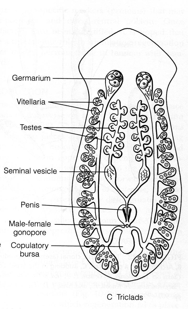
[2] From: Ruppert, Fox, Barnes,
''Invertebrate Zoology'',
2004. COPYRIGHTED
source: Ruppert, Fox, Barnes,
"Invertebrate Zoology", 2004.
(SEleM) evolves in a bilaterian.

[1] Example of the coleom's from 3
organisms UNKNOWN
source: http://4.bp.blogspot.com/_5DLPaU
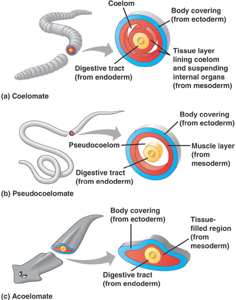
qdg2g/TBBz3rcLDOI/AAAAAAAAAGA/Z34_-_usSc
w/s1600/3927715.jpg

[2] From NATURAL HISTORY
COLLECTIONS OF THE UNIVERSITY OF
EDINBURGH Formation of the coelom or
body cavity Acoelomates lack a
body cavity. In pseudocoelomates,
the coelom is formed from a persistent
embryonic cavity. In schizocoelous
coelomates, the coelom is formed by
splits in the embryonic mesoderm, the
middle layer of the body. In
enterocoelous coelomates, the coelom
forms within pouches of the gut
wall. UNKNOWN
source: http://www.nhc.ed.ac.uk/images/c
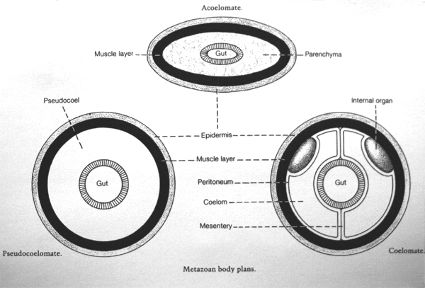
ollections/invertebrates/intros/LgCoelom
.jpg
vessels and blood evolve in a
bilaterian.
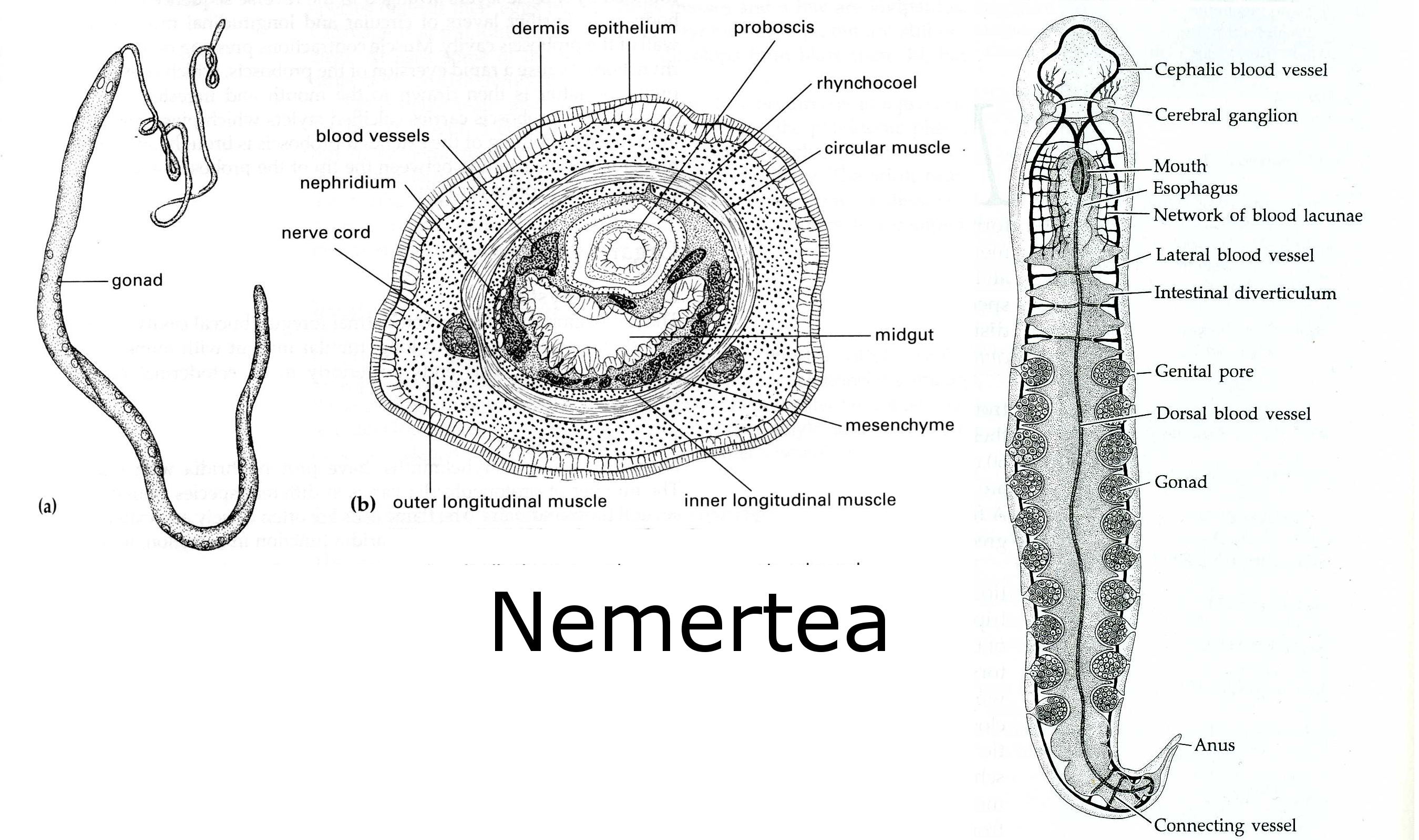
[1] D. T. Anderson, ''Invertebrate
Zoology'', Oxford University Press,
Second Edition, 2001 AND Fig 11.1G
from: Brusca and Brusca,
''Invertebrates'', 2003,
p320. COPYRIGHTED
source: D. T. Anderson, "Invertebrate
Zoology", Oxford University Press,
Second Edition, 2001Brusca and Brusca,
"Invertebrates", 2003, p320.

[2] From: D. T. Anderson,
''Invertebrate Zoology'', Oxford
University Press, Second Edition,
2001 COPYRIGHTED
source: D. T. Anderson, "Invertebrate
Zoology", Oxford University Press,
Second Edition, 2001
Ancestor of all Ecdysozoa
{eK-DiS-u-ZOu} and Lophotrochozoa
{LuFoTroKoZOu}.
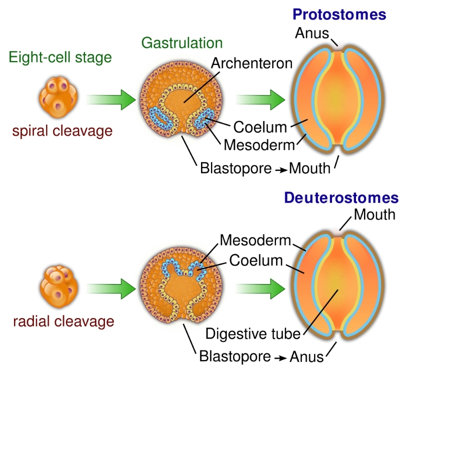
[1] English: This diagram is showing
the difference of the two major types
of coelomates: the protostomes
(molluscs, annelids, arthropods, ...)
and deuterostomes (echinoderms,
vertebrates, ...). These groups differ
in several characteristics of early
development; In deuterostomes blastula
devisions is called ''radial cleavage''
because it occurs parallel or
perpendicular to the major polar axis.
In protostomes the cleavage is called
''spirale'' because division planes are
oriented obliquely to the polar major
axis. During gastrulation, protostomes
embryos' mouth was given first by the
blastopore while the anus was formed
later and vis versa for the
deuterostomes. As examples :
Squids are protostomes. Sea
urchins are deuterostomes. Date
14 October 2009 Source Own
work Author WYassineMrabetTalk✉ CC
source: http://upload.wikimedia.org/wiki
pedia/commons/thumb/6/65/Protovsdeuteros
tomes.svg/1000px-Protovsdeuterostomes.sv
g.png
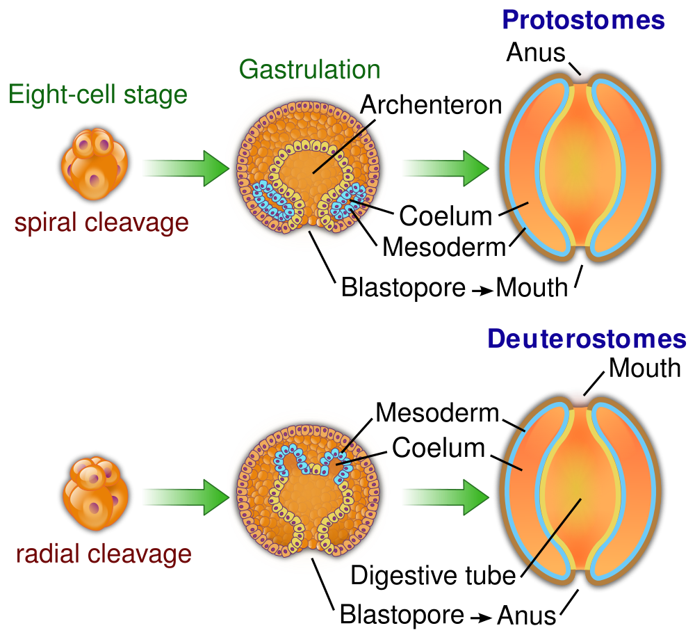
[2] English: This diagram is showing
the difference of the two major types
of coelomates: the protostomes
(molluscs, annelids, arthropods, ...)
and deuterostomes (echinoderms,
vertebrates, ...). These groups differ
in several characteristics of early
development; In deuterostomes blastula
devisions is called ''radial cleavage''
because it occurs parallel or
perpendicular to the major polar axis.
In protostomes the cleavage is called
''spirale'' because division planes are
oriented obliquely to the polar major
axis. During gastrulation, protostomes
embryos' mouth was given first by the
blastopore while the anus was formed
later and vis versa for the
deuterostomes. As examples :
Squids are protostomes. Sea
urchins are deuterostomes. Date
14 October 2009 Source Own
work Author WYassineMrabetTalk✉ CC
source: http://upload.wikimedia.org/wiki
pedia/commons/thumb/6/65/Protovsdeuteros
tomes.svg/1000px-Protovsdeuterostomes.sv
g.png
Ancestor of all Echinoderms (iKIniDRMS
}, Hemichordates, and Chordates.
[1] English: This diagram is showing
the difference of the two major types
of coelomates: the protostomes
(molluscs, annelids, arthropods, ...)
and deuterostomes (echinoderms,
vertebrates, ...). These groups differ
in several characteristics of early
development; In deuterostomes blastula
devisions is called ''radial cleavage''
because it occurs parallel or
perpendicular to the major polar axis.
In protostomes the cleavage is called
''spirale'' because division planes are
oriented obliquely to the polar major
axis. During gastrulation, protostomes
embryos' mouth was given first by the
blastopore while the anus was formed
later and vis versa for the
deuterostomes. As examples :
Squids are protostomes. Sea
urchins are deuterostomes. Date
14 October 2009 Source Own
work Author WYassineMrabetTalk✉ CC
source: http://upload.wikimedia.org/wiki
pedia/commons/thumb/6/65/Protovsdeuteros
tomes.svg/1000px-Protovsdeuterostomes.sv
g.png
[2] English: This diagram is showing
the difference of the two major types
of coelomates: the protostomes
(molluscs, annelids, arthropods, ...)
and deuterostomes (echinoderms,
vertebrates, ...). These groups differ
in several characteristics of early
development; In deuterostomes blastula
devisions is called ''radial cleavage''
because it occurs parallel or
perpendicular to the major polar axis.
In protostomes the cleavage is called
''spirale'' because division planes are
oriented obliquely to the polar major
axis. During gastrulation, protostomes
embryos' mouth was given first by the
blastopore while the anus was formed
later and vis versa for the
deuterostomes. As examples :
Squids are protostomes. Sea
urchins are deuterostomes. Date
14 October 2009 Source Own
work Author WYassineMrabetTalk✉ CC
source: http://upload.wikimedia.org/wiki
pedia/commons/thumb/6/65/Protovsdeuteros
tomes.svg/1000px-Protovsdeuterostomes.sv
g.png
Guizhou Province, South China
[1] Figure 1 from: Li, C.-W.; et al.
(2007). ''Ciliated protozoans from the
Precambrian Doushantuo Formation,
Wengan, South China''. Geological
Society, London, Special Publications
286: 151–156.
doi:10.1144/SP286.11. http://dx.doi.org
/10.1144/SP286.11
{Ciliates_Fossils_Precambrian_Li_580my
bn.pdf} COPYRIGHTED
source: http://dx.doi.org/10.1144/SP286.
11

[2] Figure 1 from: Li, C.-W.; et al.
(2007). ''Ciliated protozoans from the
Precambrian Doushantuo Formation,
Wengan, South China''. Geological
Society, London, Special Publications
286: 151–156.
doi:10.1144/SP286.11. http://dx.doi.org
/10.1144/SP286.11
{Ciliates_Fossils_Precambrian_Li_580my
bn.pdf} COPYRIGHTED
source: http://dx.doi.org/10.1144/SP286.
11
{KE-ToG-nutu} (Arrow Worms).
Earliest teeth. Animals start to eat
other animals.

[1] Chaetognatha UNKNOWN
source: http://content5.eol.org/content/
2010/08/09/03/74200_large.jpg
[2] Description Chatognath
Spadella cephaloptera Date
Unkown Source Own
work Author
Zatelmar Permission (Reusing
this file) See below. GNU
source: http://upload.wikimedia.org/wiki
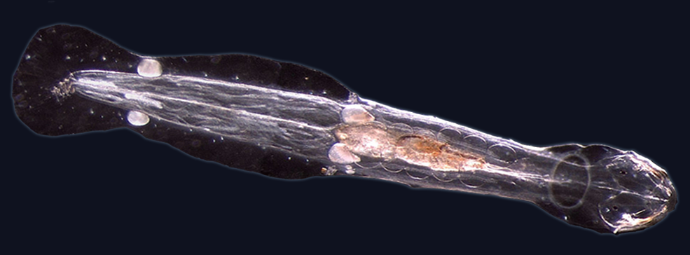
pedia/commons/8/8e/Chaetoblack.png
(pterobranchs {TARuBrANKS}, acorn
worms).
The free swimming younger form of
Pterobranch may evolve into tunicates
and then the first fish.

[1] Description Eichelwurm, Exemplar
aus der Sammlung des Institutes für
Zoologie, FU Berlin. GNU
FDL Date Source Foto:
de:Benutzer:Necrophorus Author User
Necrophorus on
de.wikipedia Permission (Reusing
this file) Released under the GNU Free
Documentation License. GNU
source: http://upload.wikimedia.org/wiki
pedia/commons/thumb/7/72/Eichelwurm.jpg/
1024px-Eichelwurm.jpg
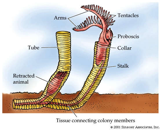
[2] Pterobranchs Resembling slugs
with hairy, branching tentacles,
Pterobranchs filter food from the water
and form colonies of “clones,” much
like coral polyps, often secreting a
network of hard tubing. Individual
zooids can crawl about freely within
the colony, but are connected to one
another by thin “cables,” quickly
retracting if disturbed. What makes the
Pterobranchs even stranger than corals
is that these slimy, slithering weirdos
are “hemichordates,” closer to us
vertebrates than to invertebrates like
worms and jellyfish. Read more:
http://www.toptenz.net/top-10-colonial-o
rganisms.php#ixzz1lJRtH61S COPYRIGHTED
source: http://www.toptenz.net/wp-conten
t/uploads/2011/10/Pterobranch-colonial-o
rganisms.jpg
evolves.
Chordates are a very large group that
include all tunicates {TUNiKiTS},
fishes, amphibians, reptiles, mammals,
and birds.
Chordates evolve upside down compared
to invertebrates, having nerve chord on
back and heart on front.

[1] from adelaide, australia UNKNOWN
source: http://news.bbc.co.uk/1/hi/sci/t
ech/3208583.stm
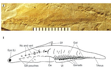
[2] [t Note that this is a vertebrate
- not a pre-vertebrate
chordate] Portion of figure
from: D.-G. Shu, S. Conway Morris, J.
Han, Z.-F. Zhang, K. Yasui, P. Janvier,
L. Chen, X.-L. Zhang, J.-N. Liu, Y. Li
and H.-Q. Liu, ''Head and backbone of
the Early Cambrian vertebrate
Haikouichthys'', Nature 421,
526-529(30 January
2003) http://www.nature.com/nature/jour
nal/v421/n6922/full/nature01264.html CO
PYRIGHTED
source: https://nature.com/journal/v421/
n6922/images/nature01264-f1.2.jpg
Tunicates {TUNiKiTS} evolve (sea
squirts).

[1] Description Clavelina
moluccensis, the bluebell
tunicate English: Tunicate colony.
(Clavelina moluccensis) Date
04/17/05 Source Own
work Author Nhobgood CC
source: http://upload.wikimedia.org/wiki
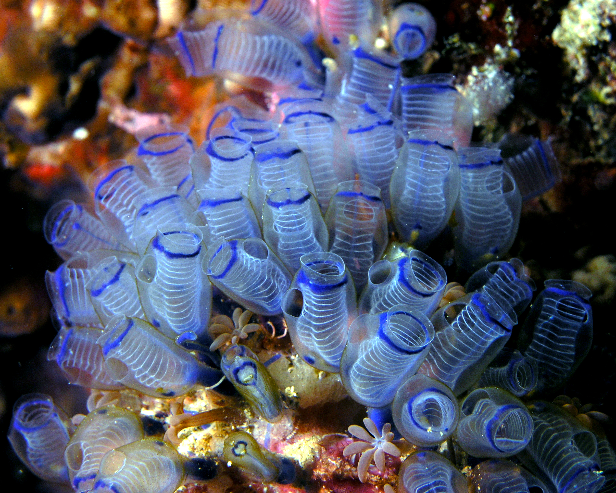
pedia/commons/9/98/Bluebell_tunicates_Ni
ck_Hobgood.jpg

[2] Timeline of phylogeny of animals,
figure 6 from: S. Blair Hedges, ''The
origin and evolution of model
organisms'', Nature Reviews Genetics 3,
838-849 (November
2002) http://www.nature.com/nrg/journal
/v3/n11/full/nrg929.html {Hedges_2002.p
df} a) The relationships and
divergence times (millions of years ago
(Mya) plusminus one standard error) of
selected model animals are shown, based
on recent multigene and multiprotein
studies51, 61, 84. The fossil
divergence time of birds and mammals
(310 Mya) was used to calibrate the
molecular clock. Branch lengths are not
proportional to time. b ) The
relationships and numbers of living
species, from a diversity of sources in
most of the main groups. COPYRIGHTED
source: http://www.nature.com/nrg/journa
l/v3/n11/images/nrg929-f6.jpg
Ningqiang County, Shaanxi
Province

[1] Cloudina COPYRIGHTED
source: http://palaeos.com/proterozoic/n
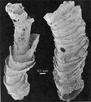
eoproterozoic/ediacaran/images/Cloudina.
jpg
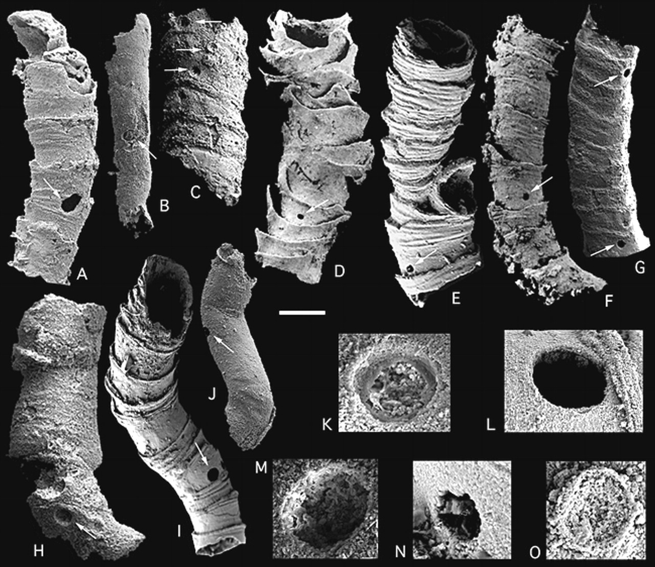
[2] Cloudina from: HONG HUA, BRIAN R.
PRATT, and LU-YI ZHANG, ''Borings in
Cloudina Shells: Complex Predator-Prey
Dynamics in the Terminal
Neoproterozoic'', PALAIOS, October
2003, v. 18, p. 454-459,
doi:10.1669/0883-1351(2003)018<0454:BICSCP>2.0.CO;2
http://palaios.geoscienceworld.org/citmg
r?gca=palaios;18/4-5/454 COPYRIGHTED
source: http://palaios.geoscienceworld.o
rg/content/vol18/issue4-5/images/large/i
0883-1351-018-04-0454-f03.jpeg
{eK-DiS-u-ZOu} (animals that molt {lose
their outer skin} as they grow).

[1] Description English: Life
restoration of Ottoia in natural
environment with nearby
Haplophrentis. Date 11-29-08 Source
Own work Author Smokeybjb GNU
source: http://upload.wikimedia.org/wiki
pedia/commons/1/10/Ottoia_burrowing.jpg
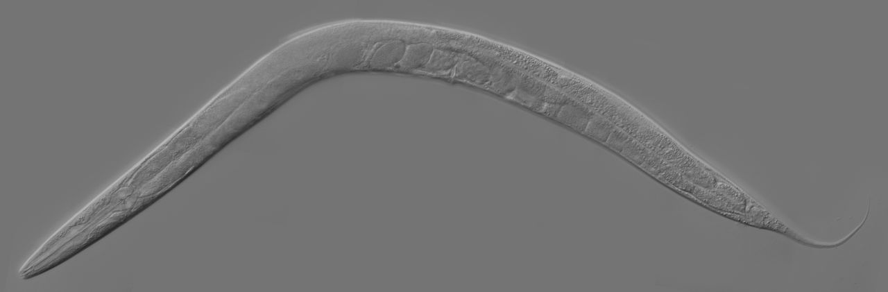
[2] Description
en:category:Caenorhabditis
elegans Date 2006-09-06 (original
upload date) (Original text :
09/05/2006) Source Originally from
en.wikipedia; description page is/was
here. (Original text : Donated by
Zeynep F. Altun) Author Original
uploader was Kbradnam at
en.wikipedia (Original text : Zeynep
F. Altun, Editor of
www.wormatlas.org) Permission (Reusing
this file) CC-BY-SA-2.5. CC
source: http://upload.wikimedia.org/wiki
pedia/commons/thumb/c/cc/Adult_Caenorhab
ditis_elegans.jpg/1280px-Adult_Caenorhab
ditis_elegans.jpg
{Lu-Fo-Tro-Ku-ZO-u} evolve.

[1] A rotifer. The cilia around
this rotifer's mouth are unusually
long; they reach as far as the strand
of spirogyra to the right. 10×
objective, 15× eyepiece. The numbered
ticks on the scale are 122 µM apart.
COPYRIGHTED
source: http://www.sciencephoto.com/imag
e/121893/530wm/C0058380-Rotifer_SEM-SPL.
jpg

[2] Description Clams Date
Source Own work Author
Marlith CC
source: http://upload.wikimedia.org/wiki
pedia/commons/8/8f/Clams.JPG

[1] Lancelet (Branchiostoma
lanceolatum) Description
Branchiostoma lanceolatum (Pallas,
1774) English: Amphioxus from course
sandy sediments (600µm) on the Belgian
continental shelf. Length: ~22
mm. Geo-location not applicable as the
picture was taken in the
lab. Français : Branchiostoma
lanceolatum, un céphalochordé,
récolté dans des sédiments de sable
grossier (600µm) sur le Plateau
continental belge. Longueur totale: 22
mm environ. Date 1997 Source
Own work Author (Hans
Hillewaert) CC
source: http://upload.wikimedia.org/wiki
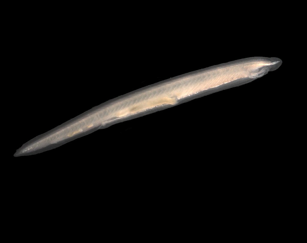
pedia/commons/4/47/Branchiostoma_lanceol
atum.jpg
{laNSleTS}. First liver and kidney.
[1] Lancelet (Branchiostoma
lanceolatum) Description
Branchiostoma lanceolatum (Pallas,
1774) English: Amphioxus from course
sandy sediments (600µm) on the Belgian
continental shelf. Length: ~22
mm. Geo-location not applicable as the
picture was taken in the
lab. Français : Branchiostoma
lanceolatum, un céphalochordé,
récolté dans des sédiments de sable
grossier (600µm) sur le Plateau
continental belge. Longueur totale: 22
mm environ. Date 1997 Source
Own work Author (Hans
Hillewaert) CC
source: http://upload.wikimedia.org/wiki
pedia/commons/4/47/Branchiostoma_lanceol
atum.jpg

[2] Lancelet COPYRIGHTED
source: http://kentsimmons.uwinnipeg.ca/
16cm05/1116/34-04b-Lancelet.jpg

[1] Description English: Priapulid
worm Priapulus caudatus in a Petry
dish. The specimen was found in the
intertidal of the Russian coast of the
Barents Sea. Русский:
Приапулида Priapulus caudatus
в чашке Петри. Особь
найдена в
приливно-отливной
зоне на российском
побережье Баренцева
моря. Date between 2005 and
2007 Source kindly granted by the
author Author Dmitry
Aristov Permission (Reusing this
file) See below. CC
source: http://upload.wikimedia.org/wiki
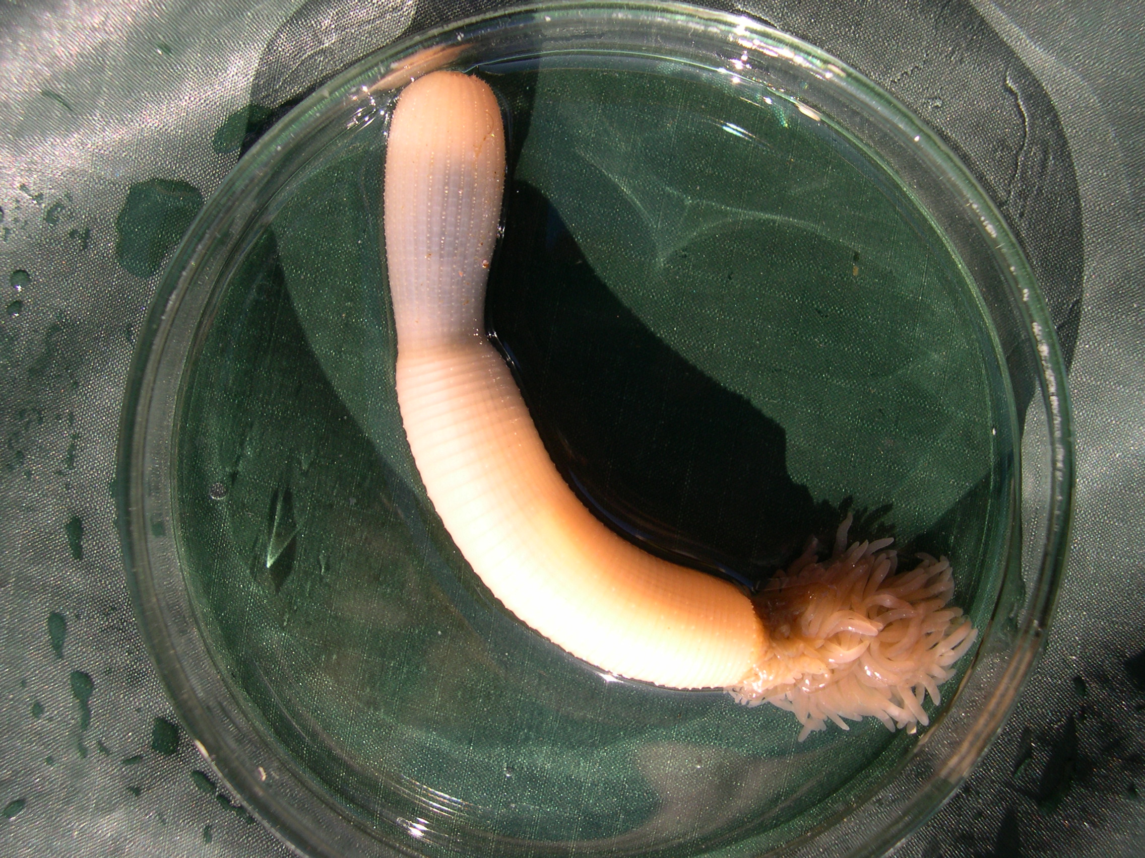
pedia/commons/6/62/Priapulus_caudatus.jp
g
[2] Giribet, G. (2008). Assembling the
lophotrochozoan (=spiralian) tree of
life. Philosophical Transactions of the
Royal Society B: Biological Sciences ,
363 (1496), 1513-1522. URL
http://dx.doi.org/10.1098/rstb.2007.2241
http://rstb.royalsocietypublishing.org
/content/363/1496/1513 COPYRIGHTED
source: http://rstb.royalsocietypublishi
ng.org/content/363/1496/1513
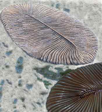
[1] Dikinsonia grew to a length of as
much as two feet (60 cm), which made it
one of the larger complex organisms of
the Vendian. It's body is segmented
with midline symmetry dividing it's
body. Its body may have been denser
than modern jellyfish or worms. [Atlas
of Prehistoric World, Discovery
Books Reconstruction of Dickinsonia,
based on images from Atlas of the
Prehistoric World, Discovery Channel
Books and Kingfisher Illustrated
Dinosaur Encyclopedia UNKNOWN
source: http://paleontology.edwardtbabin
ski.us/vendian/dickinsonia.jpg

[2] Spriggina Spriggina was
definitely a predator of the seas of
that time. UNKNOWN
source: http://www.museum.toulouse.fr/IM
G/jpg/spriginna_72dpi_680.jpg
"Cambrian explosion"), the rapid
diversification of multicellular
animals between 542 and 530 million
years ago that results in the
appearance of many of the major phyla
of animals. An increase of animals with
shells.
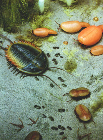
[1] Artist drawing of the bottom of the
Cambrian shallow sea floor, showing
trilobites (imagine these crawling
around on the Cambrian sea floor at
Devil's Lake state park 550 m.y. ago!)
(above). UNKNOWN
source: http://www.geology.wisc.edu/home
pages/g100s2/public_html/Geologic_Time/L
3_Cambrian_Life_More.jpg
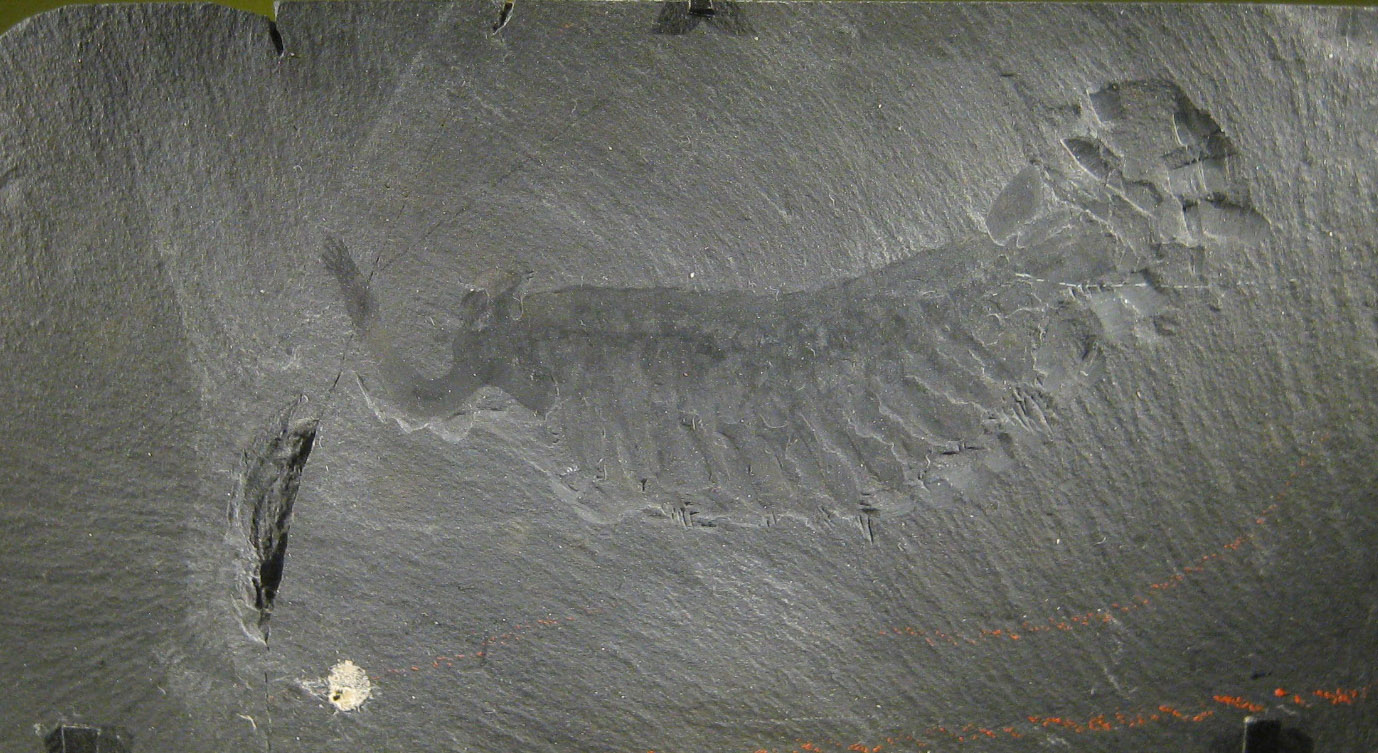
[2] Description English: Fossil
specimen of Opabinia regalis from the
Burgess shale on display at the
Smithsonian in Washington, DC. This
appears to be the exact specimen
pictured in Fig. 42 of 'The Crucible of
Creation: The Burgess Shale and the
Rise of Animals', by Simon Conway
Morris, Oxford University Press,
1998. Date 12 April 2009 (original
upload date) Source Transferred
from en.wikipedia; transferred to
Commons by User:FunkMonk using
CommonsHelper. Author Original
uploader was Jstuby at en.wikipedia PD
source: http://upload.wikimedia.org/wiki
pedia/commons/5/50/Opabinia_smithsonian.
JPG
Platyhelminthes {PlaTEheLmiNtEZ}
(flatworms).

[1] Description English: The
flatworm Pseudoceros dimidiatus. North
Horn, Osprey Reef, Coral Sea. Date
August 9, 2005 Source
Flickr Author Richard
Ling CC
source: http://upload.wikimedia.org/wiki
pedia/commons/1/1e/Pseudoceros_dimidiatu
s.jpg

[2] Two turbellarians mating by penis
fencing. Each has two penises, the
white spikes on the undersides of their
heads. Description English: Two
Individuals of Pseudobiceros bedfordi
about to have a Sperm Battle. –
Species of the flatworm genus
Pseudobiceros are hermaphroditic and
have two penises that are used to
inject sperm into the partner. P.
bedfordi is exceptional in that it
applies sperm onto the partner's skin
rather than injecting it. Deutsch:
Zwei Plattwürmer (Pseudobiceros
bedfordi) vor der Begattung. Der
doppelte Penis ist bei beiden
Individuen gut sichtbar. Date
Published: 2004-06-15 Source
Whitfield J: Everything You Always
Wanted to Know about Sexes. PLoS Biol
2/6/2004: e183.
doi:10.1371/journal.pbio.0020183.g001,
photo page Author Photo courtesy
of Nico Michiels. CC
source: http://upload.wikimedia.org/wiki
pedia/commons/3/38/Flatworm_sex.png
{rADEOlaREo}.
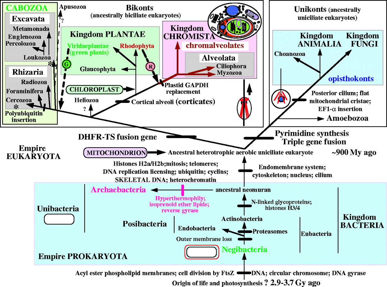
[1] FIG. 2. The tree of life based on
molecular, ultrastructural and
palaeontological evidence. Contrary to
widespread assumptions, the root is
among the eubacteria, probably within
the double-enveloped Negibacteria, not
between eubacteria and archaebacteria
(Cavalier-Smith, 2002b); it may lie
between Eobacteria and other
Negibacteria (Cavalier-Smith, 2002b).
The position of the eukaryotic root has
been nearly as controversial, but is
less hard to establish: it probably
lies between unikonts and bikonts (Lang
et al., 2002; Stechmann and
Cavalier-Smith, 2002, 2003). For
clarity the basal eukaryotic kingdom
Protozoa is not labelled; it comprises
four major groups (alveolates, cabozoa,
Amoebozoa and Choanozoa) plus the small
bikont phylum Apusozoa of unclear
precise position; whether Heliozoa are
protozoa as shown or chromists is
uncertain (Cavalier-Smith, 2003b).
Symbiogenetic cell enslavement occurred
four or five times: in the origin of
mitochondria and chloroplasts from
different negibacteria, of
chromalveolates by the enslaving of a
red alga (Cavalier-Smith, 1999, 2003;
Harper and Keeling, 2003) and in the
origin of the green plastids of
euglenoid (excavate) and chlorarachnean
(cercozoan) algae-a green algal cell
was enslaved either by the ancestral
cabozoan (arrow) or (less likely) twice
independently within excavates and
Cercozoa (asterisks) (Cavalier-Smith,
2003a). The upper thumbnail sketch
shows membrane topology in the
chimaeric cryptophytes (class
Cryptophyceae of the phylum Cryptista);
in the ancestral chromist the former
food vacuole membrane fused with the
rough endoplasmic reticulum placing the
enslaved cell within its lumen (red) to
yield the complex membrane topology
shown. The large host nucleus and the
tiny nucleomorph are shown in blue,
chloroplast green and mitochondrion
purple. In chlorarachneans (class
Chlorarachnea of phylum Cercozoa) the
former food vacuole membrane remained
topologically distinct from the ER to
become an epiplastid membrane and so
did not acquire ribosomes on its
surface, but their membrane topology is
otherwise similar to the cryptophytes.
The other sketches portray the four
major kinds of cell in the living world
and their membrane topology. The upper
ones show the contrasting ancestral
microtubular cytoskeleton (ciliary
roots, in red) of unikonts (a cone of
single microtubules attaching the
single centriole to the nucleus, blue)
and bikonts (two bands of microtubules
attached to the posterior centriole and
an anterior fan of microtubules
attached to the anterior centriole).
The lower ones show the single plasma
membrane of unibacteria (posibacteria
plus archaebacteria), which were
ancestral to eukaryotes and the double
envelope of negibacteria, which were
ancestral to mitochondria and
chloroplasts (which retained the outer
membrane, red).
source: http://aob.oxfordjournals.org/cg
i/content/full/95/1/147/FIG2
[2] Fig. 1. A consensus phylogeny of
eukaryotes. The vast majority of
characterized eukaryotes, with the
notable exception of major subgroups of
amoebae, can now be assigned to one of
eight major groups. Opisthokonts (basal
flagellum) have a single basal
flagellum on reproductive cells and
flat mitochondrial cristae (most
eukaryotes have tubular ones).
Eukaryotic photosynthesis originated in
Plants; theirs are the only plastids
with just two outer membranes.
Heterokonts (different flagellae) have
a unique flagellum decorated with
hollow tripartite hairs (stramenopiles)
and, usually, a second plain one.
Cercozoans are amoebae with filose
pseudopodia, often living with in tests
(hard outer shells), some very
elaborate (foraminiferans). Amoebozoa
are mostly naked amoebae (lacking
tests), often with lobose pseudopodia
for at least part of their life cycle.
Alveolates have systems of cortical
alveoli directly beneath their plasma
membranes. Discicristates have discoid
mitochondrial cristae and, in some
cases, a deep (excavated) ventral
feeding groove. Amitochondrial
excavates lack substantial molecular
phylogenetic support, but most have an
excavated ventral feeding groove, and
all lack mitochondria. The tree shown
is based on a consensus of molecular
(1-4) and ultrastructural (16, 17) data
and includes a rough indication of new
ciPCR ''taxa'' (broken black lines)
(7-11). An asterisk preceding the taxon
name indicates probable paraphyletic
group.
source: http://www.sciencemag.org/cgi/co
ntent/full/300/5626/1703
{FOraMiniFRu}.
[1] FIG. 2. The tree of life based on
molecular, ultrastructural and
palaeontological evidence. Contrary to
widespread assumptions, the root is
among the eubacteria, probably within
the double-enveloped Negibacteria, not
between eubacteria and archaebacteria
(Cavalier-Smith, 2002b); it may lie
between Eobacteria and other
Negibacteria (Cavalier-Smith, 2002b).
The position of the eukaryotic root has
been nearly as controversial, but is
less hard to establish: it probably
lies between unikonts and bikonts (Lang
et al., 2002; Stechmann and
Cavalier-Smith, 2002, 2003). For
clarity the basal eukaryotic kingdom
Protozoa is not labelled; it comprises
four major groups (alveolates, cabozoa,
Amoebozoa and Choanozoa) plus the small
bikont phylum Apusozoa of unclear
precise position; whether Heliozoa are
protozoa as shown or chromists is
uncertain (Cavalier-Smith, 2003b).
Symbiogenetic cell enslavement occurred
four or five times: in the origin of
mitochondria and chloroplasts from
different negibacteria, of
chromalveolates by the enslaving of a
red alga (Cavalier-Smith, 1999, 2003;
Harper and Keeling, 2003) and in the
origin of the green plastids of
euglenoid (excavate) and chlorarachnean
(cercozoan) algae-a green algal cell
was enslaved either by the ancestral
cabozoan (arrow) or (less likely) twice
independently within excavates and
Cercozoa (asterisks) (Cavalier-Smith,
2003a). The upper thumbnail sketch
shows membrane topology in the
chimaeric cryptophytes (class
Cryptophyceae of the phylum Cryptista);
in the ancestral chromist the former
food vacuole membrane fused with the
rough endoplasmic reticulum placing the
enslaved cell within its lumen (red) to
yield the complex membrane topology
shown. The large host nucleus and the
tiny nucleomorph are shown in blue,
chloroplast green and mitochondrion
purple. In chlorarachneans (class
Chlorarachnea of phylum Cercozoa) the
former food vacuole membrane remained
topologically distinct from the ER to
become an epiplastid membrane and so
did not acquire ribosomes on its
surface, but their membrane topology is
otherwise similar to the cryptophytes.
The other sketches portray the four
major kinds of cell in the living world
and their membrane topology. The upper
ones show the contrasting ancestral
microtubular cytoskeleton (ciliary
roots, in red) of unikonts (a cone of
single microtubules attaching the
single centriole to the nucleus, blue)
and bikonts (two bands of microtubules
attached to the posterior centriole and
an anterior fan of microtubules
attached to the anterior centriole).
The lower ones show the single plasma
membrane of unibacteria (posibacteria
plus archaebacteria), which were
ancestral to eukaryotes and the double
envelope of negibacteria, which were
ancestral to mitochondria and
chloroplasts (which retained the outer
membrane, red).
source: http://aob.oxfordjournals.org/cg
i/content/full/95/1/147/FIG2
[2] Fig. 1. A consensus phylogeny of
eukaryotes. The vast majority of
characterized eukaryotes, with the
notable exception of major subgroups of
amoebae, can now be assigned to one of
eight major groups. Opisthokonts (basal
flagellum) have a single basal
flagellum on reproductive cells and
flat mitochondrial cristae (most
eukaryotes have tubular ones).
Eukaryotic photosynthesis originated in
Plants; theirs are the only plastids
with just two outer membranes.
Heterokonts (different flagellae) have
a unique flagellum decorated with
hollow tripartite hairs (stramenopiles)
and, usually, a second plain one.
Cercozoans are amoebae with filose
pseudopodia, often living with in tests
(hard outer shells), some very
elaborate (foraminiferans). Amoebozoa
are mostly naked amoebae (lacking
tests), often with lobose pseudopodia
for at least part of their life cycle.
Alveolates have systems of cortical
alveoli directly beneath their plasma
membranes. Discicristates have discoid
mitochondrial cristae and, in some
cases, a deep (excavated) ventral
feeding groove. Amitochondrial
excavates lack substantial molecular
phylogenetic support, but most have an
excavated ventral feeding groove, and
all lack mitochondria. The tree shown
is based on a consensus of molecular
(1-4) and ultrastructural (16, 17) data
and includes a rough indication of new
ciPCR ''taxa'' (broken black lines)
(7-11). An asterisk preceding the taxon
name indicates probable paraphyletic
group.
source: http://www.sciencemag.org/cgi/co
ntent/full/300/5626/1703
{ne-mR-TEu} (ribbon worms).

[1] Description English: Basiodiscus
mexicanus was photographed at Los
Arcos, near Puerto Vallarta,
Mexico Date Source University
of California Museum of Paleology:
Introduction to the Nemertini Author
Chris Meyer and Allen
Collins Permission (Reusing this
file) See below. PD
source: http://upload.wikimedia.org/wiki
pedia/commons/4/49/Nemertea_Basiodiscus_
mexicanus.png
[2] Timeline of phylogeny of animals,
figure 6 from: S. Blair Hedges, ''The
origin and evolution of model
organisms'', Nature Reviews Genetics 3,
838-849 (November
2002) http://www.nature.com/nrg/journal
/v3/n11/full/nrg929.html {Hedges_2002.p
df} a) The relationships and
divergence times (millions of years ago
(Mya) plusminus one standard error) of
selected model animals are shown, based
on recent multigene and multiprotein
studies51, 61, 84. The fossil
divergence time of birds and mammals
(310 Mya) was used to calibrate the
molecular clock. Branch lengths are not
proportional to time. b ) The
relationships and numbers of living
species, from a diversity of sources in
most of the main groups. COPYRIGHTED
source: http://rstb.royalsocietypublishi
ng.org/content/363/1496/1513
{ToRDiGRADZ}.

[1] Description Willow Gabriel and
Bob Goldstein,
http://tardigrades.bio.unc.edu/ Date
2007-05-20 (original upload
date) CC
source: http://28.media.tumblr.com/tumbl
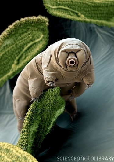
r_limfh2NXtC1qc6j5yo1_400.jpg
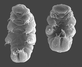
[2] from Giribet 2007
source: http://upload.wikimedia.org/wiki
pedia/commons/6/65/Hypsibiusdujardini.jp
g
{oniKoFereNS}.

[1] Euperipatoides kanangrensis on a
eucalyptus log, in which it normally
resides. Description English:
Cropped version of File:Euperipatoides
kanangrensis.jpg Date 13 October
2009 CC
source: http://upload.wikimedia.org/wiki
pedia/commons/6/67/Euperipatoides_kanang
rensis_crop.jpg
[2] Figure from: Giribet, G. (2008).
Assembling the lophotrochozoan
(=spiralian) tree of life.
Philosophical Transactions of the Royal
Society B: Biological Sciences , 363
(1496), 1513-1522. URL
http://dx.doi.org/10.1098/rstb.2007.2241
http://rstb.royalsocietypublishing.org
/content/363/1496/1513 COPYRIGHTED
source: http://rstb.royalsocietypublishi
ng.org/content/363/1496/1513
bilaterians.

[1] From: Ruppert, Fox, Barnes,
''Invertebrate Zoology'',
2004. COPYRIGHTED
source: Ruppert, Fox, Barnes,
"Invertebrate Zoology", 2004.

[2] From: Ruppert, Fox, Barnes,
''Invertebrate Zoology'',
2004. COPYRIGHTED
source: Ruppert, Fox, Barnes,
"Invertebrate Zoology", 2004.
Mollusks includes snails, clams,
mussels, and the cephalopods: squids
and octopuses.

[1] From: Ruppert, E.E., Fox, R.S.,
and Barnes, R.D. (2004). Invertebrate
Zoology (7 ed.). Brooks / Cole. pp.
284–291. ISBN 0030259827. PD
source: http://en.wikipedia.org/wiki/Mol
lusca
[2] Description Clams Date
Source Own work Author
Marlith CC
source: http://upload.wikimedia.org/wiki
pedia/commons/8/8f/Clams.JPG
worms).
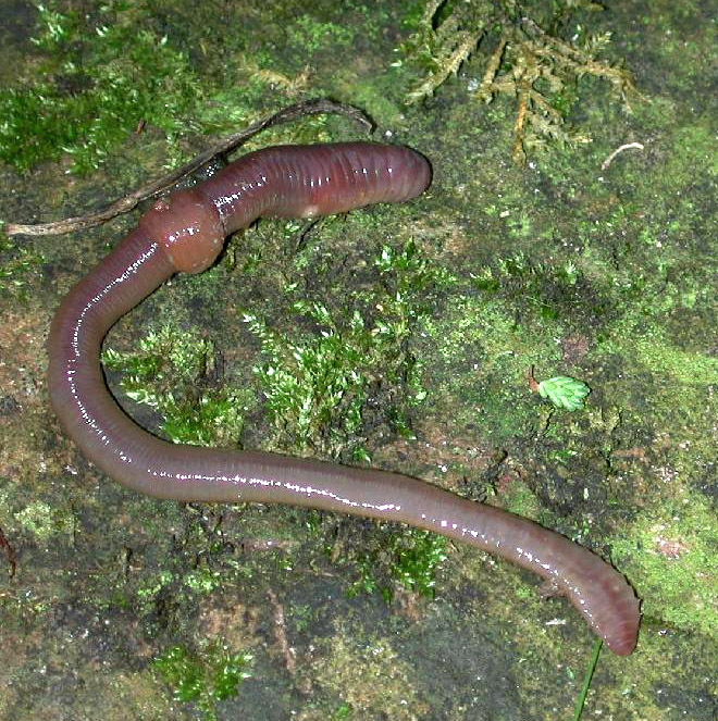
[1] An earthworm's clitellum they have
a unique reproductive organ, the
ring-shaped clitellum (''pack saddle'')
round their bodies, which produces a
cocoon that stores and nourishes
fertilized eggs until they
hatch Description Regenwurm mit
Clitellum - (sattelförmige Verdickung
im vorderen Drittel).Das Sekret der
Clitellum-Drüsen dient u. a. zur
Bildung dieses Ei-Kokons. Français :
Ver de terre (Oligochaeta,
Lumbricina) Svenska: Daggmask
(Lumbricus spec.) Русский:
Дождевой червь (род
Лумбрикус) Date Source
first upload in de wikipedia on
09:58, 16. Feb 2005 by Michael
Linnenbach Author Michael
Linnenbach GNU
source: http://upload.wikimedia.org/wiki
pedia/commons/3/30/Regenwurm1.jpg
[2] Figure from: Giribet, G. (2008).
Assembling the lophotrochozoan
(=spiralian) tree of life.
Philosophical Transactions of the Royal
Society B: Biological Sciences , 363
(1496), 1513-1522. URL
http://dx.doi.org/10.1098/rstb.2007.2241
http://rstb.royalsocietypublishing.org
/content/363/1496/1513 COPYRIGHTED
source: http://rstb.royalsocietypublishi
ng.org/content/363/1496/1513
(crustaceans, insects).

[1] Extinct and modern
arthropods English: Arthropoda
collage. From left to right and from
top to bottom: Kolihapeltis,
Stylonurus, Scorpion, Crab, Centipede,
Butterfly CC
source: http://upload.wikimedia.org/wiki
pedia/commons/8/80/Arthropoda.jpg
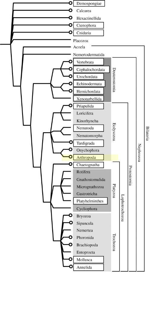
[2] Figure from: Giribet, G. (2008).
Assembling the lophotrochozoan
(=spiralian) tree of life.
Philosophical Transactions of the Royal
Society B: Biological Sciences , 363
(1496), 1513-1522. URL
http://dx.doi.org/10.1098/rstb.2007.2241
http://rstb.royalsocietypublishing.org
/content/363/1496/1513 COPYRIGHTED
source: http://rstb.royalsociorg/content
/363/1496/1513
contains most fishes, and all
amphibians, reptiles, mammals, and
birds.
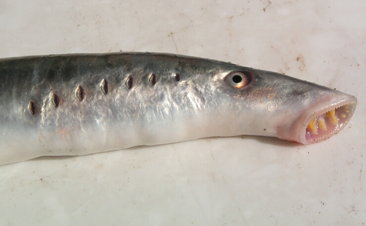
[1] Description Lampetra
fluviatilis from the german
northsea Date 2004 Source
Germany Author
M.Buschmann Permission (Reusing
this file) Author is owner CC
source: http://upload.wikimedia.org/wiki
pedia/commons/3/3f/Lampetra_fluviatilis.
jpg

[2] Description Clockwise,
starting from top left: 1. Fire
Salamander (Salamandra salamandra) 2.
Saltwater Crocodile (Crocodylus
porosus) 3. Southern Cassowary
(Casusarius casuarius) 4.
Black-and-rufus Giant Elephant Shrew
(Rhynchocyon petersi) 5. Ocean Sunfish
(Mola mola) Date CC
source: http://upload.wikimedia.org/wiki
pedia/commons/e/ec/Vertebrates.png
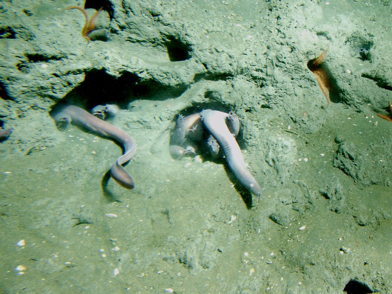
[1] Description English: Pacific
hagfish (Eptatretus stoutii) in a hole
at 150 meters depth. Latitude 37 58 N.,
Longitude 123 27 W. Location:
California, Cordell Bank National
Marine Sanctuary. Date Last Updated:
September 30, 2009. Source
http://www.photolib.noaa.gov/htmls/sanc
1692.htm
http://www.photolib.noaa.gov/bigs/sanc16
92.jpg Author Linda Snook, National
Oceanic and Atmospheric Administration
(NOAA) / Cordell Bank National Marine
Sanctuary (CBNMS) PD
source: http://upload.wikimedia.org/wiki
pedia/commons/thumb/5/52/Eptatretus_stou
tii.jpg/1280px-Eptatretus_stoutii.jpg
[2] Description Lampetra
fluviatilis from the german
northsea Date 2004 Source
Germany Author
M.Buschmann Permission (Reusing
this file) Author is owner CC
source: http://upload.wikimedia.org/wiki
pedia/commons/3/3f/Lampetra_fluviatilis.
jpg
(KeliSuroTo) (horseshoe crabs, mites,
spiders, scorpions).
Sweden

[1] Description English: Horseshoe
crab dorsal and ventral Italiano:
Limulus polyphemus dorsale e
ventrale Date 10 April 2009 Source
Own work Author Ricce PD
source: http://upload.wikimedia.org/wiki
pedia/commons/thumb/4/48/Limulo_dorsale_
e_ventrale.jpg/1280px-Limulo_dorsale_e_v
entrale.jpg

[2] taken from en:Image:Horseshoe crab
female.jpg Dead female horseshoe crab
from NOAA Photo Library: Image ID:
line2632, America's Coastlines
Collection Location: Patuxent River,
Maryland Photo Date: 2002 August
17 Photographer: Mary Hollinger,
NESDIS/NODC biologist, NOAA PD
source: http://upload.wikimedia.org/wiki
pedia/commons/1/1b/Horseshoe_crab_female
.jpg
(iKIniDRMS } (sea cucumbers, sea
urchins, sand dollars, star fish).

[1] Kachemak Bay National Estuarine
Research Reserve. A beautiful array of
starfish , sea urchins and mussel
shells in the rocky intertidal zone of
Kachemak Bay. Image ID: nerr0878,
NOAA National Estuarine Research
Reserve Collection from NOAA:
http://www.photolib.noaa.gov/nerr/nerr08
78.htm PD
source: http://upload.wikimedia.org/wiki
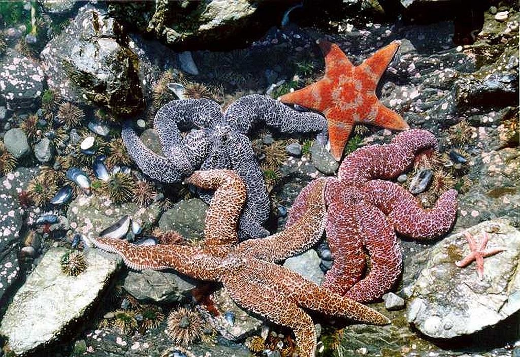
pedia/commons/thumb/e/e9/Nerr0878.jpg/10
24px-Nerr0878.jpg

[2] Description English: The first
in a sequence of three photos that show
a brittle star flipping itself
rightside-up. Date 1 May
2011 Source Own work Author
Alexcooper1 CC
source: http://upload.wikimedia.org/wiki
pedia/commons/thumb/c/c8/A_brittle_star_
flipping_itself_rightside-up.jpg/1024px-
A_brittle_star_flipping_itself_rightside
-up.jpg
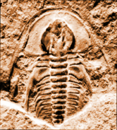
[1] example of earliest trilobites
(e.g., Fallotaspis longa) UNKNOWN
source: http://www.trilobites.info/biost
ratfallon.jpg
[2] Niles Eldredge, ''Trilobites and
Evolutionary Patterns'', p305-332 in
Anthony Hallam, ''Patterns of evolution
as illustrated by the fossil record,
Volume 5'', 1977,
p322. http://books.google.com/books?id=
q7GjDIyyWegC COPYRIGHTED
source: http://books.google.com/books?id
=q7GjDIyyWegC
crabs, lobsters, barnicles).
England
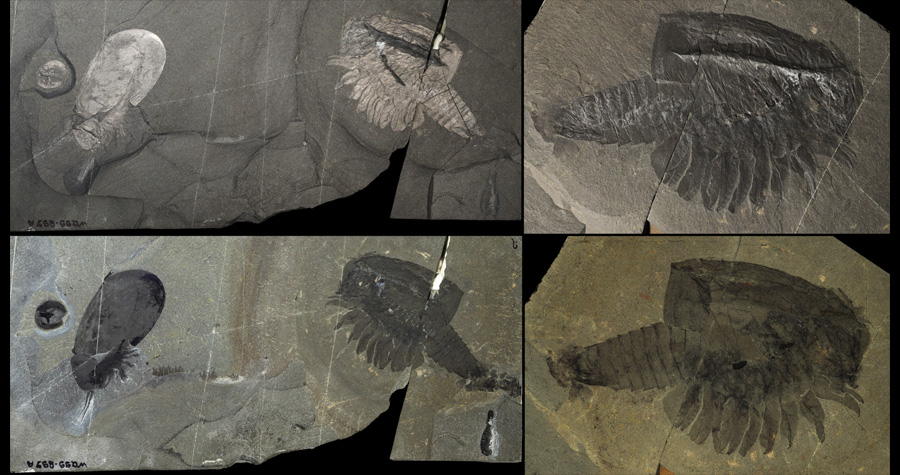
[1] Canadaspis perfecta (ROM 61119) –
Part and counterpart. Complete specimen
showing phosphatized gut diverticulae
and posterior dark stain (probably
representing decay fluids), lateral
view. Left images, complete slab (part)
showing associated species; Yohoia
tenuis (bottom right), Waptia
fieldensis (left, partially covered by
a disarticulated carapace of
Canadaspis), Burgessia bella (far
left). Right images, details of the
counterpart. Specimen length = 72 mm.
Specimen dry – direct light (top
row), dry – polarized light (bottom
left), wet – polarized light (bottom
right). Walcott Quarry. © Royal
Ontario Museum. Photos: Jean-Bernard
Caron COPYRIGHTED
source: http://www.burgess-shale.rom.on.
ca/images/zoomify/canadaspis-rom-61119.j
pg

[2] 3D model of Canadaspis
perfecta. COPYRIGHTED
source: http://burgess-shale.rom.on.ca/v
ideo/fossil-gallery/0b1-canadaspis-turnt
able.jpg
(centipedes and millipedes).
deposits)(Wheeler Formation) Utah, USA
and (Ust-Majan formation) East
Siberia|(earliest fossils) Shropshire,
England
[1] Description Lithobius
forficatus Deutsch: Steinläufer Date
9 August 2005 Source Own
work Author Darkone CC
source: http://upload.wikimedia.org/wiki
pedia/commons/7/79/Steinl%C3%A4ufer_%28L
ithobius_forficatus%29_3.jpg
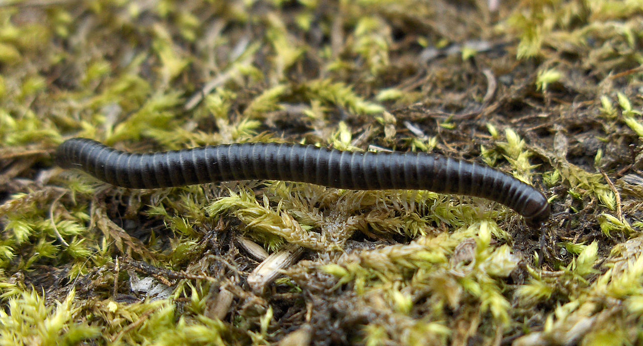
[2] Description Tachypodoiulus
niger Date 2007-06-28 Source Own
work Author Stemonitis CC
source: http://upload.wikimedia.org/wiki
pedia/commons/thumb/8/86/Tachypodoiulus_
niger_1.jpg/1280px-Tachypodoiulus_niger_
1.jpg
the number of genera {JeN-R-u} will
quadruple.

[1] Recreation of life during the
Ordovician UNKNOWN
source: http://ferrebeekeeper.files.word
press.com/2010/11/ordovician.jpg
[2] A second peak time in the
abundance of shell-surviving life forms
was in the Upper Ordovician (by this
time also, the first larger
vertebrates, fossil fish, had
appeared). Below are two illustrations:
the first, an artist' conception of
marine invertebrate life in the late
Ordovician; the second, a typical slab
of Ordovician limestone (from Indiana)
containing the fossil types listed in
its caption: PD
source: http://rst.gsfc.nasa.gov/Sect20/
ordovicsea.jpg
Bryophyta {BrIoFiTo}, (Liverworts,
Hornworts, Mosses).

[1] Phaeoceros laevis (L.) Prosk. gnu
source: http://en.wikipedia.org/wiki/Ima
ge:Anthoceros_levis.jpg

[2] English: A closeup shot of moss on
a rock in Beacon Hill Park, Victoria,
Canada. Sony Alpha A100 Date 25
March 2007 Source Own
work Author KirinX at
en.wikipedia Permission (Reusing this
file) CC-BY-SA-2.5. CC
source: http://upload.wikimedia.org/wiki
pedia/commons/1/1c/Moss_closeup.jpg

[1] Gray, J., Massa, D., & Boucot, A.
J. Caradocian land plant microfossils
from libya. Geology , April 1982, 10
(4), 197-201. URL
http://dx.doi.org/10.1130/0091-7613(1982
)10<197:CLPMFL>2.0.CO;2 http://geology.gsapubs.org/
content/10/4/197.abstract?sid=dadb8801-c
fd4-4eb4-b70e-95cb217113e4 {Gray_Jane_1
98204xx.pdf} COPYRIGHTED
source: http://geology.gsapubs.org/conte
nt/10/4/197.abstract?sid=dadb8801-cfd4-4
eb4-b70e-95cb217113e4
[2] Phaeoceros laevis (L.) Prosk. gnu
source: http://en.wikipedia.org/wiki/Ima
ge:Anthoceros_levis.jpg
arthropods Myriapoda (centipedes and
millipedes).
Ontario, Canada
[1] Figure 4. Field photographs of
representative trackways. Scale bars
represent 5 cm. A: Trackway with
central drag and well-defined appendage
marks. Bottom surface. B: Trackway with
central drag and poorly defined
appendage marks. Top surface. Surface
dips to top of photograph; note downdip
offset of central drag. C: Robust
trackway with well-developed appendage
marks and no central drag. Note
push-ups of sand (arrows) associated
with appendage impressions. Figure 4
from: MacNaughton, Robert B., Jennifer
M. Cole, Robert W. Dalrymple, Simon J.
Braddy, Derek E.G. Briggs, and Terrence
D. Lukie. “First Steps on Land:
Arthropod Trackways in
Cambrian-Ordovician Eolian Sandstone,
Southeastern Ontario, Canada.”
Geology 30, no. 5 (May 2002): 391
–394. http://geology.geoscienceworld.
org/citmgr?gca=geology;30/5/391 COPYRIG
HTED
source: http://geology.geoscienceworld.o
rg/citmgr?gca=geology;30/5/391

[2] Figure 2 from: Heather M. Wilson
and Lyall I. Anderson, ''Morphology and
Taxonomy of Paleozoic Millipedes
(Diplopoda: Chilognatha: Archipolypoda)
from Scotland'', Journal of
Paleontology, Vol. 78, No. 1 (Jan.,
2004), pp.
169-184 http://www.jstor.org/stable/409
4847 {Anderson_Lyall_200401xx.pdf} COP
YRIGHTED
source: http://www.jstor.org/stable/4094
847?&Search=yes&searchText=MILLIPEDES&se
archText=TAXONOMY&searchText=MORPHOLOGY&
searchText=PALEOZOIC&list=hide&searchUri
=%2Faction%2FdoBasicSearch%3FQuery%3DMOR
PHOLOGY%2BAND%2BTAXONOMY%2BOF%2BPALEOZOI
C%2BMILLIPEDES%26acc%3Don%26wc%3Don&prev
Search=&item=2&ttl=43&returnArticleServi
ce=showFullText
Gnathostomata {no toST omoTo} First
vertebrate teeth.

[1] Image from: Palmer, D. The
Marshall Illustrated Encyclopedia of
Dinosaurs & Prehistoric Animals: A
Comprehensive Color Guide to Over 500
Species. New Line Books,
2002. COPYRIGHTED
source: Palmer, D. The Marshall
Illustrated Encyclopedia of Dinosaurs &
Prehistoric Animals: A Comprehensive
Color Guide to Over 500 Species. New
Line Books, 2002.

[2] Kardong, ''Vertebrates'', Third
Edition, 2002. COPYRIGHTED
source: Kardong, "Vertebrates", Third
Edition, 2002.
(ancestor of all sharks, rays, skates,
and sawfishes).
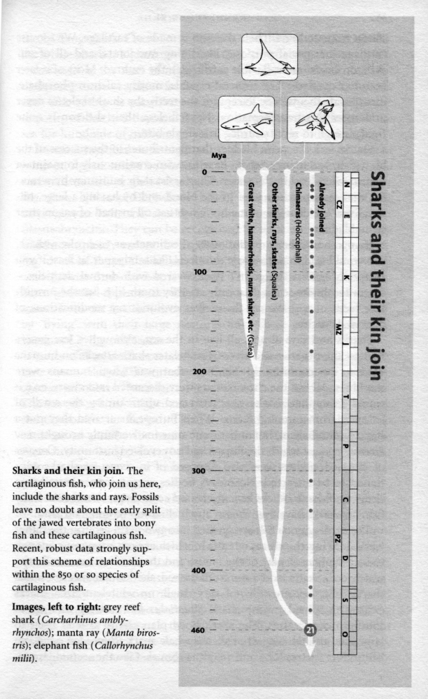
[1] Richard Dawkins, ''The Ancestor's
Tale'', (Boston, MA: Houghton Mifflin
Company, 2004), p360-363. COPYRIGHTED
source: Richard Dawkins, "The
Ancestor's Tale", (Boston, MA: Houghton
Mifflin Company, 2004), p360-363.

[2] Miller, Randall F., Richard
Cloutier, and Susan Turner. “The
Oldest Articulated Chondrichthyan from
the Early Devonian Period.” Nature
425.6957 (2003): 501–504. Web. 23 May
2012. http://www.nature.com/nature/jour
nal/v425/n6957/full/nature02001.html {M
iller_Chondrichthyans_2003.pdf} COPYRIG
HTED
source: http://www.nature.com/nature/jou
rnal/v425/n6957/full/nature02001.html
of all terrestrial fungi.

[1] Earliest Glomerales fossil fig 1
from: Redecker D, Kodner R, Graham LE.
(2000). ''Glomalean fungi from the
Ordovician''. Science 289 (5486):
1920–21. Bibcode 2000Sci...289.1920R.
doi:10.1126/science.289.5486.1920. PMID
10988069. http://www.jstor.org/stable/3
077684 COPYRIGHTED
source: http://www.jstor.org/stable/3077
684
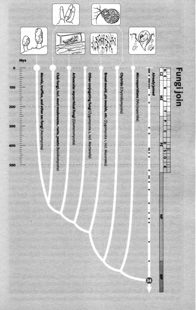
[2] Phylogenetic tree from: Richard
Dawkins, ''The Ancestor's Tale'',
(Boston, MA: Houghton Mifflin Company,
2004), p511. COPYRIGHTED
source: Richard Dawkins, "The
Ancestor's Tale", (Boston, MA: Houghton
Mifflin Company, 2004), p511.
{GlO-mi-rO-mI-KO-Tu}.

[1] Gigaspora margarita in association
with Lotus corniculatus Description
Lotus corniculatus var. japonicus
kolonisiert durch Gigaspora
margarita Date 18 September
2007 Source Own work Author
Mike Guether GNU
source: http://upload.wikimedia.org/wiki
pedia/commons/thumb/c/c7/Gigaspora_marga
rita.JPG/1024px-Gigaspora_margarita.JPG

[2] germinating Gigaspora decipiens
source: http://pages.unibas.ch/bothebel/
people/redecker/ff/glomero.htm

[1] Description Equisetum telmateia
(Equisetopsida) at Cambridge Botanic
Garden Date 18 May 2008 Source Own
work Author Rror Other versions
Derivative works of this file:
species on earth.jpg GNU
source: http://upload.wikimedia.org/wiki
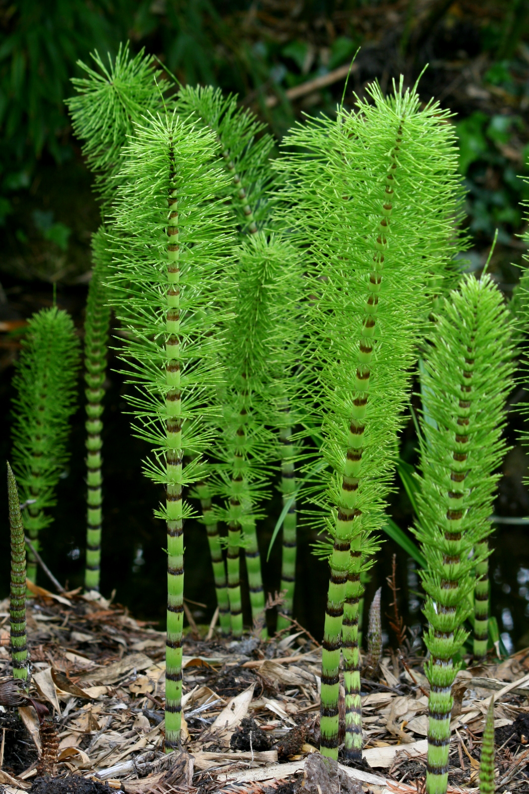
pedia/commons/7/7c/Equisetopsida.jpg

[2] Fig. 2. Chronogram showing
estimates of phylogenetic relationships
and divergence times among the major
groups of extant land plants. The
estimate of relationships is
synthesized from the following papers
in this issue: Burleigh and Mathews
(2004) , Pryer et al. (2004) , Shaw and
Renzaglia (2004) , and Soltis and
Soltis (2004) . Divergence time
estimates are mostly based on analyses
of molecular data with fossil
constraints (Wikström et al., 2001 ;
Pryer et al., 2004 ) and are augmented
by fossil evidence (Kenrick and Crane,
1997 ; Wellman et al., 2003 ).
Estimates of the number of species in
each group are from Judd et al. (2002)
and W. S. Judd (personal
communication). Groups covered by a
particular article in this special
issue are circled and connected to the
names of the article's authors. ''Other
conifers'' refers to the clade
consisting of all conifers except for
Pinaceae (see Burleigh and Mathews,
2004 ). ''Lepto. ferns'' refers to
leptosporangiate ferns fig 2
from: Jeffrey D. Palmer, Douglas E.
Soltis and Mark W. Chase, ''The plant
tree of life: an overview and some
points of view'', American Journal of
Botany. 2004;91:1437-1445., (2004).
http://www.amjbot.org/content/91/10/14
37.full {Chase_Mark_2004.pdf}
COPYRIGHTED
source: http://www.amjbot.org/content/91
/10/1437/F2.large.jpg

[1] Adapted from: Richard Dawkins,
''The Ancestor's Tale'', (Boston, MA:
Houghton Mifflin Company, 2004),
p339. COPYRIGHTED
source: Richard Dawkins, "The
Ancestor's Tale", (Boston, MA: Houghton
Mifflin Company, 2004), p339.

[2] A sturgeon
(pt:esturjāo). esturgeon noir
d'Amérique (Acipenser oxyrinchus
oxyrinchus) http://images.fws.gov/ PD
source: http://upload.wikimedia.org/wiki
pedia/commons/c/c2/Sturgeon2.jpg

[1] Image from: Palmer, D. The
Marshall Illustrated Encyclopedia of
Dinosaurs & Prehistoric Animals: A
Comprehensive Color Guide to Over 500
Species. New Line Books,
2002. COPYRIGHTED
source: Palmer, D. The Marshall
Illustrated Encyclopedia of Dinosaurs &
Prehistoric Animals: A Comprehensive
Color Guide to Over 500 Species. New
Line Books, 2002.

[2] Earliest fish with lung in
existance?[t] Nile Bichir (Polypterus
bichir bichir) from Günther, A.C.L.G.,
1880. An introduction to the study of
fishes. Today & Tomorrow's Book Agency,
New Delhi. GNU
source: http://upload.wikimedia.org/wiki
pedia/commons/e/e8/Nile_bichir.png

[1] Description Preserved
specimen of chalumnae (Also known as
Coelacanth [1]) in the Natural History
Museum, Vienna, Austria. Believed
to have been extinct for 70 million
years, this specimen was caught the 18
October of 1974, next to
Salimani/Selimani (Grande Comore,
Comoros Islands) 11°48′40.7″S
43°16′3.3″E Length: 170 cm -
Weight: 60 kg Obtained by stiching
3 HiRes images and removing the
background with image
post-processing. Date August
2007 Source Own work Author
Alberto Fernandez Fernandez GNU
source: http://upload.wikimedia.org/wiki
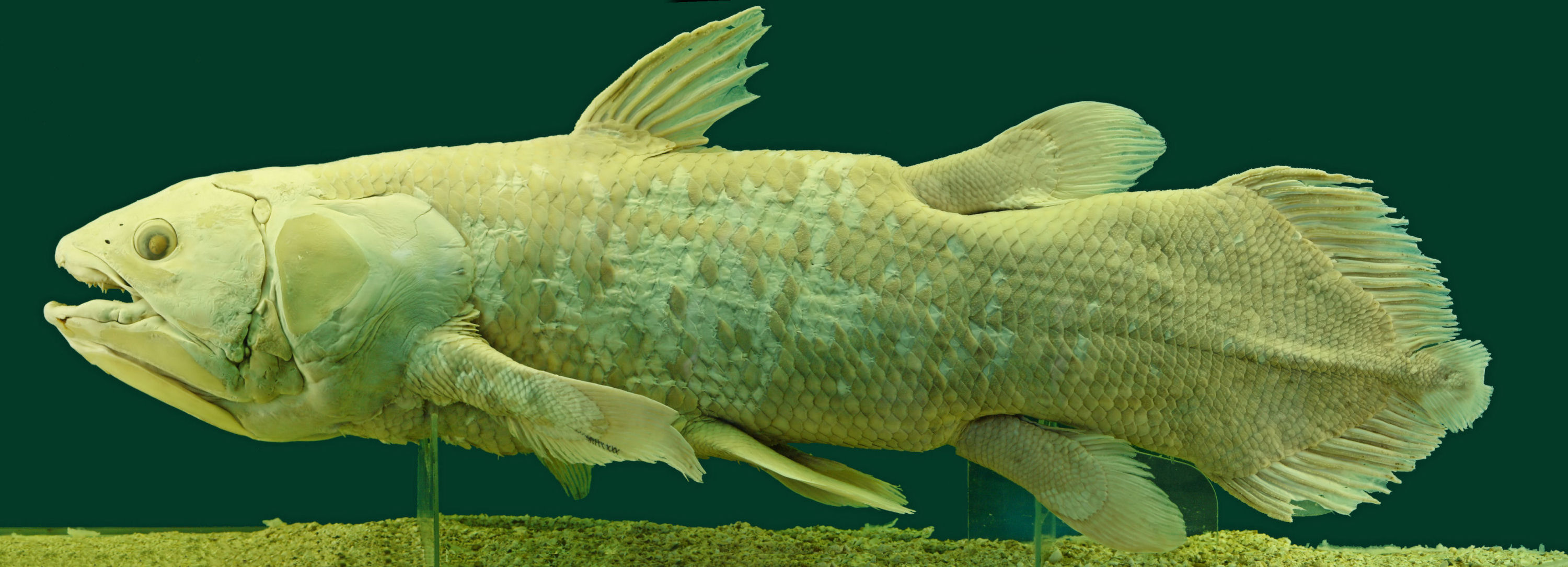
pedia/commons/f/fa/Latimeria_Chalumnae_-
_Coelacanth_-_NHMW.jpg
includes all insects).
Scotland
[1] Description Protura specimen,
taken under stereo microscope (40x).
Acerentomon sp. Date 7 December 2008,
03:13 Source Protura Uploaded
by Richard001 Author Gregor
?nidar CC
source: http://upload.wikimedia.org/wiki
_micrograph.jpg)
pedia/commons/b/bc/Protura_specimen_(Ace
rentomon_species)_micrograph.jpg

[2] Description English: Campodea
staphylinus, a dipluran. Photo by
Michel Vuijlsteke. Taken on May 9, 2006
at 4.09pm CEST in Gent, Belgium. Date
2007-07-08 (original upload
date) Source Transferred from
en.wikipedia Author Original uploader
was Mvuijlst at
en.wikipedia Permission (Reusing this
file) CC
source: http://upload.wikimedia.org/wiki
pedia/commons/2/2e/Diplura.jpg
Silverfish.
[1] Description Français : Groupe
de Petrobius maritimus sur falaise
supralittorale, Toull ar C'Hrabanoù,
Goulien, Finistère, Bretagne,
France Date 2 June 2010 Source Own
work Author Jymm PD
source: http://upload.wikimedia.org/wiki
pedia/commons/a/a4/Petrobius_maritimus_2
010-06-02.jpg

[2] Description Archaeognatha:
Machilidae, collected from Anglesey,
UK Date 2006-12-28 Source Own work
(own photo) Author
User:Stemonitis Permission (Reusing
this file) CC Attribution
ShareAlike 2.5 CC
source: http://upload.wikimedia.org/wiki
pedia/commons/4/42/Archaeognatha.jpg
{aS-KO-mI-KO-Tu} (yeasts, truffles,
Penicillium, morels {mu reLZ}).
Aberdeenshire, Scotland
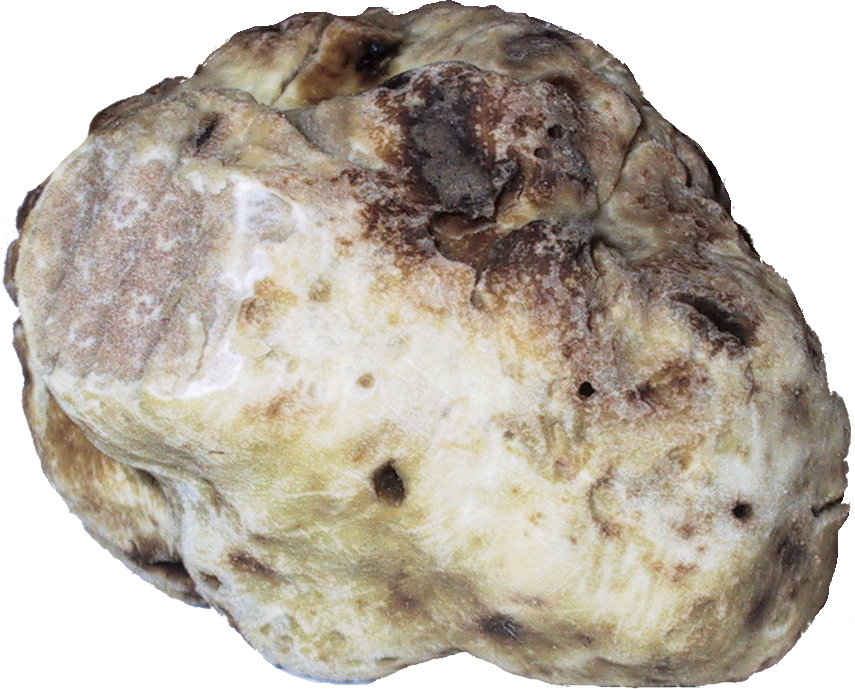
[1] white truffle
cutted photographed by
myself GNU head Permission is
granted to copy, distribute and/or
modify this document under the terms of
the GNU Free Documentation License,
Version 1.2 or any later version
published by the Free Software
Foundation; with no Invariant Sections,
no Front-Cover Texts, and no Back-Cover
Texts. A copy of the license is
included in the section entitled ''Text
of the GNU Free Documentation
License.''
source: http://upload.wikimedia.org/wiki
pedia/commons/f/fd/Truffle_washed_and_cu
tted.jpg
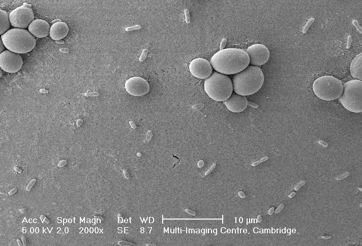
[2] EColi-Scerevisiae.jpg (50KB, MIME
type: image/jpeg) Wikimedia Commons
logo This is a file from the Wikimedia
Commons. The description on its
description page there is shown
below. Escherichia coli (little
forms) & Saccharomyces cerevisiae (big
forms) by MEB Public domain This file
has been released into the public
domain by the copyright holder, its
copyright has expired, or it is
ineligible for copyright. This applies
worldwide. brewer's yeast/baker's
yeast
source: http://en.wikipedia.org/wiki/Ima
ge:EColi-Scerevisiae.jpg
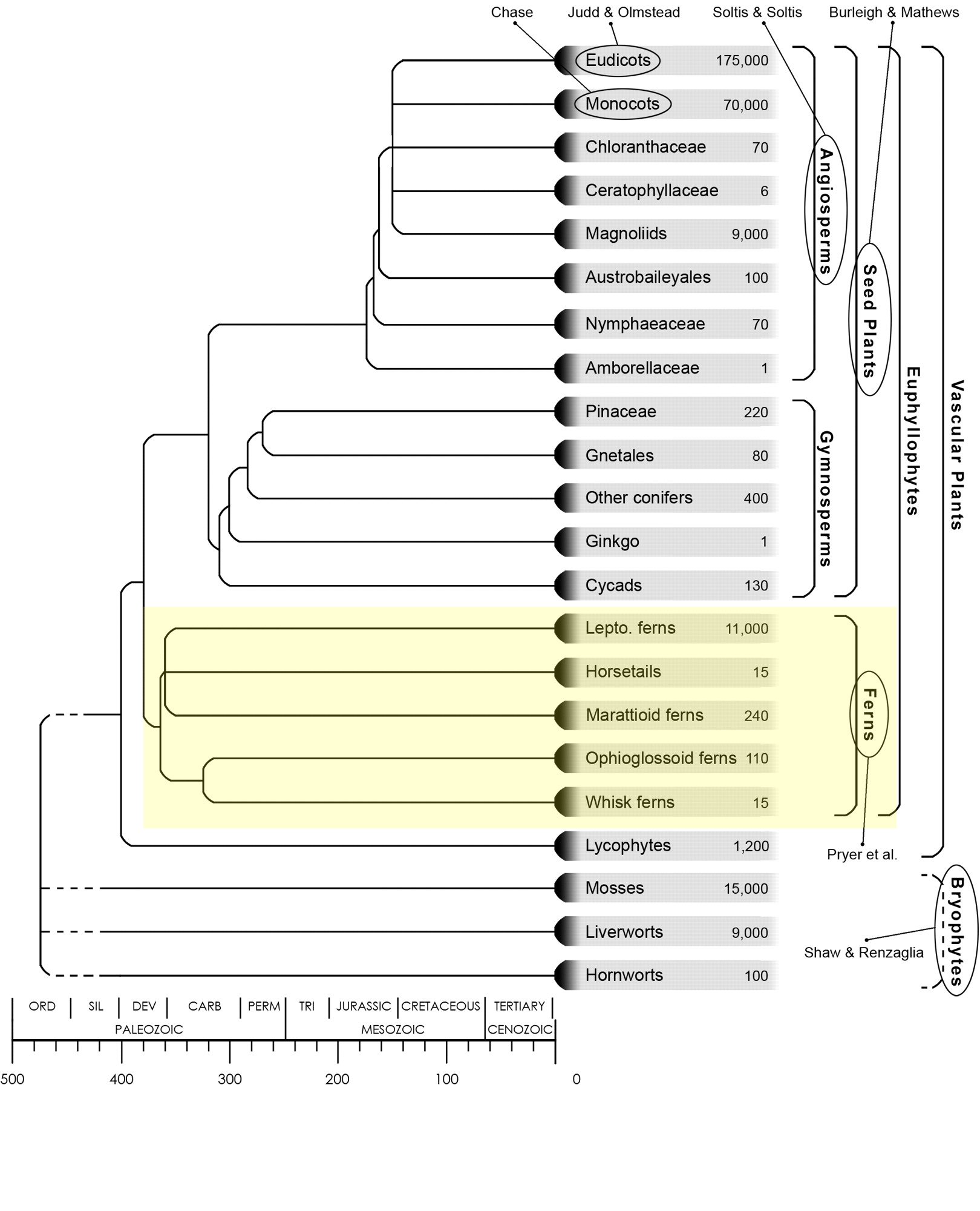
[1] Fig. 2. Chronogram showing
estimates of phylogenetic relationships
and divergence times among the major
groups of extant land plants. The
estimate of relationships is
synthesized from the following papers
in this issue: Burleigh and Mathews
(2004) , Pryer et al. (2004) , Shaw and
Renzaglia (2004) , and Soltis and
Soltis (2004) . Divergence time
estimates are mostly based on analyses
of molecular data with fossil
constraints (Wikström et al., 2001 ;
Pryer et al., 2004 ) and are augmented
by fossil evidence (Kenrick and Crane,
1997 ; Wellman et al., 2003 ).
Estimates of the number of species in
each group are from Judd et al. (2002)
and W. S. Judd (personal
communication). Groups covered by a
particular article in this special
issue are circled and connected to the
names of the article's authors. ''Other
conifers'' refers to the clade
consisting of all conifers except for
Pinaceae (see Burleigh and Mathews,
2004 ). ''Lepto. ferns'' refers to
leptosporangiate ferns fig 2
from: Jeffrey D. Palmer, Douglas E.
Soltis and Mark W. Chase, ''The plant
tree of life: an overview and some
points of view'', American Journal of
Botany. 2004;91:1437-1445., (2004).
http://www.amjbot.org/content/91/10/14
37.full {Chase_Mark_2004.pdf}
COPYRIGHTED
source: http://www.amjbot.org/content/91
/10/1437/F2.large.jpg

[2] Fig. 2. Chronogram showing
estimates of phylogenetic relationships
and divergence times among the major
groups of extant land plants. The
estimate of relationships is
synthesized from the following papers
in this issue: Burleigh and Mathews
(2004) , Pryer et al. (2004) , Shaw and
Renzaglia (2004) , and Soltis and
Soltis (2004) . Divergence time
estimates are mostly based on analyses
of molecular data with fossil
constraints (Wikström et al., 2001 ;
Pryer et al., 2004 ) and are augmented
by fossil evidence (Kenrick and Crane,
1997 ; Wellman et al., 2003 ).
Estimates of the number of species in
each group are from Judd et al. (2002)
and W. S. Judd (personal
communication). Groups covered by a
particular article in this special
issue are circled and connected to the
names of the article's authors. ''Other
conifers'' refers to the clade
consisting of all conifers except for
Pinaceae (see Burleigh and Mathews,
2004 ). ''Lepto. ferns'' refers to
leptosporangiate ferns fig 2
from: Jeffrey D. Palmer, Douglas E.
Soltis and Mark W. Chase, ''The plant
tree of life: an overview and some
points of view'', American Journal of
Botany. 2004;91:1437-1445., (2004).
http://www.amjbot.org/content/91/10/14
37.full {Chase_Mark_2004.pdf}
COPYRIGHTED
source: http://www.amjbot.org/content/91
/10/1437/F2.large.jpg
USA
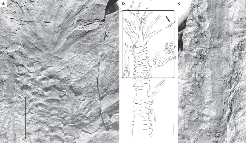
[1] a, General view of the crown
portion, showing longitudinal ranks of
branch bases on the trunk proximally,
and attached branches with digitate
ramification and speckled surface
pattern distally. Scale bar, 20 cm. b,
Line drawing of the specimen as
recovered including trunk and crown;
the box shows the portion in a, and the
arrow indicates the branch in c. Scale
bar, 10 cm. c, Close-up of a distal
branch showing speckled texture and
lateral appendages. Scale bar, 20
mm. figure 1 from: William E. Stein1,
Frank Mannolini2, Linda VanAller
Hernick2, Ed Landing2 & Christopher M.
Berry3, ''Giant cladoxylopsid trees
resolve the enigma of the Earth's
earliest forest stumps at Gilboa'',
Nature 446, 904-907 (19 April
2007) http://www.nature.com/nature/jour
nal/v446/n7138/full/nature05705.html CO
PYRIGHTED
source: http://www.nature.com/nature/jou
rnal/v446/n7138/images/nature05705-f1.2.
jpg
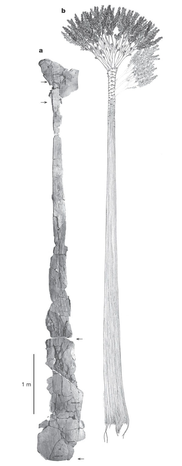
[2] a, Composite image of large trunk
specimen, a cast with upper and lower
counterparts, NYSM 17040. Arrows at the
distal end (top) correspond to the
region in Fig. 3a; arrows at the
proximal end (bottom) correspond to the
region in Fig. 3b. b, Line drawing
showing the architecture of Wattieza
attached to Eospermatopteris. The
length of the trunk is not firmly
established, so the minimum tree height
is shown. Light branches right, also in
Fig. 1a right, appear in life position
but are not definitively attached.
Scale bar, 1 m for both panels. figure
2 from: William E. Stein1, Frank
Mannolini2, Linda VanAller Hernick2, Ed
Landing2 & Christopher M. Berry3,
''Giant cladoxylopsid trees resolve the
enigma of the Earth's earliest forest
stumps at Gilboa'', Nature 446, 904-907
(19 April
2007) http://www.nature.com/nature/jour
nal/v446/n7138/full/nature05705.html CO
PYRIGHTED
source: http://www.nature.com/nature/jou
rnal/v446/n7138/images/nature05705-f2.2.
jpg
arthropod insect. Ancestor of all
winged insects (Pterygota {TARiGOTu})
(Mayflies, Dragonflies, Damselflies).
southeastern Massachusetts and Upper
Silesian Basin, Czech Republic

[1] English: A female subimago of March
Brown (Rhithrogena germanica) of family
Heptageniidae. Mayflies are insects
which belong to the Order Ephemeroptera
(from the Greek ephemeros, short-lived
and pteron, wing, referring to the
short life span of adults). They have
been placed into an ancient group of
insects termed the Paleoptera, which
also contains the dragonflies and
damselflies. They are aquatic insects
whose immature stage (called naiad or,
colloquially, nymph) usually lasts one
year in fresh water. The rests on Rough
Horsetail or Scouringrush Horsetail
(Equisetum hyemale) Date 8 January
2008 Source Own work Author Richard
Bartz, Munich aka Makro Freak
Image:MFB.jpg CC
source: http://upload.wikimedia.org/wiki
pedia/commons/4/49/Rhithrogena_germanica
_subimago_on_Equisetum_hyemale.jpg

[2] FIGURE 2—Preliminary hypothesis
of phylogenetic relationships among
major and interesting groups of living
and extinct hexapods and
basal pterygote Insecta. Numbers refer
to synapomorphies (see Table 1); empty
boxes are homoplasious synapomorphies.
Some significant fossils
are-CSCO-3h--F3.large denoted by
circled letters (see Table 2), but many
fossils are not listed for most groups.
Thick lines indicate the approximate
chronology of lineages. The number of
lineages depicted for paraphyletic
lineages
(‘‘Protodonata,’’‘‘Protortho
ptera,’’ Blattaria [Blattoptera])
are arbitrary, and simply indicate
multiple, unresolved lineages. The
names of orders with freshwater aquatic
larvae are shaded (a presumed ancestral
habit). Relationships are based on
Kristensen (1975, 1991, 1999), Willmann
(1997, 1999), Grimaldi (1997, for
Dictyoptera), Engel and Grimaldi (2000,
Zoraptera and related orders), and
others. Figure 2 from: Grimaldi, D.
2001. Insect evolutionary history from
Handlirsch to Hennig, and beyond.
Journal of Paleontology
75:1152-1160. http://jpaleontol.geoscie
nceworld.org/content/75/6/1152
AND www.online-keys.net/sciaroidea/2000
_/Grimaldi_2001_insect_evolution_history
.pdf COPYRIGHTED
source: www.online-keys.net/sciaroidea/2
000_/Grimaldi_2001_insect_evolution_hist
ory.pdf
(organisms with four feet), the
amphibians. First limbs (arms and legs)
and fingers.
equator)
[1] Timeline of phylogeny of animals,
figure 6 from: S. Blair Hedges, ''The
origin and evolution of model
organisms'', Nature Reviews Genetics 3,
838-849 (November
2002) http://www.nature.com/nrg/journal
/v3/n11/full/nrg929.html {Hedges_2002.p
df} a) The relationships and
divergence times (millions of years ago
(Mya) plusminus one standard error) of
selected model animals are shown, based
on recent multigene and multiprotein
studies51, 61, 84. The fossil
divergence time of birds and mammals
(310 Mya) was used to calibrate the
molecular clock. Branch lengths are not
proportional to time. b ) The
relationships and numbers of living
species, from a diversity of sources in
most of the main groups. COPYRIGHTED
source: http://www.nature.com/nrg/journa
l/v3/n11/images/nrg929-f6.jpg
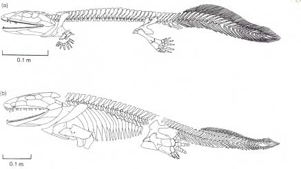
[2] Reconstructions of (a)
Acanthostega and (b) Ichthyostega, from
Benton, 1997. COPYRIGHTED
source: http://palaeo.gly.bris.ac.uk/Pal
aeofiles/Fossilgroups/Amphibia/amphibpic
s/ichthyostega.jpg
(an amphibian).
equator)

[1] Yes, it's time for the Palaeozoic
scenes of Life before Man. Or some of
them, anyway - if you really want to
see a load of trilobites, you're
probably Richard Fortey, and I will
ignore any comments that claim
otherwise. (Also, I can't include
everything - otherwise we'd have to
rename this blog Love in the Time of
Burian, which sounds a bit rubbish.) My
bias is most definitely towards
vertebrates and, in particular,
tetrapods, and the below scene -
featuring Ichthyostega - marks their
first appearance in the book. This
painting is perhaps unique in this book
as it combines the elements of the
animal-free landscapes with, well, some
animals. Burian's skill is in making
this scene, filled as it is with flora
so utterly different to what we are
accustomed to seeing today, look as if
he just took a casual stroll out into
the country to paint it. UNKNOWN
source: http://3.bp.blogspot.com/-Zdvegv
1Fny4/UCk-Z929irI/AAAAAAAABM8/_7c21BO7T1
s/s1600/Ichthyostega.jpg
[2] Timeline of phylogeny of animals,
figure 6 from: S. Blair Hedges, ''The
origin and evolution of model
organisms'', Nature Reviews Genetics 3,
838-849 (November
2002) http://www.nature.com/nrg/journal
/v3/n11/full/nrg929.html {Hedges_2002.p
df} a) The relationships and
divergence times (millions of years ago
(Mya) plusminus one standard error) of
selected model animals are shown, based
on recent multigene and multiprotein
studies51, 61, 84. The fossil
divergence time of birds and mammals
(310 Mya) was used to calibrate the
molecular clock. Branch lengths are not
proportional to time. b ) The
relationships and numbers of living
species, from a diversity of sources in
most of the main groups. COPYRIGHTED
source: http://www.nature.com/nrg/journa
l/v3/n11/images/nrg929-f6.jpg
{Bo-SiDEO-mI-KO-Tu} (most mushrooms,
rusts, club fungi).
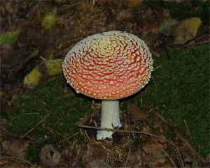
[1] Amanita muscaria
(Homobasidiomycetes)
source: http://en.wikipedia.org/wiki/Ima
ge:Agaricales.jpg

[2] Basidiomycete Life Cycle tjv
source: http://botit.botany.wisc.edu/ima
ges/332/Basidiomycota/General_basidio/Ba
sidiomycete_Life_Cycle_tjv.php?highres=t
rue
eggintoni, Coseley Lagerstätte)
Staffordshire, UK
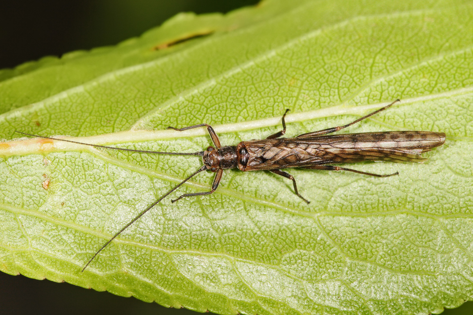
[1] Stonefly in the genus Dinotoperla.
Taken in Swifts Creek, Victoria in
November 2007 GNU
source: http://upload.wikimedia.org/wiki
pedia/commons/e/e6/Stonefly_-_dinotoperl
a.jpg

[2] Nymph of unidentified
stonefly Description Deutsch:
Steinfliegenlarve Date 16 June
2006 Source Own work Author
böhringer friedrich CC
source: http://upload.wikimedia.org/wiki
pedia/commons/thumb/b/b1/SteinfliegenLar
ve2.JPG/1280px-SteinfliegenLarve2.JPG
Ancestor of all seed plants.
[1] Henry N. Andrews, ''Early Seed
Plants'', Science, New Series, Vol.
142, No. 3594 (Nov. 15, 1963), pp.
925-931. http://www.jstor.org/stable/17
11577 COPYRIGHTED
source: http://www.jstor.org/stable/1711
577
[2] Fig. 2. Chronogram showing
estimates of phylogenetic relationships
and divergence times among the major
groups of extant land plants. The
estimate of relationships is
synthesized from the following papers
in this issue: Burleigh and Mathews
(2004) , Pryer et al. (2004) , Shaw and
Renzaglia (2004) , and Soltis and
Soltis (2004) . Divergence time
estimates are mostly based on analyses
of molecular data with fossil
constraints (Wikström et al., 2001 ;
Pryer et al., 2004 ) and are augmented
by fossil evidence (Kenrick and Crane,
1997 ; Wellman et al., 2003 ).
Estimates of the number of species in
each group are from Judd et al. (2002)
and W. S. Judd (personal
communication). Groups covered by a
particular article in this special
issue are circled and connected to the
names of the article's authors. ''Other
conifers'' refers to the clade
consisting of all conifers except for
Pinaceae (see Burleigh and Mathews,
2004 ). ''Lepto. ferns'' refers to
leptosporangiate ferns fig 2
from: Jeffrey D. Palmer, Douglas E.
Soltis and Mark W. Chase, ''The plant
tree of life: an overview and some
points of view'', American Journal of
Botany. 2004;91:1437-1445., (2004).
http://www.amjbot.org/content/91/10/14
37.full {Chase_Mark_2004.pdf}
COPYRIGHTED
source: http://www.amjbot.org/content/91
/10/1437/F2.large.jpg
Paddlefish.

[1] Adapted from: Richard Dawkins,
''The Ancestor's Tale'', (Boston, MA:
Houghton Mifflin Company, 2004),
p339. COPYRIGHTED
source: Richard Dawkins, "The
Ancestor's Tale", (Boston, MA: Houghton
Mifflin Company, 2004), p339.
[2] Adapted from: Richard Dawkins,
''The Ancestor's Tale'', (Boston, MA:
Houghton Mifflin Company, 2004),
p339. COPYRIGHTED
source: Richard Dawkins, "The
Ancestor's Tale", (Boston, MA: Houghton
Mifflin Company, 2004), p339.
(Cockroaches, Termites).

[1] Description English: Juvenile,
Madagascar hissing cockroach at the
Atlanta Botanical Garden. Taken
9/23/2007. Date 25 September 2007
(original upload date) Source
Transferred from en.wikipedia;
transferred to Commons by
User:Sreejithk2000 using
CommonsHelper. Author Original
uploader was Almabes at
en.wikipedia Permission (Reusing this
file) Released into the public
domain (by the author). Other versions
Derivative works of this file:
Female Madagascar hissing
cockroach2.jpg PD
source: http://upload.wikimedia.org/wiki
pedia/commons/4/46/Female_Madagascar_his
sing_cockroach.JPG

[2] Figure 4.11. German Cockroaches,
Various Stages and Ages PD
source: http://www.cdc.gov/nceh/publicat
ions/books/housing/Graphics/chapter_04/F
igure4.11.jpg
Amniota {aMnEOtu} (ancestor of
reptiles, mammals and birds).
Start of
vertebrate internal fertilization.
Lothian, Scotland
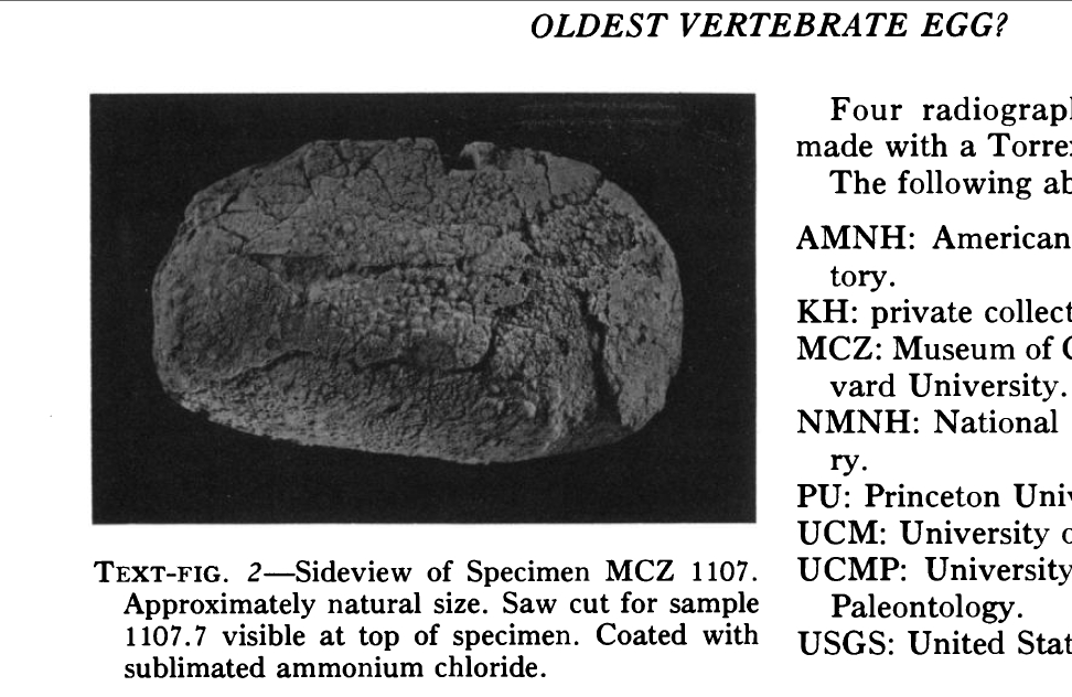
[1] Figure 2 from: [t Note that this
egg is only of Permian age: 299-251
mybn] Karl F. Hirsch, ''The Oldest
Vertebrate Egg?'', Journal of
Paleontology, Vol. 53, No. 5 (Sep.,
1979), pp.
1068-1084. http://www.jstor.org/stable/
1304086 COPYRIGHTED
source: http://www.jstor.org/stable/1304
086

[2] Prothero, ''Bringing Fossils To
Life'', 2004. COPYRIGHTED
source: Prothero, "Bringing Fossils To
Life", 2004. COPYRIGHTED
the Sauropsida {SOR-roP-SiDu} (which
includes reptiles and birds) and the
Synapsida {Si-naP-Si-Du} (which
includes mammals).
(Cumberland group, Joggins formation)
Joggins, Nova Scotia, Canada
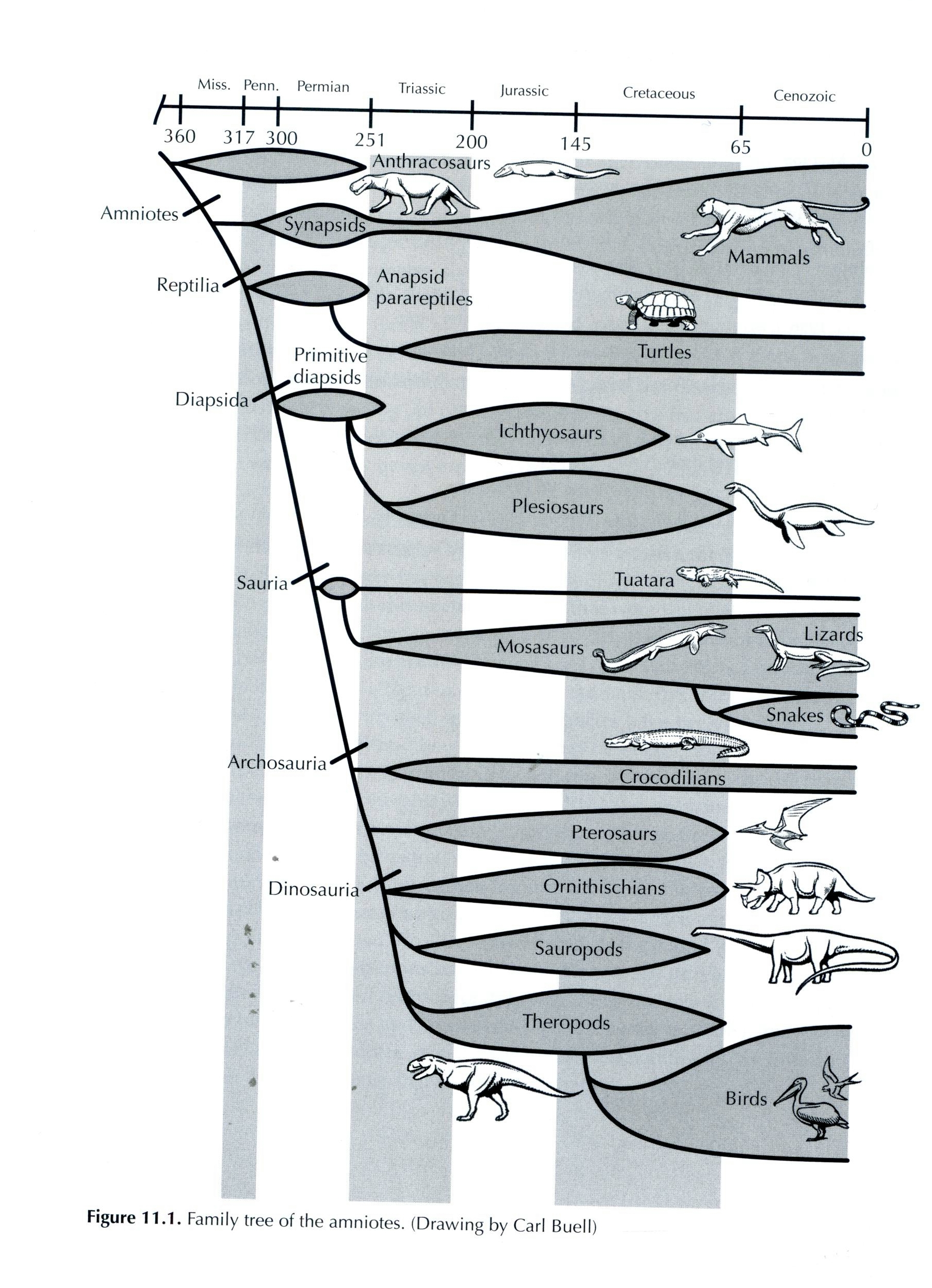
[1] Prothero, ''Evolution What the
Fossils Say and Why It Matters'', 2007,
p232. COPYRIGHTED
source: Prothero, "Evolution What the
Fossils Say and Why It Matters", 2007,
p232.
[2] Prothero, ''Bringing Fossils To
Life'', 2004. COPYRIGHTED
source: Prothero, "Bringing Fossils To
Life", 2004. COPYRIGHTED

[1] Description Eocaecilia
micropodia, an early caecilian from the
Lower Jurassic of Arizona, pencil
drawing Date 22 August
2007 Source Own work Author
Nobu Tamura
email:nobu.tamura@yahoo.com
www.palaeocritti.com Permission (Reusi
ng this file) See below. GNU
source: http://upload.wikimedia.org/wiki
pedia/commons/2/27/Eocaecilia_BW.jpg
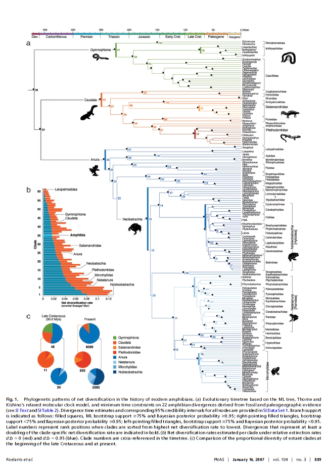
[2] Figure 1 from: Roelants, K.,
Gower, D. J., Wilkinson, M., Loader, S.
P., Biju, S. D., Guillaume, K., Moriau,
L., & Bossuyt, F. (2007). Global
patterns of diversification in the
history of modern amphibians.
Proceedings of the National Academy of
Sciences , 104 (3), 887-892. URL
http://dx.doi.org/10.1073/pnas.060837810
4 COPYRIGHTED
source: http://dx.doi.org/10.1073/pnas.0
608378104
of all Cycads, Ginkgos and the
Conifers: Pine, Fir, Spruce, Redwood,
Cedar, Juniper, and Cypress).

[1] Fig. 2. Chronogram showing
estimates of phylogenetic relationships
and divergence times among the major
groups of extant land plants. The
estimate of relationships is
synthesized from the following papers
in this issue: Burleigh and Mathews
(2004) , Pryer et al. (2004) , Shaw and
Renzaglia (2004) , and Soltis and
Soltis (2004) . Divergence time
estimates are mostly based on analyses
of molecular data with fossil
constraints (Wikström et al., 2001 ;
Pryer et al., 2004 ) and are augmented
by fossil evidence (Kenrick and Crane,
1997 ; Wellman et al., 2003 ).
Estimates of the number of species in
each group are from Judd et al. (2002)
and W. S. Judd (personal
communication). Groups covered by a
particular article in this special
issue are circled and connected to the
names of the article's authors. ''Other
conifers'' refers to the clade
consisting of all conifers except for
Pinaceae (see Burleigh and Mathews,
2004 ). ''Lepto. ferns'' refers to
leptosporangiate ferns fig 2
from: Jeffrey D. Palmer, Douglas E.
Soltis and Mark W. Chase, ''The plant
tree of life: an overview and some
points of view'', American Journal of
Botany. 2004;91:1437-1445., (2004).
http://www.amjbot.org/content/91/10/14
37.full {Chase_Mark_2004.pdf}
COPYRIGHTED
source: http://www.amjbot.org/content/91
/10/1437/F2.large.jpg
[2] Fig. 2. Chronogram showing
estimates of phylogenetic relationships
and divergence times among the major
groups of extant land plants. The
estimate of relationships is
synthesized from the following papers
in this issue: Burleigh and Mathews
(2004) , Pryer et al. (2004) , Shaw and
Renzaglia (2004) , and Soltis and
Soltis (2004) . Divergence time
estimates are mostly based on analyses
of molecular data with fossil
constraints (Wikström et al., 2001 ;
Pryer et al., 2004 ) and are augmented
by fossil evidence (Kenrick and Crane,
1997 ; Wellman et al., 2003 ).
Estimates of the number of species in
each group are from Judd et al. (2002)
and W. S. Judd (personal
communication). Groups covered by a
particular article in this special
issue are circled and connected to the
names of the article's authors. ''Other
conifers'' refers to the clade
consisting of all conifers except for
Pinaceae (see Burleigh and Mathews,
2004 ). ''Lepto. ferns'' refers to
leptosporangiate ferns fig 2
from: Jeffrey D. Palmer, Douglas E.
Soltis and Mark W. Chase, ''The plant
tree of life: an overview and some
points of view'', American Journal of
Botany. 2004;91:1437-1445., (2004).
http://www.amjbot.org/content/91/10/14
37.full {Chase_Mark_2004.pdf}
COPYRIGHTED
source: http://www.amjbot.org/content/91
/10/1437/F2.large.jpg
grasshoppers).
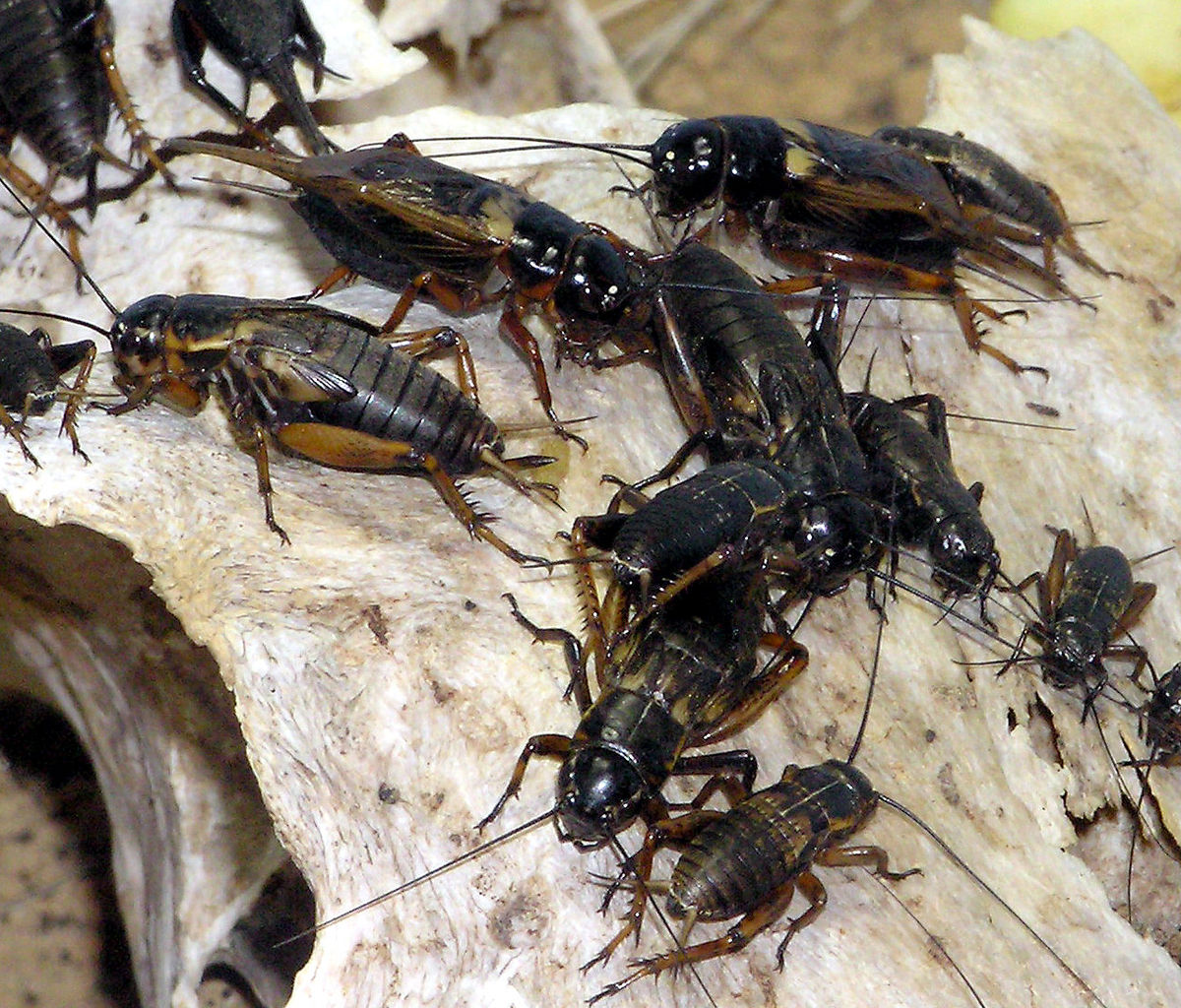
[1] African Field cricket Gryllus
bimaculatus at Bristol Zoo, Bristol,
England. Photographed by Adrian
Pingstone in February 2005 and released
to the public domain. PD
source: http://upload.wikimedia.org/wiki
pedia/commons/thumb/2/27/African.field.c
ricket.arp.jpg/1200px-African.field.cric
ket.arp.jpg
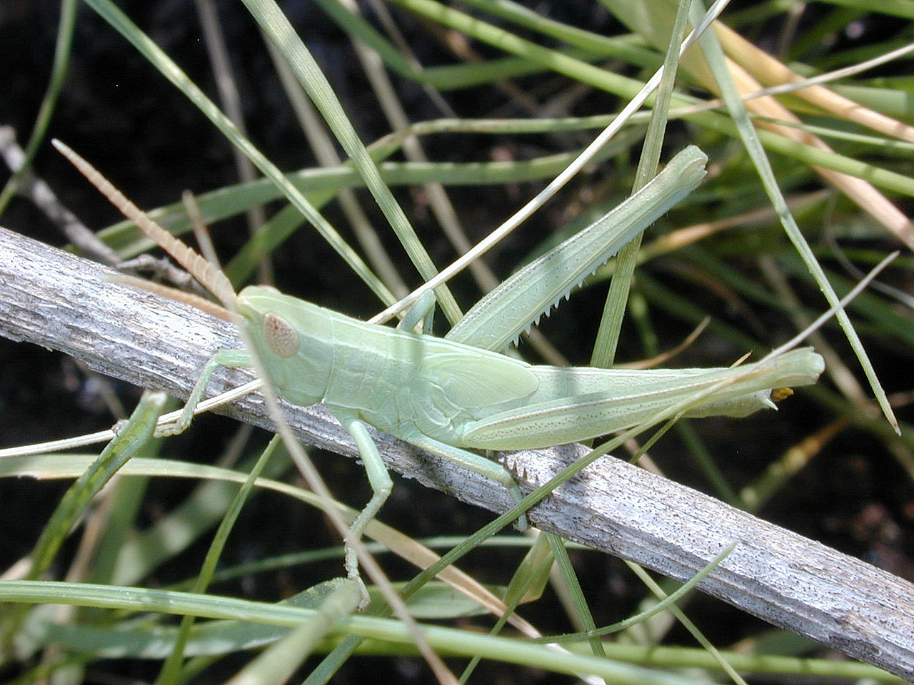
[2] Description
grasshopper Source self
made Date unknown Author
Stephen Friedt PD
source: http://upload.wikimedia.org/wiki
pedia/en/thumb/3/3c/Grasshopper_%2827%29
.JPG/1280px-Grasshopper_%2827%29.JPG
Nova Scotia, Canada
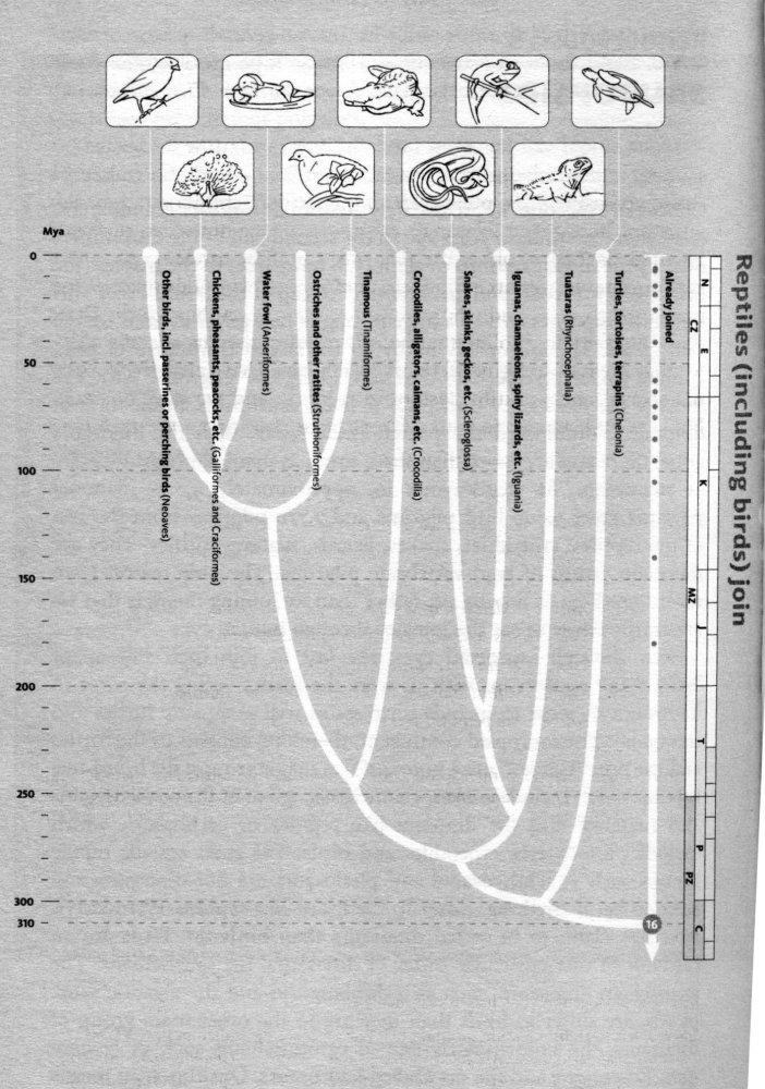
[1] from: Richard Dawkins, ''The
Ancestor's Tale'', (Boston, MA:
Houghton Mifflin Company, 2004), p262.
COPYRIGHTED
source: Richard Dawkins, "The
Ancestor's Tale", (Boston, MA: Houghton
Mifflin Company, 2004), p262.

[2] Description English: Reptilia
(reptiles), based on:
File:Buberel cayman 3.jpg
File:Crotalus adamanteus (5).jpg
File:Karettschildkroete 01.jpg
File:Henry at Invercargill.jpg All
of them are either under a free licence
already in Wikicommons or in the public
domain Date 3/2/09 Source
Compilation made by myself Author
see respective profiles of
photos PD
source: http://upload.wikimedia.org/wiki
pedia/commons/6/67/Reptiles.jpg
cicadas, aphids).

[1] Description Tibicen
linnei English: Annual cicada. Date
22 June 2003 Source Own work
http://www.cirrusimage.com/homoptera_cic
ada_T_linnei.htm Author Bruce
Marlin CC
source: http://upload.wikimedia.org/wiki
pedia/commons/thumb/f/fb/Tibicen_linnei.
jpg/1142px-Tibicen_linnei.jpg
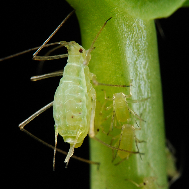
[2] Description English: Pea aphids
extracting sap from the stem and leaves
of garden peas. Date February
2010 Source PLoS Biology, February
2010 direct link to the image
description Author Shipher Wu
(photograph) and Gee-way Lin (aphid
provision), National Taiwan
University CC
source: http://upload.wikimedia.org/wiki
pedia/commons/2/20/Acyrthosiphon_pisum_%
28pea_aphid%29-PLoS.jpg
HOlOmeTABoluS} insects (ancestor of
beetles, bees, true flies, and
butterflies). Complete metamorphosis.

[1] Description wespenpoppen in
verschillende ontwikkelstadia Eigen
foto's Date 2005-06-13 (original
upload date) Source Originally from
nl.wikipedia; description page is/was
here. Author Original uploader was
Asaf at
nl.wikipedia Permission (Reusing this
file) SELF2 GNU
source: http://upload.wikimedia.org/wiki
pedia/commons/7/79/Ontwikkelstadia_wespe
npoppen.jpg

[2] Miomoptera- viewed by many as
stem-group Holometabola. UNKNOWN
source: http://wdict.net/img/miomoptera,
2.jpg

[1] Richard Dawkins, ''The Ancestor's
Tale'', (Boston, MA: Houghton Mifflin
Company, 2004), 303. COPYRIGHTED
source: Richard Dawkins, "The
Ancestor's Tale", (Boston, MA: Houghton
Mifflin Company, 2004), 303.

[2] Description English: A green
frog on a palm frond. Date 18
October 2003 Source Burning
Well Author Leon Brooks PD
source: http://upload.wikimedia.org/wiki
pedia/commons/8/8d/Frog_on_palm_frond.jp
g
(Beetles).
deposit) Mazon Creek, Illinois,
USA
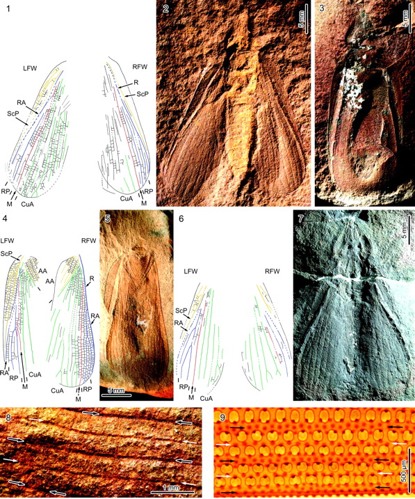
[1] Figure 1. 1–7, Adiphlebia lacoana
Scudder, 1885. 1, 2, holotype specimen
(USNM 38143), reconstruction of the
wing venation (1), and photograph
(negative imprint, light-mirrored,
composite; 2); 3, specimen USNM 38140,
photograph (negative imprint,
light-mirrored, composite); 4,5,
specimen FMNH PE 3416, reconstruction
of the wing venation (forewings
separated; 4) and photograph (negative
imprint, composite; 5); 6, 7, specimen
FMNH PE 60291, reconstruction of the
wing venation (6) and photograph
(positive imprint; 7); 8, 9, details of
forewing main and intercalary veins
(black and white arrows, respectively)
in Adiphlabia lacoana (specimen FMNH PE
3416, right forewing; 8) and
Tetraphalerus bruchi Heller, 1913 (♀,
ventral view; 9). Abbreviations: LFW,
left forewing; RFW, right forewing;
ScP, posterior Subcosta; R, Radius; RA,
anterior Radius; RP, posterior Radius;
M, Media; CuA, anterior Cubitus; CuP,
posterior Cubitus; AA: anterior anal
vein. Color-coding: Subcosta, yellow;
Radius, blue; Media, red; Cubitus,
green; Analis, yellow. from Béthoux,
Olivier. “The Earliest Beetle
Identified.” Journal of Paleontology
83.6 (2009):
931–937. http://www.bioone.org/doi/ab
s/10.1666/08-158.1 COPYRIGHTED
source: http://jpaleontol.geoscienceworl
d.org/content/vol83/issue6/images/large/
i0022-3360-83-6-931-f01.jpeg

[2] {ULSF: Early Permian fossil
beetles see {Kukalová (1969), in
particular pl. 1; Ponomarenko (1969),
in particular figs. 16, 31, 32, 36, 40
41, 43, 44} and representatives of the
beetle sub-order Archostemata,
represented nowadays, exhibit
intercalary veins (Fig. 1.9) similar to
those exhibited by A.
lacoana} Archostemata is the smallest
suborder of beetles, consisting of
fewer than fifty known species
organized into five families.
Archostemata is an ancient lineage with
a number of primitive characteristics.
They are similar in morphology to the
first beetles, which appear in the
fossil record approximately 250 million
years ag Description
Tenomerga mucida (Chevrolat, 1829)
(Coleoptera: Cupedidae) - female.
Loc: Yokohama, kanagawa, japan.
ja: ナガヒラタムシ(鞘翅目:
ナガヒラタムシ科)のメス。�
��浜市内。産卵管をさかんに�
�し入れし、朽木の割れ目に挿
し込もうとしていたことから�
��産卵に来ていたものと思わ�
�る。 Date 13 July 2005 Source
my own file Author me PD
source: http://upload.wikimedia.org/wiki
pedia/commons/6/68/Tenomerga_mucida01.jp
g
wasps).
[1] {ULSF: Xyelidae saw flies are the
most primitive of the
hymenoptera} Hymenoptera, Xyelidae,
dorsal - Macroxyela ferruginea -
Female Ames - Tullamore, Story County,
Iowa, USA April 30, 2008 Size: 11
mm It's a big one. (11 mm includes the
ovipositor) Oak hickory maple basswood
woodland malaise, April 23-30,
2008. Photo - still floating in
alcohol. Copyright © 2008 MJ
Hatfield COPYRIGHTED Fig. 2
Placement of fossil evidence for the
earliest Holometabola within a
phylogenetic context. Geologic time
line at left is after Ogg, et al.
(2008); note that the Mississippian is
equivalent to the Early Carboniferous
and Pennsylvanian equivalent to the
Late Carboniferous. Earliest reliable
occurrences of taxa (solid dots,
followed by a thick black line) are
after various sources mentioned in the
text; major localities for the initial
diversification of the Holometabola
are: Elmo, Kansas, the “insect bed”
of the Wellington Formation from the
Artinskian Stage of the Early Permian;
Calhoun, the Calhoun Coal Member of the
Mattoon Formation, from the Kasimovian
Stage of the Late Pennsylvanian; Mazon
Creek of the Francis Creek Shale Member
of the Carbondale Formation, from the
Moscovian Stage of the Middle
Pennsylvanian; and the Terril Shale at
Pas-de-Calais, Bruay-la-Bussière,
France, from the Bashkirian Stage of
the Early Pennsylvanian. The horizontal
stippled bar at bottom represents the
initial diversification and the
earliest fossil occurrences of
holometabolan insects in the fossil
record. Labandeira, Conrad C.
“Evidence for an Earliest Late
Carboniferous Divergence Time and the
Early Larval Ecology and
Diversification of Major Holometabola
Lineages.” Entomologica Americana
117.1 & 2 (2011):
9–21. http://www.bioone.org/doi/abs/1
0.1664/10-RA-011.1 COPYRIGHTED
source: http://bugguide.net/images/raw/S
H8RHHPR0H7RDZHZULYLULRZ2LLZTLSZBLQZKH4RH
H7ZVL4RVL0ZALSZBLXZKH8RVLXZHHPRLHQRLH.jp
g

[2] Macroxyela ferruginea
Trusted Creative Commons Attribution
Non Commercial Share Alike 3.0 (CC
BY-NC-SA 3.0) ©
SusanneSchulmeister Source:
Morphbank Image Repository
COPYRIGHTED
source: http://www.bioone.org/na101/home
/literatum/publisher/bioone/journals/con
tent/nynt.1/2011/19475144-117.1/10-ra-01
1.1/production/images/large/i1947-5144-1
17-1-9-f02.jpeg
[1] Fig. 1. A consensus phylogeny of
eukaryotes. The vast majority of
characterized eukaryotes, with the
notable exception of major subgroups of
amoebae, can now be assigned to one of
eight major groups. Opisthokonts (basal
flagellum) have a single basal
flagellum on reproductive cells and
flat mitochondrial cristae (most
eukaryotes have tubular ones).
Eukaryotic photosynthesis originated in
Plants; theirs are the only plastids
with just two outer membranes.
Heterokonts (different flagellae) have
a unique flagellum decorated with
hollow tripartite hairs (stramenopiles)
and, usually, a second plain one.
Cercozoans are amoebae with filose
pseudopodia, often living with in tests
(hard outer shells), some very
elaborate (foraminiferans). Amoebozoa
are mostly naked amoebae (lacking
tests), often with lobose pseudopodia
for at least part of their life cycle.
Alveolates have systems of cortical
alveoli directly beneath their plasma
membranes. Discicristates have discoid
mitochondrial cristae and, in some
cases, a deep (excavated) ventral
feeding groove. Amitochondrial
excavates lack substantial molecular
phylogenetic support, but most have an
excavated ventral feeding groove, and
all lack mitochondria. The tree shown
is based on a consensus of molecular
(1-4) and ultrastructural (16, 17) data
and includes a rough indication of new
ciPCR ''taxa'' (broken black lines)
(7-11). An asterisk preceding the taxon
name indicates probable paraphyletic
group COPYRIGHTED
source: http://www.sciencemag.org/cgi/co
ntent/full/300/5626/1703

[2] Pacific Rockweed (Fucus distichus)
in Olympic National Park Cropped from
PhotoCD image, from Kodak ISO 800 film,
taken by k.lee June 2004, hereby
released under GFDL.
source: http://en.wikipedia.org/wiki/Ima
ge:Pacific_rockweed%2C_Olympic_National_
Park%2C_USA.jpg
[1] Fig. 1. A consensus phylogeny of
eukaryotes. The vast majority of
characterized eukaryotes, with the
notable exception of major subgroups of
amoebae, can now be assigned to one of
eight major groups. Opisthokonts (basal
flagellum) have a single basal
flagellum on reproductive cells and
flat mitochondrial cristae (most
eukaryotes have tubular ones).
Eukaryotic photosynthesis originated in
Plants; theirs are the only plastids
with just two outer membranes.
Heterokonts (different flagellae) have
a unique flagellum decorated with
hollow tripartite hairs (stramenopiles)
and, usually, a second plain one.
Cercozoans are amoebae with filose
pseudopodia, often living with in tests
(hard outer shells), some very
elaborate (foraminiferans). Amoebozoa
are mostly naked amoebae (lacking
tests), often with lobose pseudopodia
for at least part of their life cycle.
Alveolates have systems of cortical
alveoli directly beneath their plasma
membranes. Discicristates have discoid
mitochondrial cristae and, in some
cases, a deep (excavated) ventral
feeding groove. Amitochondrial
excavates lack substantial molecular
phylogenetic support, but most have an
excavated ventral feeding groove, and
all lack mitochondria. The tree shown
is based on a consensus of molecular
(1-4) and ultrastructural (16, 17) data
and includes a rough indication of new
ciPCR ''taxa'' (broken black lines)
(7-11). An asterisk preceding the taxon
name indicates probable paraphyletic
group COPYRIGHTED
source: http://www.sciencemag.org/cgi/co
ntent/full/300/5626/1703
[2] Figure 1. Phylogenetic hypothesis
of the eukaryotic lineage based on
ultrastructural and molecular data.
Organisms are divided into three main
groups distinguished by mitochondrial
cristal shape (either discoidal,
flattened or tubular). Unbroken lines
indicate phylogenetic relationships
that are firmly supported by available
data; broken lines indicate
uncertainties in phylogenetic
placement, resolution of which will
require additional data. Color coding
of organismal genus names indicates
mitochondrial genomes that have been
completely (Table 1), almost completely
(Jakoba, Naegleria and
Thraustochytrium) or partially (*)
sequenced by the OGMP (red), the FMGP
(black) or other groups (green). Names
in blue indicate those species whose
mtDNAs are currently being sequenced by
the OGMP or are future candidates for
complete sequencing. Amitochondriate
retortamonads are positioned at the
base of the tree, with broken arrows
denoting the endosymbiotic origin(s) of
mitochondria from a Rickettsia-like
eubacterium. Macrophar.,
Macropharyngomonas. COPYRIGHTED
source: http://nar.oxfordjournals.org/cg
i/content/full/26/4/865
growing animal.
[1] Description English: Life
restoration of Purlovia maxima. Based
on figures 8-10 of ''Permian and
Triassic therocephals (Eutherapsida) of
Eastern Europe'' by M. F. Ivakhnenko
(Paleontological Journal 45 (9):
981-1144). Date 8 January
2012 Source Own
work Author Smokeybjb CC
source: http://upload.wikimedia.org/wiki
pedia/commons/a/a5/Purlovia_maxima.jpg
[2] Description Bauria , a
therocephalian therapsid from the early
Middle Triassic of South Africa, pencil
drawing Date 20 February
2007 Source Own work Author
Nobu Tamura
email:nobu.tamura@yahoo.com
www.palaeocritti.com GNU
source: http://upload.wikimedia.org/wiki
pedia/commons/c/c0/Bauria_BW.jpg
flies, single pair of wings: ancestor
of mosquito, gnat, fruit fly, and house
fly).
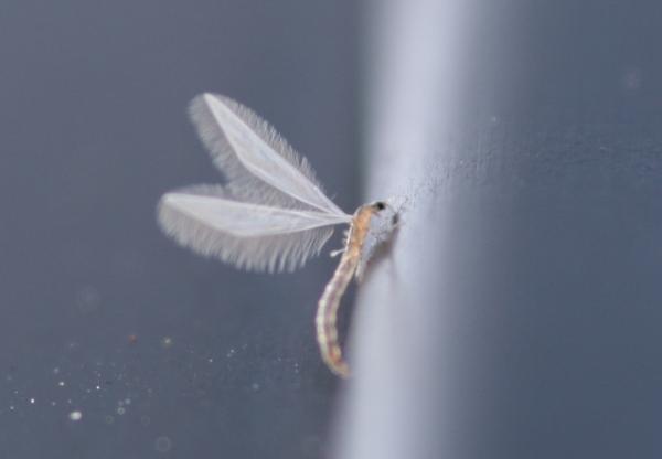
[1] Nymphomyia alba adult UNKNOWN
source: http://whyevolutionistrue.files.
wordpress.com/2011/03/nymphomyia-alba.jp
g

[2] Nymphomyia alba larva UNKNOWN
source: http://whyevolutionistrue.files.
wordpress.com/2011/03/nymphomyia.jpg
history.
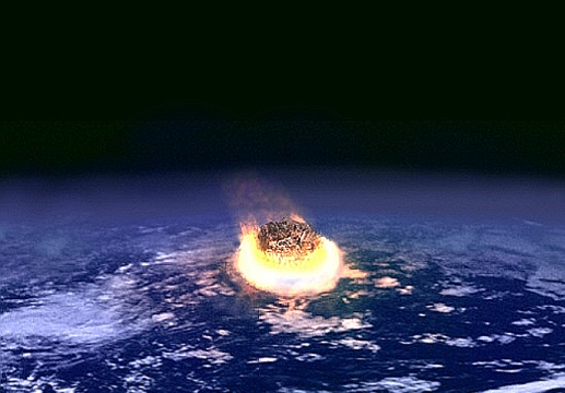
[1] Description English:
Description: Illustration of an
en:impact event. Source Made by
Fredrik. Cloud texture from public
domain NASA image. PD
source: http://upload.wikimedia.org/wiki
pedia/commons/c/cb/Impact_event.jpg
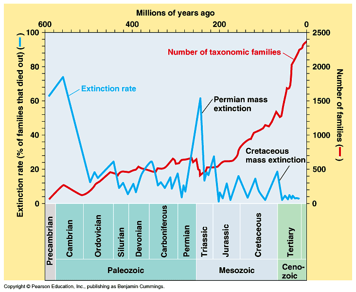
[2] Timeline of mass extinctions.
COPYRIGHTED Benjamin
Cummings. COPYRIGHTED
source: http://io.uwinnipeg.ca/~simmons/
16cm05/1116/16macro.htm
forms.

[1] 260 Ma Late Permian UNKNOWN
source: http://jan.ucc.nau.edu/~rcb7/260
_Permian_2globes.jpg

[2] In geologic terms, a plate is a
large, rigid slab of solid rock. The
word tectonics comes from the Greek
root ''to build.'' Putting these two
words together, we get the term plate
tectonics, which refers to how the
Earth's surface is built of plates. The
theory of plate tectonics states that
the Earth's outermost layer is
fragmented into a dozen or more large
and small plates that are moving
relative to one another as they ride
atop hotter, more mobile material.
Before the advent of plate tectonics,
however, some people already believed
that the present-day continents were
the fragmented pieces of preexisting
larger landmasses
(''supercontinents''). The diagrams
below show the break-up of the
supercontinent Pangaea (meaning ''all
lands'' in Greek), which figured
prominently in the theory of
continental drift -- the forerunner to
the theory of plate tectonics. PD
source: http://pubs.usgs.gov/gip/dynamic
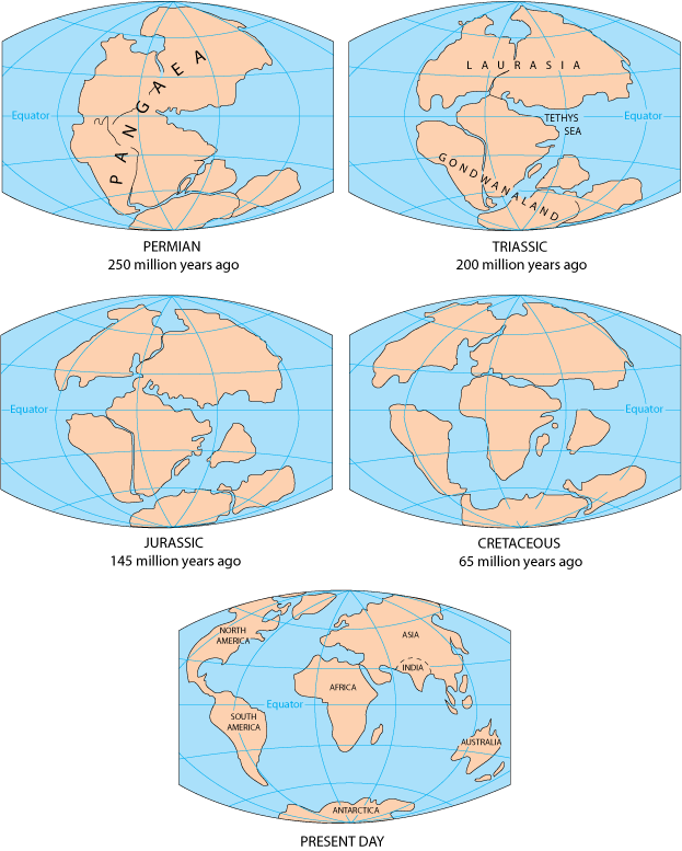
/graphics/Fig2-5globes.gif
(Coccolithophores {KoK-o-lit-O-FORZ}).
[1] Fig. 1. A consensus phylogeny of
eukaryotes. The vast majority of
characterized eukaryotes, with the
notable exception of major subgroups of
amoebae, can now be assigned to one of
eight major groups. Opisthokonts (basal
flagellum) have a single basal
flagellum on reproductive cells and
flat mitochondrial cristae (most
eukaryotes have tubular ones).
Eukaryotic photosynthesis originated in
Plants; theirs are the only plastids
with just two outer membranes.
Heterokonts (different flagellae) have
a unique flagellum decorated with
hollow tripartite hairs (stramenopiles)
and, usually, a second plain one.
Cercozoans are amoebae with filose
pseudopodia, often living with in tests
(hard outer shells), some very
elaborate (foraminiferans). Amoebozoa
are mostly naked amoebae (lacking
tests), often with lobose pseudopodia
for at least part of their life cycle.
Alveolates have systems of cortical
alveoli directly beneath their plasma
membranes. Discicristates have discoid
mitochondrial cristae and, in some
cases, a deep (excavated) ventral
feeding groove. Amitochondrial
excavates lack substantial molecular
phylogenetic support, but most have an
excavated ventral feeding groove, and
all lack mitochondria. The tree shown
is based on a consensus of molecular
(1-4) and ultrastructural (16, 17) data
and includes a rough indication of new
ciPCR ''taxa'' (broken black lines)
(7-11). An asterisk preceding the taxon
name indicates probable paraphyletic
group
source: http://www.sciencemag.org/cgi/co
ntent/full/300/5626/1703
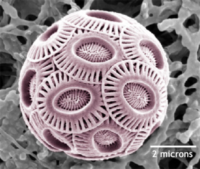
[2] Emiliania huxleyi, a
coccolithophore. Photo courtesy Dr.
Markus Geisen - photographer, and The
Natural History Museum. PD
source: http://en.wikipedia.org/wiki/Ima
ge:Emiliania_huxleyi_3.jpg
Formation) Valley of the Moon,
Ischigualasto Provinvial Park,
northwestern Argestina
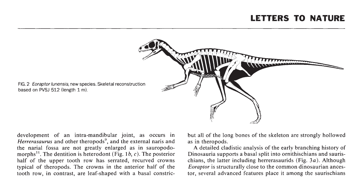
[1] Figure 2 from: Sereno, Paul C. et
al. “Primitive dinosaur skeleton from
Argentina and the early evolution of
Dinosauria.” Nature 361.6407 (1993) :
64-66. http://www.nature.com/nature/jou
rnal/v361/n6407/abs/361064a0.html COPYR
IGHTED
source: http://www.nature.com/nature/jou
rnal/v361/n6407/abs/361064a0.html
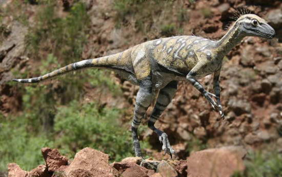
[2] Eoraptor was a genus of small,
slender theropod native to northwest
Argentina. It was quite possibly the
earliest theropod genus and has not
been classified in any family.
UNKNOWN
source: http://images.wikia.com/deadtime
s/images/a/a2/Eoraptor.jpg
gland.
Kalgary, Crosby County, Texas,
USA

[1] Figure 6 from: Spencer G. Lucas
and Zhexi Luo, ''Adelobasileus from the
Upper Triassic of West Texas: The
Oldest Mammal'', Journal of Vertebrate
Paleontology, Vol. 13, No. 3 (Sep. 23,
1993), pp. 309-334 Published by:
Taylor & Francis, Ltd. on behalf of The
Society of Vertebrate
Paleontology Article Stable URL:
http://www.jstor.org/stable/4523514 COP
YRIGHTED
source: http://www.jstor.org/stable/4523
514
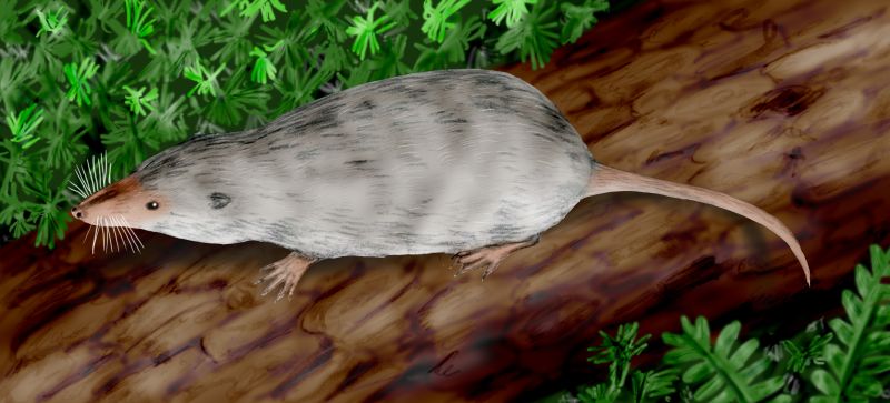
[2] [t Note that this image is not
clearly from a scholarly
source] Description English:
Adelobasileus cromptoni, a mammaliaform
from the Late Triassic of Texas.
Digital. Date 9 September
2008 Source Own work Author
Nobu Tamura
email:nobu.tamura@yahoo.com
www.palaeocritti.com Permission (Reusi
ng this file) See below. GNU
source: http://upload.wikimedia.org/wiki
pedia/commons/2/2f/Adelobasileus_BW.jpg
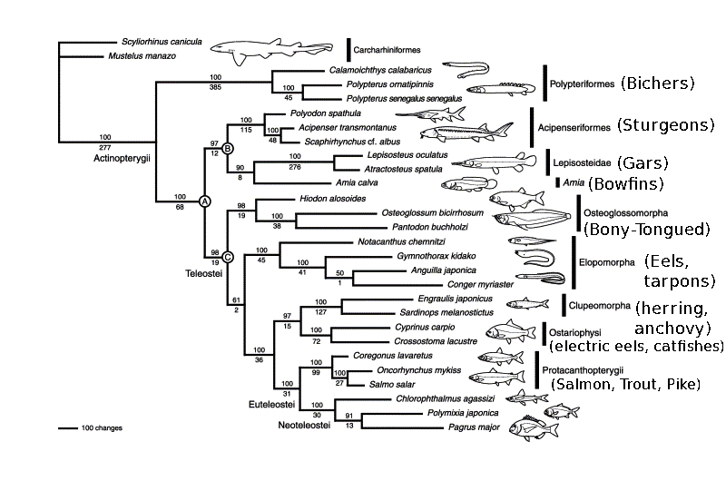
[1] Fig. 2. The single
most-parsimonious (MP) tree derived
from unweighted analysis of mitogenomic
data comprising concatenated nucleotide
sequences from 12 protein-coding
(excluding the ND6 gene and third codon
positions) and 22 transfer RNA (tRNA)
genes (stem regions only) from all 28
species examined. Tree length, 12,709
steps; consistency index, 0.355;
retention index, 0.471; and rescaled
consistency index, 0.167. Numbers above
and below internal branches indicate
jackknife values obtained for 500
replicates using the heuristic search
option in PAUP*4.0b10 (Swofford, 2002)
with 20 random-addition sequences being
performed in each replication and decay
indices, respectively. The scale
indicates 100 changes. from: Inoue,
JG, Miya, M, Tsukamoto, K, Nishida, M
(2003) ''Basal actinopterygian
relationships: A mitogenomic
perspective on the phylogeny of the
ldquoancient fish.rdquo'' Mol
Phylogenet Evol 26:
110-120 http://www.sciencedirect.com/sc
ience/article/pii/S1055790302003317 COP
YRIGHTED
source: http://www.sciencedirect.com/cac
he/MiamiImageURL/B6WNH-475B9D7-6-1K/0?wc
hp=dGLbVlz-zSkzk

[2] Arapaima gigas at the Smithsonian
Zoo. GNU
source: http://upload.wikimedia.org/wiki
pedia/commons/b/b1/Arapaima_gigas.jpg
[1] Richard Dawkins, ''The Ancestor's
Tale'', (Boston, MA: Houghton Mifflin
Company, 2004), 262. COPYRIGHTED
source: Richard Dawkins, "The
Ancestor's Tale", (Boston, MA: Houghton
Mifflin Company, 2004), 262.

[2] English: Odontochelys
semitestacea, from the Late Triassic of
China, the oldest known turtle.
Digital. 中文(简体):
半甲齿龟,已知最为古老的乌�
��,于2007年在中国贵州境内发�
��。(三维模拟图) Date 4
December 2008 Source Own
work Author Nobu Tamura
email:nobu.tamura@yahoo.com
www.palaeocritti.com GNU
source: http://upload.wikimedia.org/wiki
pedia/commons/3/39/Odontochelys_BW.jpg
(Pterosaur).

[1] Eudimorphon and Peteinosaurus
from: Wellnhofer, ''Pterosaurs'',
1991, p60-61. COPYRIGHTED
source: Wellnhofer, "Pterosaurs", 1991,
p60-61.
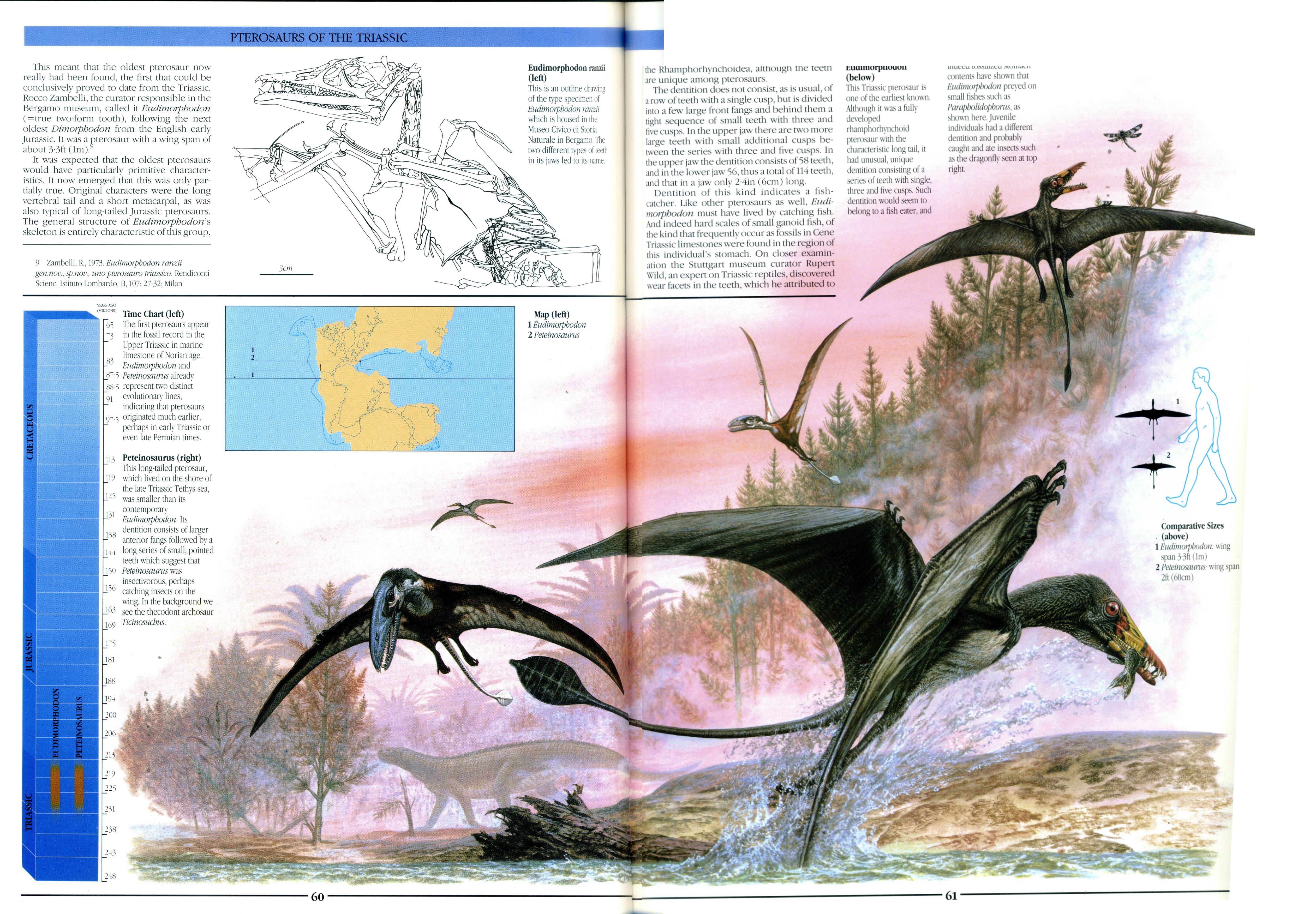
[2] Eudimorphon and Peteinosaurus
from: Wellnhofer, ''Pterosaurs'',
1991, p60-61. COPYRIGHTED
source: Wellnhofer, "Pterosaurs", 1991,
p60-61.
spiny lizards.
[1] Richard Dawkins, ''The Ancestor's
Tale'', (Boston, MA: Houghton Mifflin
Company, 2004), 262. COPYRIGHTED
source: Richard Dawkins, "The
Ancestor's Tale", (Boston, MA: Houghton
Mifflin Company, 2004), 262.

[2] Description Iguana sp. Foto
tomada en el Zoo de Madrid. Date
Summer 2007 Source Own
work Author Manuel de Corselas
ARS SUMMUM, Centro para el Estudio y
Difusión Libres de la Historia del
Arte PD
source: http://upload.wikimedia.org/wiki
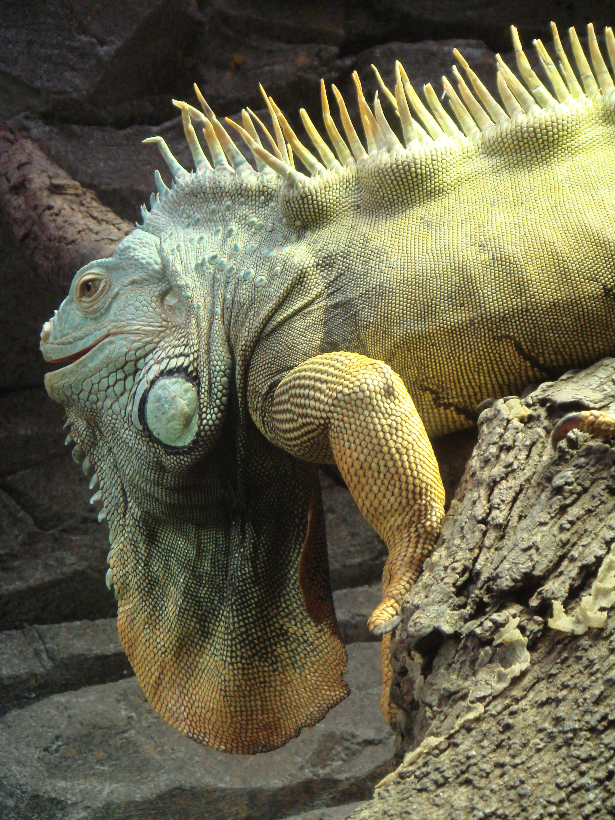
pedia/commons/5/57/AA_Iguana_Fot_Ars_Sum
mum.JPG
geckos.
[1] Richard Dawkins, ''The Ancestor's
Tale'', (Boston, MA: Houghton Mifflin
Company, 2004), 262. COPYRIGHTED
source: Richard Dawkins, "The
Ancestor's Tale", (Boston, MA: Houghton
Mifflin Company, 2004), 262.

[2] Description Deutsch:
Versteinerung eines Archaeophis proavus
Massalongo - aus Monte Bolca. Museum
für Naturkunde (Berlin). English:
Fossil of a Archaeophis proavus
Massalongo, Monte Bolca. Museum für
Naturkunde (Berlin). Date 22 July
2007 Source Own work Author
Raymond - Raimond
Spekking Permission (Reusing this
file) See
below. Attribution (required by the
license) © Raimond Spekking /
CC-BY-SA-3.0 CC
source: http://upload.wikimedia.org/wiki
pedia/commons/f/f1/Naturkundemuseum_Berl
in_-_Archaeophis_proavus_Massalongo_-_Mo
nte_Bolca.jpg

[1] Adapted from: Richard Dawkins,
''The Ancestor's Tale'', (Boston, MA:
Houghton Mifflin Company, 2004),
p339. COPYRIGHTED
source: Richard Dawkins, "The
Ancestor's Tale", (Boston, MA: Houghton
Mifflin Company, 2004), p339.
[2] Adapted from: Richard Dawkins,
''The Ancestor's Tale'', (Boston, MA:
Houghton Mifflin Company, 2004),
p339. COPYRIGHTED
source: Richard Dawkins, "The
Ancestor's Tale", (Boston, MA: Houghton
Mifflin Company, 2004), p339.
caimans {KAmeNS}.
[1] Richard Dawkins, ''The Ancestor's
Tale'', (Boston, MA: Houghton Mifflin
Company, 2004), p262. COPYRIGHTED
source: Richard Dawkins, "The
Ancestor's Tale", (Boston, MA: Houghton
Mifflin Company, 2004), p262.

[2] Nile crocodile, taken at the Le
Bonheur Crocodile Farm near
Stellenbosch, South Africa. CC
source: http://upload.wikimedia.org/wiki
pedia/commons/8/81/NileCrocodile.jpg
[1] Adapted from: Richard Dawkins,
''The Ancestor's Tale'', (Boston, MA:
Houghton Mifflin Company, 2004),
p339. COPYRIGHTED
source: Richard Dawkins, "The
Ancestor's Tale", (Boston, MA: Houghton
Mifflin Company, 2004), p339.
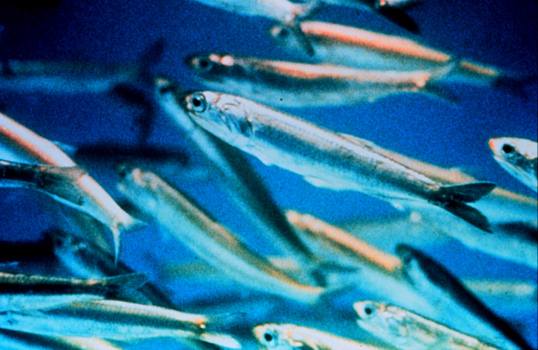
[2] Description Northern
anchovies are important prey for marine
mammals and game fish Image ID:
nur00009, National Undersearch Research
Program (NURP) Collection Location:
Pacific Ocean. Credit: OAR/National
Undersea Research Program
(NURP) Downloaded from:
http://www.photolib.noaa.gov/htmls/nur00
009.htm Note: Another image from this
collection had fish described as
northern anchovies, with the scientific
name Engraulis mordax, or Californian
anchovy. The species may be
misidentified. Date 2006-12-08
(original upload date) PD
source: http://upload.wikimedia.org/wiki
pedia/commons/0/0f/Anchovy_closeup.jpg
Laurasia and Gondwana. The northern
part, Laurasia will form North America
and Europe. The southern part, Gondwana
will form South America and Africa.

[1] 200 Ma Early Jurassic UNKNOWN
source: http://jan.ucc.nau.edu/~rcb7/200
_Jurassic_2globes.jpg
[2] In geologic terms, a plate is a
large, rigid slab of solid rock. The
word tectonics comes from the Greek
root ''to build.'' Putting these two
words together, we get the term plate
tectonics, which refers to how the
Earth's surface is built of plates. The
theory of plate tectonics states that
the Earth's outermost layer is
fragmented into a dozen or more large
and small plates that are moving
relative to one another as they ride
atop hotter, more mobile material.
Before the advent of plate tectonics,
however, some people already believed
that the present-day continents were
the fragmented pieces of preexisting
larger landmasses
(''supercontinents''). The diagrams
below show the break-up of the
supercontinent Pangaea (meaning ''all
lands'' in Greek), which figured
prominently in the theory of
continental drift -- the forerunner to
the theory of plate tectonics. PD
source: http://pubs.usgs.gov/gip/dynamic
/graphics/Fig2-5globes.gif
(moths, butterflies, caterpillars).

[1] Description Photograph of a male
Monarch butterfly (Danaus plexippus en
). This butterfly was stationary on a
leaf with his wings outstretched in an
attempt to show off and attract a mate.
The picture was taken in the butterfly
house at the Tyler Arboretum. Camera
and Exposure Details: Camera: Nikon
D50 Lens: Nikon Nikkor ED AF-S DX
18-55mm f/3.5-5.6G Exposure: 55mm
(82.5mm in 35mm equivalent) f/9 @ 1/125
s. Date 9 September 2006 Source Own
work (Own Picture) Author Photo
(c)2006 Derek Ramsey
(Ram-Man) Permission (Reusing this
file) You may NOT use this image
on your own web site or anywhere else
unless you release this image and any
derivative works (which may include the
web page or other medium where this
image is used, if it is not considered
a ''collective work'') by following the
terms of the following license. Any
other use will be considered a breach
of copyright law. Please do not copy
this image illegally by ignoring the
terms of the license, as it is not in
the public domain. If you would like
special permission to use, license, or
purchase the image or prints of the
image, or for use in any other fashion
or would simply like a copy of the
original file, please contact me or
email me first to ask. Please see the
non-legalese usage guide for more
information. Note: While you are not
required to do so by the license,
please consider letting me know when
you reuse one of my photograph images,
as a courtesy. GNU
source: http://upload.wikimedia.org/wiki
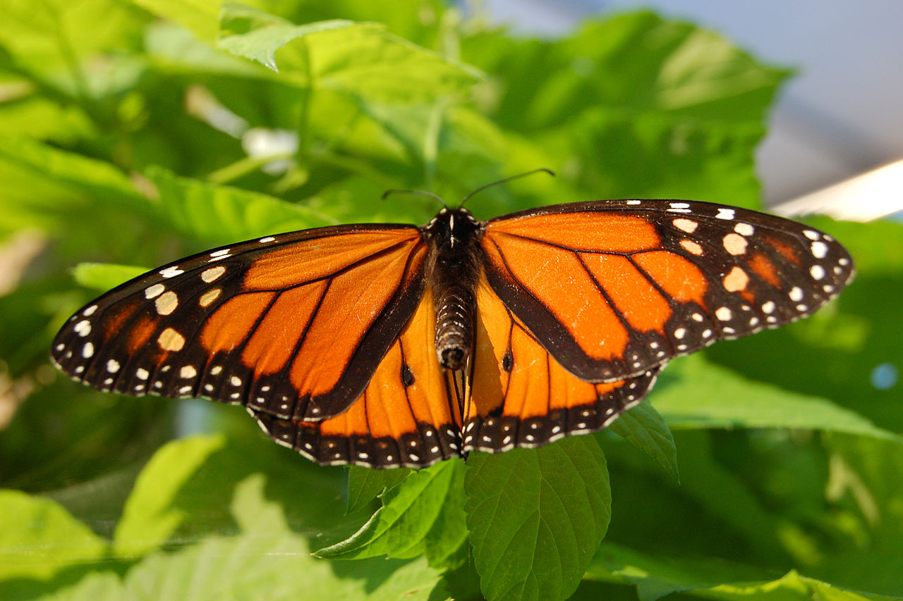
pedia/commons/thumb/a/ab/Monarch_Butterf
ly_Showy_Male_3000px.jpg/1280px-Monarch_
Butterfly_Showy_Male_3000px.jpg

[2] Description Photograph of a
female Monarch Butterflyen (Danaus
plexippus en ) laying an egg on a
Mexican Milkweeden (Asclepias
curassavica en 'Silky Gold'). The
picture was taken in Aston Township,
Pennsylvania. Camera and Exposure
Details: Camera: Nikon D50 Lens:
Sigma 70mm f/2.8 EX DG Macro Exposure:
70mm (105mm in 35mm equivalent) f/8 @
1/160 s. (200 ISO) Date Friday,
August 8, 2008 Source Own
Picture. Author Photo by and (c)2009
Derek Ramsey
(Ram-Man) Permission (Reusing this
file) You may NOT use this image
on your own web site or anywhere else
unless you release this image and any
derivative works (which may include the
web page or other medium where this
image is used, if it is not considered
a ''collective work'') by following the
terms of the following license. Any
other use will be considered a breach
of copyright law. Please do not copy
this image illegally by ignoring the
terms of the license, as it is not in
the public domain. If you would like
special permission to use, license, or
purchase the image or prints of the
image, or for use in any other fashion
or would simply like a copy of the
original file, please contact me or
email me first to ask. Please see the
non-legalese usage guide for more
information. Note: While you are not
required to do so by the license,
please consider letting me know when
you reuse one of my photograph images,
as a courtesy. GNU
source: http://upload.wikimedia.org/wiki
pedia/commons/thumb/7/7a/Monarch_Butterf
ly_Danaus_plexippus_Laying_Eggs.jpg/1096
px-Monarch_Butterfly_Danaus_plexippus_La
ying_Eggs.jpg
Monotremes {moNeTrEMZ} evolve.
Guinea

[1] From: Richard Dawkins, ''The
Ancestor's Tale'', (Boston, MA:
Houghton Mifflin Company, 2004),
239. COPYRIGHTED
source: Richard Dawkins, "The
Ancestor's Tale", (Boston, MA: Houghton
Mifflin Company, 2004), 239.

[2] Description Photo: model of
Steropodon galmani at the Australian
Museum, Sydney. Date 20 April
2008 Source Own work Author
Matt Martyniuk
(Dinoguy2) Permission (Reusing this
file) See below. Other versions
Derivative works of this file:
Prototheria collage.png GNU
source: http://upload.wikimedia.org/wiki
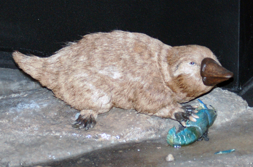
pedia/commons/f/f8/Steropodon_model_aus.
jpg
[1] Adapted from: Richard Dawkins,
''The Ancestor's Tale'', (Boston, MA:
Houghton Mifflin Company, 2004),
p339. COPYRIGHTED
source: Richard Dawkins, "The
Ancestor's Tale", (Boston, MA: Houghton
Mifflin Company, 2004), p339.
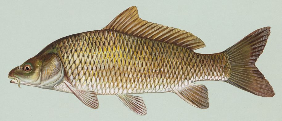
[2] Source:
http://en.wikipedia.org/wiki/Image:Commo
n_carp.jpg Common carp (Cyprinus
carpio). Public domain image from USFWS
National Image Library. Created by
Duane Raver. PD
source: http://upload.wikimedia.org/wiki
pedia/commons/a/a8/Common_carp.jpg
[1] Adapted from: Richard Dawkins,
''The Ancestor's Tale'', (Boston, MA:
Houghton Mifflin Company, 2004),
p339. COPYRIGHTED
source: Richard Dawkins, "The
Ancestor's Tale", (Boston, MA: Houghton
Mifflin Company, 2004), p339.
[2] Fig. 2. The single
most-parsimonious (MP) tree derived
from unweighted analysis of mitogenomic
data comprising concatenated nucleotide
sequences from 12 protein-coding
(excluding the ND6 gene and third codon
positions) and 22 transfer RNA (tRNA)
genes (stem regions only) from all 28
species examined. Tree length, 12,709
steps; consistency index, 0.355;
retention index, 0.471; and rescaled
consistency index, 0.167. Numbers above
and below internal branches indicate
jackknife values obtained for 500
replicates using the heuristic search
option in PAUP*4.0b10 (Swofford, 2002)
with 20 random-addition sequences being
performed in each replication and decay
indices, respectively. The scale
indicates 100 changes. from: Inoue,
JG, Miya, M, Tsukamoto, K, Nishida, M
(2003) ''Basal actinopterygian
relationships: A mitogenomic
perspective on the phylogeny of the
ldquoancient fish.rdquo'' Mol
Phylogenet Evol 26:
110-120 http://www.sciencedirect.com/sc
ience/article/pii/S1055790302003317 COP
YRIGHTED
source: http://www.sciencedirect.com/cac
he/MiamiImageURL/B6WNH-475B9D7-6-1K/0?wc
hp=dGLbVlz-zSkzk
[1] Richard Dawkins, ''The Ancestor's
Tale'', (Boston, MA: Houghton Mifflin
Company, 2004), 303. COPYRIGHTED
source: Richard Dawkins, "The
Ancestor's Tale", (Boston, MA: Houghton
Mifflin Company, 2004), 303.
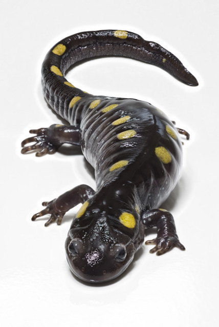
[2] Description central
Pennsylvania Spotted Salamander
(Ambystoma maculatum) Source
self-made Date 25 March
2008 Author Camazine (talk) Scott
Camazine web.mac.com/camazine CC
source: http://upload.wikimedia.org/wiki
pedia/en/b/b2/SpottedSalamander.jpg
Dragonfish.
[1] Adapted from: Richard Dawkins,
''The Ancestor's Tale'', (Boston, MA:
Houghton Mifflin Company, 2004),
p339. COPYRIGHTED
source: Richard Dawkins, "The
Ancestor's Tale", (Boston, MA: Houghton
Mifflin Company, 2004), p339.
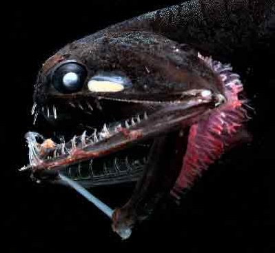
[2] Description English: This
deep-sea fish, Photostomias guernei,
has a built-in bioluminescent
''flashlight'' it uses to help it see
in the dark. Date 1999 Source
Photostomias.jpg Author
derivative work: Una Smith
Photostomias.jpg: Edith
Widder/HBOI PD
source: http://upload.wikimedia.org/wiki
pedia/commons/6/63/Photostomias2.jpg
[1] Richard Dawkins, ''The Ancestor's
Tale'', (Boston, MA: Houghton Mifflin
Company, 2004), p262. COPYRIGHTED
source: Richard Dawkins, "The
Ancestor's Tale", (Boston, MA: Houghton
Mifflin Company, 2004), p262.

[2] Description English:
Archaeopteryx lithographica, specimen
displayed at the Museum für Naturkunde
in Berlin. (This image shows the
original fossil - not a
cast.) Deutsch: Archaeopteryx
lithographica, Exemplar im Museum für
Naturkunde in Berlin. (Dieses Bild
zeigt das Original-Fossil, keinen
Abguss.) Date 5 July 2009 Source
Own work Author H. Raab
(User:Vesta) CC
source: http://upload.wikimedia.org/wiki
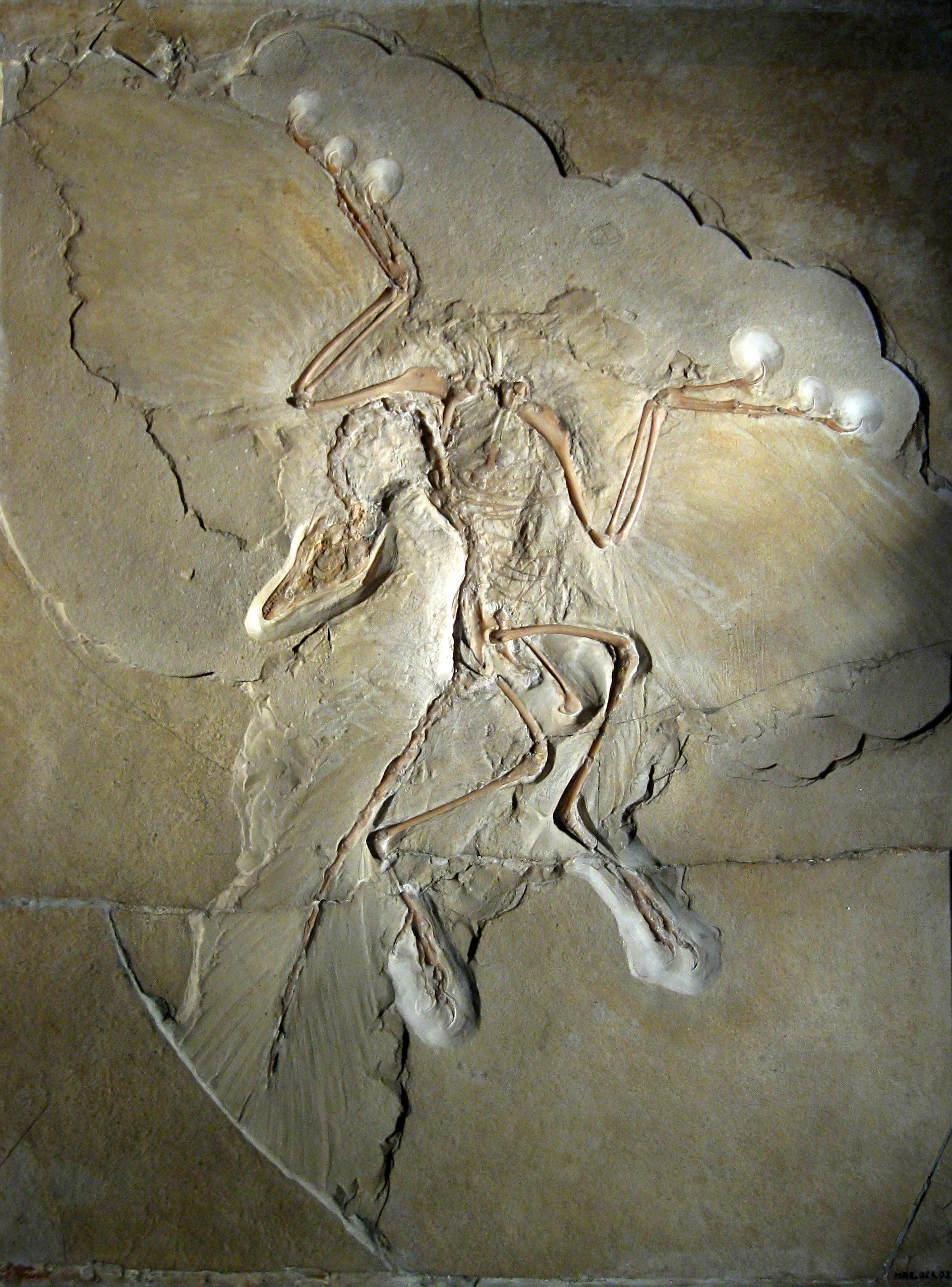
pedia/commons/9/9d/Archaeopteryx_lithogr
aphica_%28Berlin_specimen%29.jpg
flowering plant. Almost all grains,
beans, nuts, fruits, vegetables, herbs
and spices come from plants with
flowers.
China
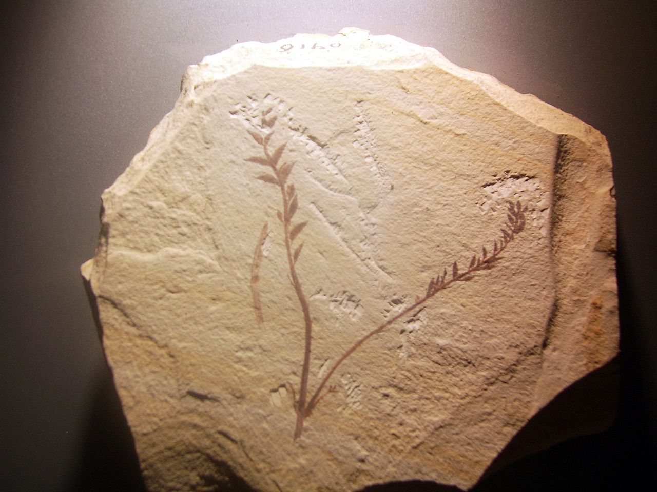
[1] Description
辽宁古果(Archaefructus
liaoningensis),为迄今发现的最
早的花(早白垩纪),于北京�
��然博物馆 Date 17:15, 18 October
2006 (UTC) Source Own work Author
Shizhao CC
source: http://upload.wikimedia.org/wiki
pedia/commons/thumb/7/7f/Archaefructus_l
iaoningensis.jpg/1280px-Archaefructus_li
aoningensis.jpg

[2] Figure 2 from: Sun, G. , Dilcher,
D. L. , Zheng, S.-L. & Zhou, Z.-K. In
search of the first flower: A Jurassic
angiosperm, Archaefructus, from
northeast China. Science 282,
1692–1695
(1998). http://www.sciencemag.org/conte
nt/282/5394/1692
AND http://www.jstor.org/stable/2896858
COPYRIGHTED
source: Sun, G. , Dilcher, D. L. ,
Zheng, S.-L. & Zhou, Z.-K. In search of
the first flower: A Jurassic
angiosperm, Archaefructus, from
northeast China. Science 282,
1692–1695
(1998). http://www.sciencemag.org/conte
nt/282/5394/1692http://www.jstor.org/sta
ble/2896858
"Amborella".

[1] N Wikstrom, V Savolainen, MW Chase,
''Evolution of the angiosperms:
calibrating the family tree'', Proc
Biol Sci. 2001 Nov
7;268(1482):2211-20., (2001).
http://rspb.royalsocietypublishing.org
/content/268/1482/2211.abstract COPYRIG
HTED
source: http://rspb.royalsocietypublishi
ng.org/content/268/1482/2211.abstract
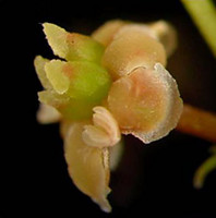
[2] Photo of Amborella trichopoda
(Amborellaceae; photo © Sangtae Kim).
source: http://tolweb.org/tree?group=ang
iosperms
{niM-FE-A-lEZ} (water lilies).

[1] Nymphaea alba Nymphaea alba -
image taken on 29 August 2004 in the
outdoor botanical garden of Technion -
Haifa, Israel public domain
source: http://en.wikipedia.org/wiki/Nym
phaeaceae
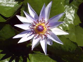
[2] Nymphaea colorata from
Africa presume is gnu or pd
source: same
and breast.
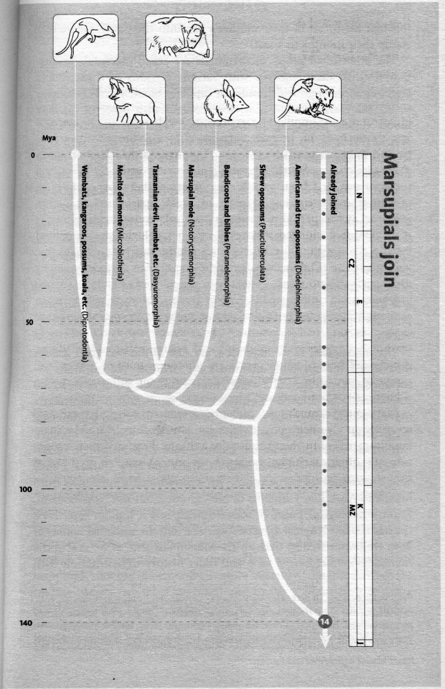
[1] From: Richard Dawkins, ''The
Ancestor's Tale'', (Boston, MA:
Houghton Mifflin Company, 2004),
p231. COPYRIGHTED
source: Richard Dawkins, "The
Ancestor's Tale", (Boston, MA: Houghton
Mifflin Company, 2004), p231.
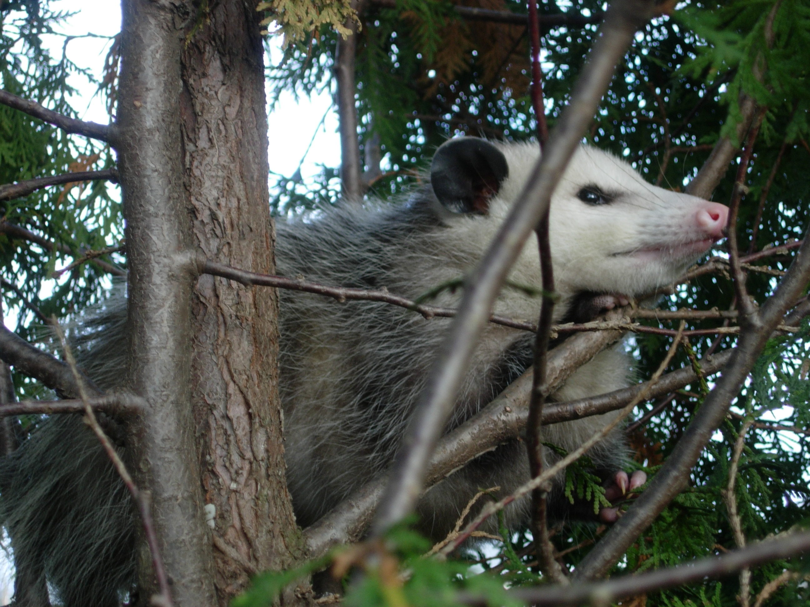
[2] Description English: Virginia
Opossum (Didelphis virginiana) in a
juniper tree in northeastern
Ohio. Date 27 December
2008 Source Own work Author
Wilson44691 Permission (Reusing
this file) See below. Other versions
PD
source: http://upload.wikimedia.org/wiki
pedia/commons/6/6a/Possum122708.JPG
evolve (magnolia, nutmeg, avocado,
cinnamon, black pepper).

[1] Magnolia This photo is a part of
the Wikipedia:Plant photo collection
I. Downloaded URL:
http://tencent.homestead.com/files/magno
lia.jpg Warning sign This image has
no source information. Source
information must be provided so that
the copyright status can be verified by
others. Unless the copyright status is
provided and a source is given, the
image will be deleted seven days after
this template was added (see page
history). If you just added this
template, please use {{no source
source: http://en.wikipedia.org/wiki/Mag
noliales
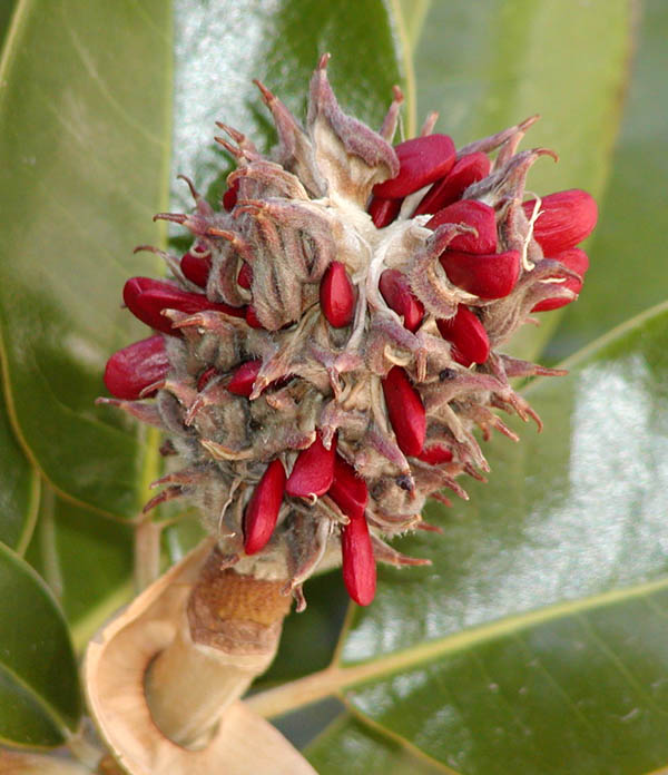
[2] ~~~~~}} (to include the date
here). Please consider using
source: same
(the largest lineage of flowers).

[1] N Wikstrom, V Savolainen, MW Chase,
''Evolution of the angiosperms:
calibrating the family tree'', Proc
Biol Sci. 2001 Nov
7;268(1482):2211-20., (2001).
http://rspb.royalsocietypublishing.org
/content/268/1482/2211.abstract COPYRIG
HTED
source: http://rspb.royalsocietypublishi
ng.org/content/268/1482/2211.abstract
[2] N Wikstrom, V Savolainen, MW
Chase, ''Evolution of the angiosperms:
calibrating the family tree'', Proc
Biol Sci. 2001 Nov
7;268(1482):2211-20., (2001).
http://rspb.royalsocietypublishing.org
/content/268/1482/2211.abstract COPYRIG
HTED
source: http://rspb.royalsocietypublishi
ng.org/content/268/1482/2211.abstract
fish, pufferfish, barracuda.
[1] Adapted from: Richard Dawkins,
''The Ancestor's Tale'', (Boston, MA:
Houghton Mifflin Company, 2004),
p339. COPYRIGHTED
source: Richard Dawkins, "The
Ancestor's Tale", (Boston, MA: Houghton
Mifflin Company, 2004), p339.

[2] Seahorse - Hippocampus
sp. Image ID reef2027, The
Coral Kingdom Collection Location
Gulf of Aqaba, Red Sea Photographer
Mr. Mohammed Al Momany, Aqaba,
Jordan Source
http://www.photolib.noaa.gov/htmls/reef2
027.htm PD
source: http://upload.wikimedia.org/wiki
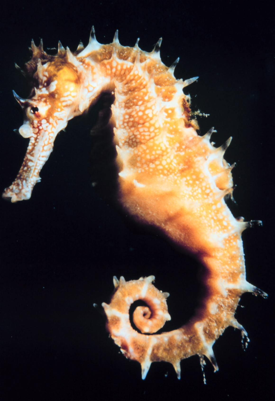
pedia/commons/4/4b/Hippocampus.jpg
[1] Adapted from: Richard Dawkins,
''The Ancestor's Tale'', (Boston, MA:
Houghton Mifflin Company, 2004),
p339. COPYRIGHTED
source: Richard Dawkins, "The
Ancestor's Tale", (Boston, MA: Houghton
Mifflin Company, 2004), p339.

[2] Fig. 2. The single
most-parsimonious (MP) tree derived
from unweighted analysis of mitogenomic
data comprising concatenated nucleotide
sequences from 12 protein-coding
(excluding the ND6 gene and third codon
positions) and 22 transfer RNA (tRNA)
genes (stem regions only) from all 28
species examined. Tree length, 12,709
steps; consistency index, 0.355;
retention index, 0.471; and rescaled
consistency index, 0.167. Numbers above
and below internal branches indicate
jackknife values obtained for 500
replicates using the heuristic search
option in PAUP*4.0b10 (Swofford, 2002)
with 20 random-addition sequences being
performed in each replication and decay
indices, respectively. The scale
indicates 100 changes. from: Inoue,
JG, Miya, M, Tsukamoto, K, Nishida, M
(2003) ''Basal actinopterygian
relationships: A mitogenomic
perspective on the phylogeny of the
ldquoancient fish.rdquo'' Mol
Phylogenet Evol 26:
110-120 http://www.sciencedirect.com/sc
ience/article/pii/S1055790302003317 COP
YRIGHTED
source: http://upload.wikimedia.org/wiki
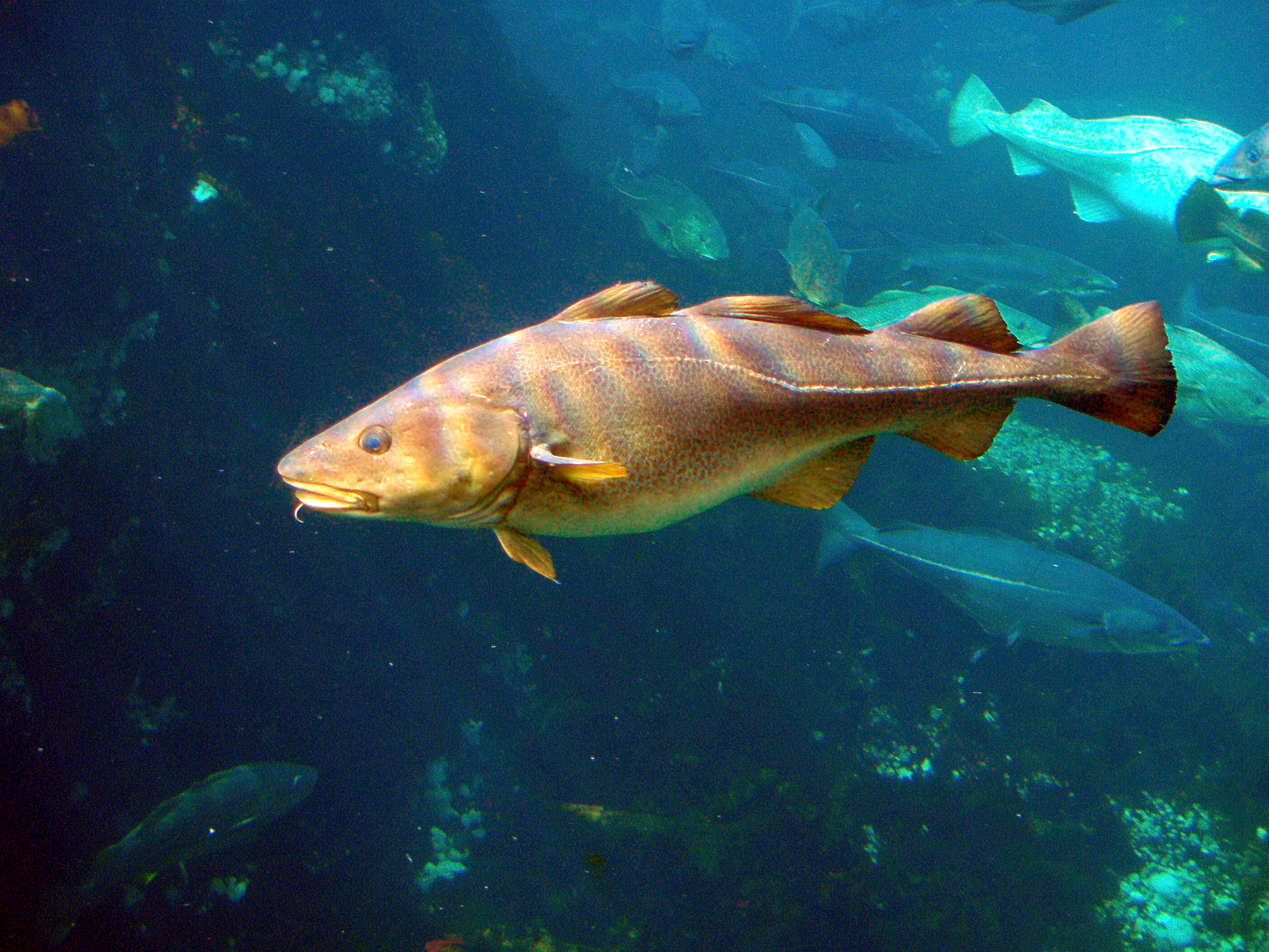
pedia/commons/c/cf/Gadus_morhua-Cod-2-At
lanterhavsparken-Norway.JPG
evolve.
County, Liaoning Province, China

[1] Description English:
Juramaia Date 30 April 2012 Source
Own work Author Nobu Tamura
http://paleoexhibit.blogspot.com/
http://spinops.blogspot.com/
http://www.palaeocritti.com CC
source: http://upload.wikimedia.org/wiki
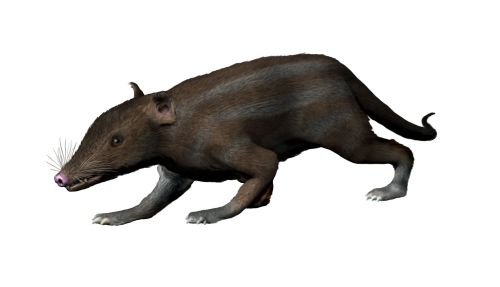
pedia/commons/1/15/Juramaia_NT.jpg

[2] Figure 1 from: Luo Z, Yuan C,
Meng Q & Ji Q (2011), ''A Jurassic
eutherian mammal and divergence of
marsupials and placentals'', Nature
476(7361): p.
42–45. http://www.nature.com/nature/j
ournal/v476/n7361/full/nature10291.html
{nature10291.pdf} COPYRIGHTED
source: http://nature.com/nature/journal
/v476/n7361/carousel/nature10291-f1.2.jp
g
(modern birds).
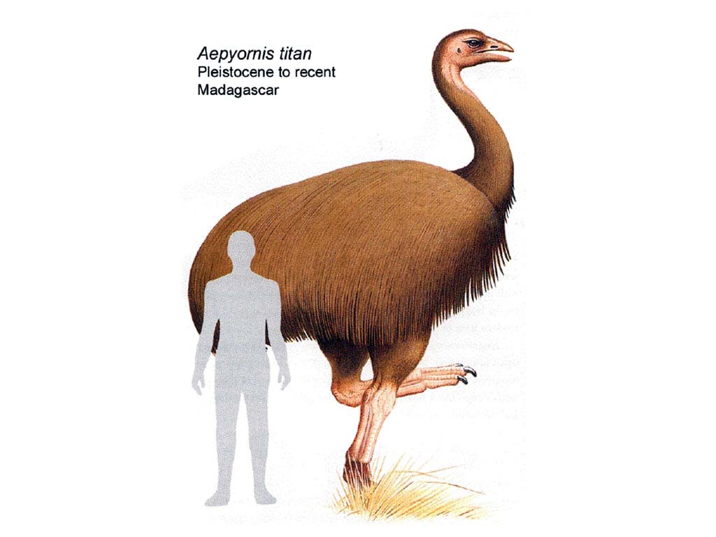
[1] aepyornis The same image appears
in: Palmer, The Marshall Illustrated
Encyclopedia of Dinosaurs and
Prehistoric Animals, 1999,
p176. COPYRIGHTED
source: http://photo.starnet.ru/Thematic
_Wallpapers/Zhizn/Dikie_zhivotnye/dinosa
urs_fossils/images/aepyornis.jpg
[2] From: Richard Dawkins, ''The
Ancestor's Tale'', (Boston, MA:
Houghton Mifflin Company, 2004),
p262. COPYRIGHTED
source: Richard Dawkins, "The
Ancestor's Tale", (Boston, MA: Houghton
Mifflin Company, 2004), p262.
plants that have a single cotyledon
(seed leaf) in the embryo.

[1] N Wikstrom, V Savolainen, MW Chase,
''Evolution of the angiosperms:
calibrating the family tree'', Proc
Biol Sci. 2001 Nov
7;268(1482):2211-20., (2001).
http://rspb.royalsocietypublishing.org
/content/268/1482/2211.abstract COPYRIG
HTED
source: http://rspb.royalsocietypublishi
ng.org/content/268/1482/2211.abstract
[2] N Wikstrom, V Savolainen, MW
Chase, ''Evolution of the angiosperms:
calibrating the family tree'', Proc
Biol Sci. 2001 Nov
7;268(1482):2211-20., (2001).
http://rspb.royalsocietypublishing.org
/content/268/1482/2211.abstract COPYRIG
HTED
source: http://rspb.royalsocietypublishi
ng.org/content/268/1482/2211.abstract
(buttercup, poppy, macadamia,
sycamore).
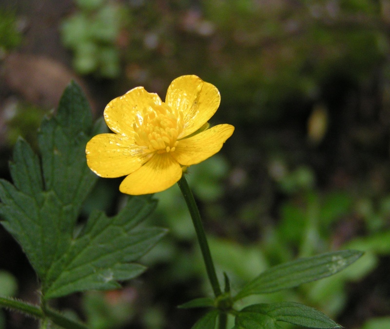
[1] Creeping butercup (Ranunculus
repens). GNU
source: http://en.wikipedia.org/wiki/Ima
ge:Creeping_butercup_close_800.jpg

[2] Clematis hybrid from
http://www.ars.usda.gov/is/graphics/phot
os/ public domain
source: http://en.wikipedia.org/wiki/Cle
matis
caper, buckwheat, rhubarb, venus
flytrap, old world pitcher plants,
beet, quinoa, spinach, grape plants).
[1] N Wikstrom, V Savolainen, MW Chase,
''Evolution of the angiosperms:
calibrating the family tree'', Proc
Biol Sci. 2001 Nov
7;268(1482):2211-20., (2001).
http://rspb.royalsocietypublishing.org
/content/268/1482/2211.abstract COPYRIG
HTED
source: http://rspb.royalsocietypublishi
ng.org/content/268/1482/2211.abstract
[2] N Wikstrom, V Savolainen, MW
Chase, ''Evolution of the angiosperms:
calibrating the family tree'', Proc
Biol Sci. 2001 Nov
7;268(1482):2211-20., (2001).
http://rspb.royalsocietypublishing.org
/content/268/1482/2211.abstract COPYRIG
HTED
source: http://rspb.royalsocietypublishi
ng.org/content/268/1482/2211.abstract
Afrotheres evolves.
[1] From: Richard Dawkins, ''The
Ancestor's Tale'', (Boston, MA:
Houghton Mifflin Company, 2004),
p225. COPYRIGHTED
source: Richard Dawkins, "The
Ancestor's Tale", (Boston, MA: Houghton
Mifflin Company, 2004), p225.
[2] Description Afrotheria Date
18 December 2007 Source
self-made, based on:
Image:Orycteropus afer.jpg
Image:Dugong.jpg Image:Elephant
Shrew.jpg Image:Manatee Looking at
the Camera.jpg Image:Taupe
doree.jpg Image:Klippschliefer
Suedafrika Hermanus.jpg
Image:Elefante Lake Manyara Park.jpg
Image:Tanrek.jpg Author
Esculapio GNU
source: http://upload.wikimedia.org/wiki
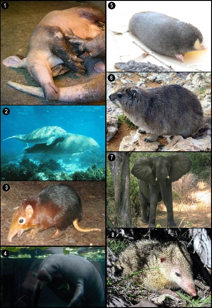
pedia/commons/f/f0/Afrotheria.jpg
emu, cassowary {KaSOwaRE}, kiwis).
[1] Richard Dawkins, ''The Ancestor's
Tale'', (Boston, MA: Houghton Mifflin
Company, 2004), p262. COPYRIGHTED
source: Richard Dawkins, "The
Ancestor's Tale", (Boston, MA: Houghton
Mifflin Company, 2004), p262.
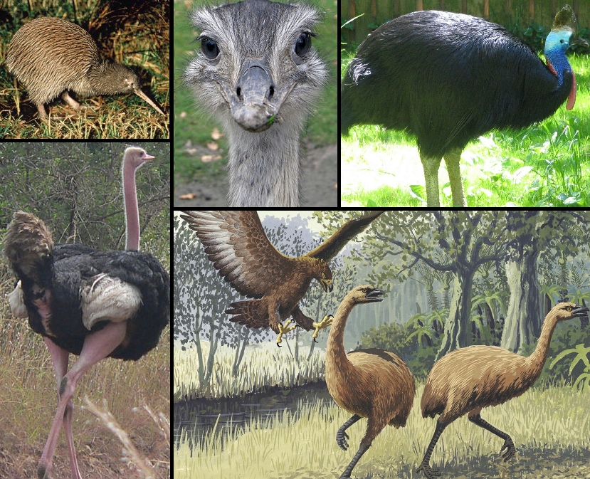
[2] Description Various Ratite
birds (clockwise from top left): Brown
kiwi Apteryx mantelli, Greater rhea,
double-wattled cassowary Casuarius
casuarius, Haast's eagle attacking New
Zealand moa, Masai ostrich
(photographed in Nairobi National Park,
Kenya). Date 19 June 2007 Source
self-made from
Image:Brown_kiwi.jpg,
Image:Nandu-Portrait 2.jpg,
Image:Casuarius_casuarius_-_double-wattl
ed_cassowary.jpg,
Image:Giant_Haasts_eagle_attacking_New_Z
ealand_moa.jpg, Image:Masai ostrich.jpg
(see original images for copyright
information). Author
Richard001 GNU
source: http://upload.wikimedia.org/wiki
pedia/commons/3/31/Ratites.PNG
{ZeNoRtreNZ} evolve (Sloths, Anteaters,
Armadillos).

[1] From: Richard Dawkins, ''The
Ancestor's Tale'', (Boston, MA:
Houghton Mifflin Company, 2004),
p220. COPYRIGHTED
source: Richard Dawkins, "The
Ancestor's Tale", (Boston, MA: Houghton
Mifflin Company, 2004), p220.

[2] Description Hoffmann's Two-toed
Sloth (Choloepus hoffmanni) in
Milwaukee County Zoological
Gardens Date 8 January
2006 Source Flickr Author
Woodsm CC
source: http://upload.wikimedia.org/wiki
pedia/commons/b/b5/Choloepus_hoffmanni.j
pg
clove, guava, allspice, eucalyptus).

[1] N Wikstrom, V Savolainen, MW Chase,
''Evolution of the angiosperms:
calibrating the family tree'', Proc
Biol Sci. 2001 Nov
7;268(1482):2211-20., (2001).
http://rspb.royalsocietypublishing.org
/content/268/1482/2211.abstract COPYRIG
HTED
source: http://rspb.royalsocietypublishi
ng.org/content/268/1482/2211.abstract

[2] A photo of the tree Staphylea
colchica taken by me in Århus, Denmark
GNU
source: http://en.wikipedia.org/wiki/Cro
ssosomatales
(beans, pea, peanut, soy, lentil).

[1] N Wikstrom, V Savolainen, MW Chase,
''Evolution of the angiosperms:
calibrating the family tree'', Proc
Biol Sci. 2001 Nov
7;268(1482):2211-20., (2001).
http://rspb.royalsocietypublishing.org
/content/268/1482/2211.abstract COPYRIG
HTED
source: http://rspb.royalsocietypublishi
ng.org/content/268/1482/2211.abstract

[2] Abrus precatorius (Black-eyed
Susan) USGS public domain
source: http://en.wikipedia.org/wiki/Abr
us
orchid, asparagus, onion, garlic,
agave, aloe, lily).
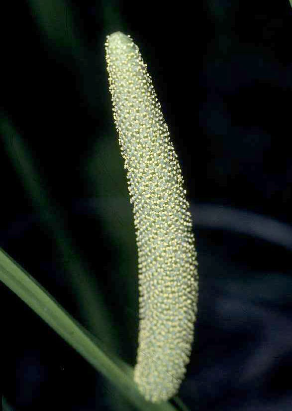
[1] Sweet Flag (Acorus calamus) -
spadix Spadix of Sweet Flag. usgs
public domain
source: http://en.wikipedia.org/wiki/Aco
rus
[2] Ivy Duckweed (Lemna
trisulca) Name Lemna
trisulca Family Lemnaceae
source: http://en.wikipedia.org/wiki/Ali
smatales
{KomelIniDZ} evolve (palms, coconut,
corn, rice, barley, oat, wheat, rye,
sugarcane, bamboo, grass, pineapple,
papyrus, turmeric {TRmRiK}, banana,
ginger).
[1] Manila dwarf coconut palm from
http://www.ars.usda.gov/is/graphics/phot
os/ Manila dwarf coconut palm
thumbnail A Manila dwarf coconut palm
on the grounds of the Tropical
Agriculture Research Station in
Mayaguez, Puerto Rico. dept of
ag public domain
source: http://en.wikipedia.org/wiki/Are
cales

[2] coconut GOV public domain
source: http://www.nps.gov/kaho/KAHOckLs
/KAHOplnt/images/IMG_03957.jpg
(kiwi, ebony, persimmon, blueberry,
cranberry, brazil nut, new world
pitcher plants, tea).
[1] N Wikstrom, V Savolainen, MW Chase,
''Evolution of the angiosperms:
calibrating the family tree'', Proc
Biol Sci. 2001 Nov
7;268(1482):2211-20., (2001).
http://rspb.royalsocietypublishing.org
/content/268/1482/2211.abstract COPYRIG
HTED
source: http://rspb.royalsocietypublishi
ng.org/content/268/1482/2211.abstract
[2] Actinidia fruit. kiwifruit. public
domain
source: http://en.wikipedia.org/wiki/Ima
ge:Aethionema_grandiflora0.jpg
celery, cilantro, carrot, parsnip,
fennel, parsley, ivy).
[1] Variegated Ground-elder (Aegopodium
podagraria L.) in flower. GNU
source: http://en.wikipedia.org/wiki/Ima
ge:Ground-elder_bloom.jpg

[2] An established spread of
variegated Ground-elder (Aegopodium
podagraria L.). GNU
source: http://en.wikipedia.org/wiki/Ima
ge:Ground-elder.jpg
(tarragon, daisy, artichoke, sunflower,
lettuce, dandelion).

[1] Ray floret, typical for flowers of
the family Asteraceae. GNU
source: http://en.wikipedia.org/wiki/Ima
ge:Ray.floret01.jpg
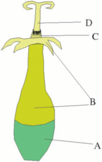
[2] disc floret, typical part of a
flower of the family Asteraceae. GNU
source: http://en.wikipedia.org/wiki/Ima
ge:Disc_floret01.jpg
{maLPiGEAlEZ} (coca, rubber tree,
cassava, poinsettia, willow, poplar,
aspen).

[1] mangosteen public domain
source: http://en.wikipedia.org/wiki/Gar
cinia
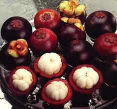
[2] Mangosteen fruit public domain
source: http://en.wikipedia.org/wiki/Man
gosteen
(horseradish, mustard, cabbage,
broccoli, radish, papaya).
[1] N Wikstrom, V Savolainen, MW Chase,
''Evolution of the angiosperms:
calibrating the family tree'', Proc
Biol Sci. 2001 Nov
7;268(1482):2211-20., (2001).
http://rspb.royalsocietypublishing.org
/content/268/1482/2211.abstract COPYRIG
HTED
source: http://rspb.royalsocietypublishi
ng.org/content/268/1482/2211.abstract

[2] Aethionema grandiflora, GFDL by
Kurt Stueber
source: http://en.wikipedia.org/wiki/Ima
ge:Aethionema_grandiflora0.jpg
hop, jackfruit, fig, strawberry, rose,
raspberry, apple, pear, plum, cherry,
peach, almond).

[1] Filipendula ulmaria, GNU
source: http://en.wikipedia.org/wiki/Fil
ipendula

[2] A display of different apples,
We've even worked on bashless
bagging-packaging systems that are used
by wholesalers to bring you apples
without bruises. US ARS public domain
source: http://en.wikipedia.org/wiki/App
le
evolve (oleander, coffee).
[1] N Wikstrom, V Savolainen, MW Chase,
''Evolution of the angiosperms:
calibrating the family tree'', Proc
Biol Sci. 2001 Nov
7;268(1482):2211-20., (2001).
http://rspb.royalsocietypublishing.org
/content/268/1482/2211.abstract COPYRIG
HTED
source: http://rspb.royalsocietypublishi
ng.org/content/268/1482/2211.abstract

[2] Anthocleista grandiflora. PD
source: http://en.wikipedia.org/wiki/Ima
ge:Anthocleista_grandiflora.jpg
evolve (bell pepper, tomato, tobacco,
potato, eggplant).
[1] N Wikstrom, V Savolainen, MW Chase,
''Evolution of the angiosperms:
calibrating the family tree'', Proc
Biol Sci. 2001 Nov
7;268(1482):2211-20., (2001).
http://rspb.royalsocietypublishing.org
/content/268/1482/2211.abstract COPYRIG
HTED
source: http://rspb.royalsocietypublishi
ng.org/content/268/1482/2211.abstract
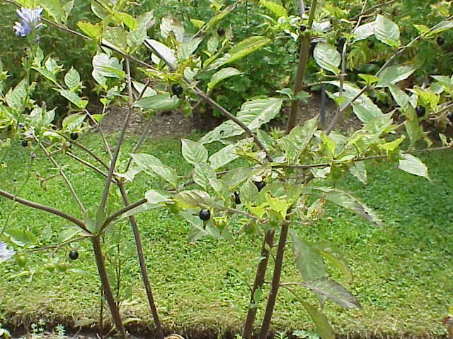
[2] Atropa belladonna. Deadly
nightshade. GFDL by Kurt Stueber
source: http://en.wikipedia.org/wiki/Ima
ge:Atropa_bella-donna1.jpg
(KYUKRBiTAlEZ} evolve (melon, cucumber,
pumpkin, squash, zucchini).

[1] White bryony (Bryonia dioica). GNU
source: http://en.wikipedia.org/wiki/Ima
ge:White_bryony_male_800.jpg

[2] watermelon public domain
source: http://en.wikipedia.org/wiki/Ima
ge:Vampire_watermelon.jpg
(Birch, Hazel {nut}, Chestnut, Beech
{nut}, Oak, Walnut, Pecan, Hickory).

[1] Alnus serrulata (Tag Alder) Male
catkins on right, mature female catkins
left Johnsonville, South Carolina GFDL
source: http://en.wikipedia.org/wiki/Ima
ge:Tagalder8139.jpg
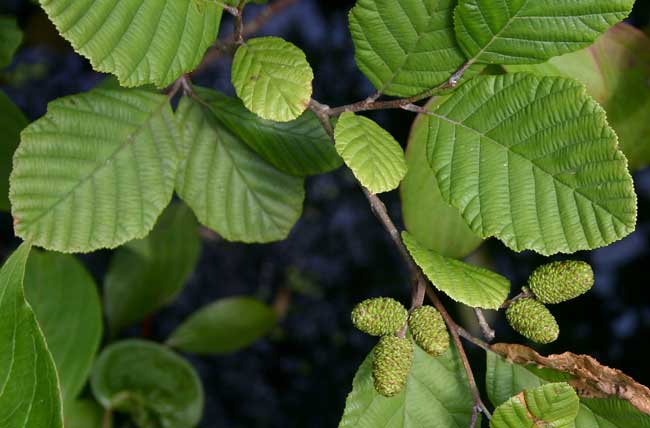
[2] Speckled Alder (Alnus incana
subsp. rugosa) - leaves GNU
source: http://en.wikipedia.org/wiki/Ima
ge:Alnus_incana_rugosa_leaves.jpg
evolve (Chicken, Turkey, Pheasant,
Peacock, Quail).
[1] Richard Dawkins, ''The Ancestor's
Tale'', (Boston, MA: Houghton Mifflin
Company, 2004), p262. COPYRIGHTED
source: Richard Dawkins, "The
Ancestor's Tale", (Boston, MA: Houghton
Mifflin Company, 2004), p262.
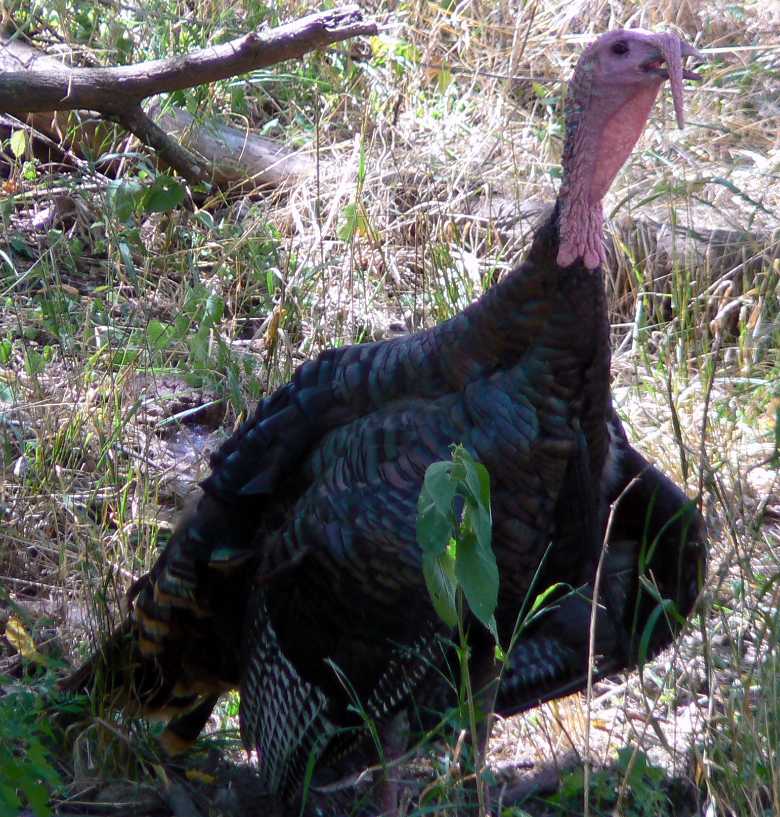
[2] Description English: Meleagris
gallopavo (Wild Turkey) Date 30
July 2006 Source Own work Author
MONGO PD
source: http://upload.wikimedia.org/wiki
pedia/commons/6/69/Meleagris_gallopavo_W
ild_Turkey.jpg
evolve (ducks, geese, swans).
[1] Richard Dawkins, ''The Ancestor's
Tale'', (Boston, MA: Houghton Mifflin
Company, 2004), p262. COPYRIGHTED
source: Richard Dawkins, "The
Ancestor's Tale", (Boston, MA: Houghton
Mifflin Company, 2004), p262.

[2] Description English: Pair of
Wood Ducks Date 18 April
2007 Source
http://flickr.com/photos/sherseydc/
1623995158/ Author
http://www.flickr.com/people/sherse
ydc/ CC
source: http://upload.wikimedia.org/wiki
pedia/commons/0/08/Pair_of_Wood_Ducks.jp
g
mammal "Laurasiatheres" evolves.

[1] From: Richard Dawkins, ''The
Ancestor's Tale'', (Boston, MA:
Houghton Mifflin Company, 2004),
p200. COPYRIGHTED
source: Richard Dawkins, "The
Ancestor's Tale", (Boston, MA: Houghton
Mifflin Company, 2004), p200.

[2] Description Mamíferos
(mammals), based on:
Image:Giraffa camelopardalis angolensis
(head).jpg Image:Golden crowned
fruit bat.jpg
Image:Hedgehog-en.jpg Image:Lion
waiting in Nambia.jpg All of them
under a free licence already in
Wikicommons Date
11-01-2008 Source
Compilation made by myself,
Authors of the photos see
below. Author Hans Hillewaert
(Giraffe); (Bat) Original uploader was
Latorilla at en.wikipedia;
(Hedgehog-en) John Mittler at
777Life.com Free Image Archive; (Lion)
yaaaay CC
source: http://upload.wikimedia.org/wiki
pedia/commons/a/a5/Mam%C3%ADferos.jpg
evolve (okra, cotton, cacao {KoKoU}).
[1] N Wikstrom, V Savolainen, MW Chase,
''Evolution of the angiosperms:
calibrating the family tree'', Proc
Biol Sci. 2001 Nov
7;268(1482):2211-20., (2001).
http://rspb.royalsocietypublishing.org
/content/268/1482/2211.abstract COPYRIG
HTED
source: http://rspb.royalsocietypublishi
ng.org/content/268/1482/2211.abstract
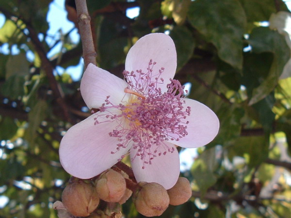
[2] Bixa orellana L., floro en Lavras,
Minas Gerais, Brazilo, GNU
source: http://en.wikipedia.org/wiki/Ima
ge:Aethionema_grandiflora0.jpg
(maple, citris, cashew, mango,
pistachio).
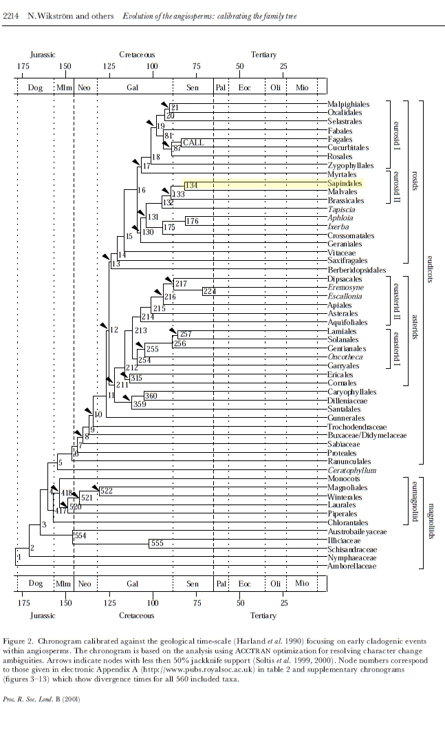
[1] N Wikstrom, V Savolainen, MW Chase,
''Evolution of the angiosperms:
calibrating the family tree'', Proc
Biol Sci. 2001 Nov
7;268(1482):2211-20., (2001).
http://rspb.royalsocietypublishing.org
/content/268/1482/2211.abstract COPYRIG
HTED
source: http://en.wikipedia.org/wiki/Ima
ge:Aethionema_grandiflora0.jpg
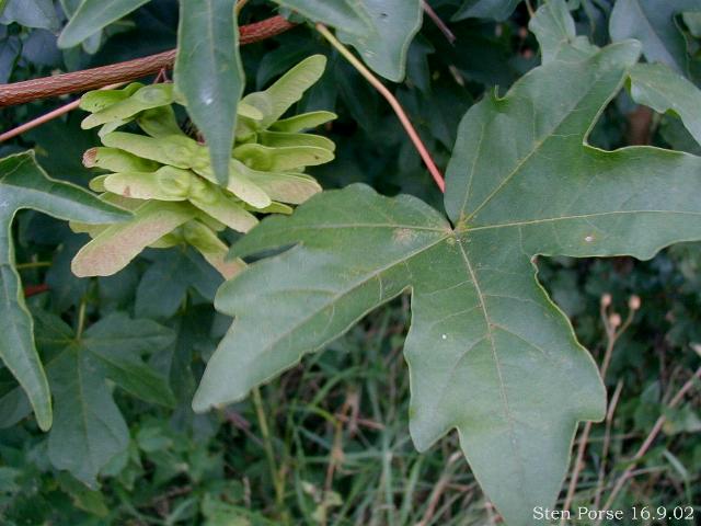
[2] Field Maple foliage and flowers,
Acer campestre. GNU
source: http://en.wikipedia.org/wiki/Ima
ge:Acer-campestre.JPG
evolves (shrews, moles, hedgehogs).

[1] From: Richard Dawkins, ''The
Ancestor's Tale'', (Boston, MA:
Houghton Mifflin Company, 2004),
p200. COPYRIGHTED
source: Richard Dawkins, "The
Ancestor's Tale", (Boston, MA: Houghton
Mifflin Company, 2004), p200.

[2] From: Richard Dawkins, ''The
Ancestor's Tale'', (Boston, MA:
Houghton Mifflin Company, 2004),
p200. COPYRIGHTED
source: Richard Dawkins, "The
Ancestor's Tale", (Boston, MA: Houghton
Mifflin Company, 2004), p200.
[1] From: Richard Dawkins, ''The
Ancestor's Tale'', (Boston, MA:
Houghton Mifflin Company, 2004),
p231. COPYRIGHTED
source: Richard Dawkins, "The
Ancestor's Tale", (Boston, MA: Houghton
Mifflin Company, 2004), p231.
[2] Description English: North
American Opossum with winter
coat. Français : Opossum de Virginie
en livrée d'hiver. Deutsch: Ein
Nordopossum (Didelphis virginiana) im
Winterfell Date 21 February
2007 Source
Wikipedia:User:Cody.pope Author
Cody Pope CC
source: http://upload.wikimedia.org/wiki
pedia/commons/2/27/Opossum_2.jpg
[1] From: Richard Dawkins, ''The
Ancestor's Tale'', (Boston, MA:
Houghton Mifflin Company, 2004),
p225. COPYRIGHTED
source: Richard Dawkins, "The
Ancestor's Tale", (Boston, MA: Houghton
Mifflin Company, 2004), p225.

[2] Description An aardvark at
Detroit Zoo Date 15 April
2008 Source Cropped from
File:Porcs formiguers (Orycteropus
afer).jpg Author MontageMan is
the author of the original image, I did
the crop Permission (Reusing this
file) See below. CC
source: http://upload.wikimedia.org/wiki
pedia/commons/8/8a/Porc_formiguer.JPG
(mint, basil, oregano, rosemary, sage,
thyme, sesame, olive, ash, lilac,
jasmine).

[1] Common Bugle (Ajuga reptans) GNU
source: http://en.wikipedia.org/wiki/Ima
ge:Ajuga-reptans01.jpg
[2] Calamintha grandiflora. GFDL by
Kurt Stueber
source: http://en.wikipedia.org/wiki/Ima
ge:Calamintha_grandiflora2.jpg
{BiLBEZ}.
[1] From: Richard Dawkins, ''The
Ancestor's Tale'', (Boston, MA:
Houghton Mifflin Company, 2004),
p231. COPYRIGHTED
source: Richard Dawkins, "The
Ancestor's Tale", (Boston, MA: Houghton
Mifflin Company, 2004), p231.

[2] Description Eastern Barred
Bandicoot (Perameles gunnii), Poimena
Reserve, Austin's Ferry, Tasmania,
Australia. The photo taken at night
with off camera flashes. Date 31
July 2010 Source Own work Author
Noodle snacks
(http://www.noodlesnacks.com/) CC
source: http://upload.wikimedia.org/wiki
pedia/commons/8/8b/Perameles_gunni.jpg
and Pikas {PIKuZ}.

[1] From: Richard Dawkins, ''The
Ancestor's Tale'', (Boston, MA:
Houghton Mifflin Company, 2004),
p187. COPYRIGHTED
source: Richard Dawkins, "The
Ancestor's Tale", (Boston, MA: Houghton
Mifflin Company, 2004), p187.
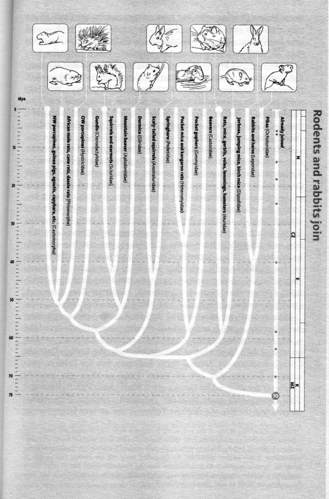
[2] From: Richard Dawkins, ''The
Ancestor's Tale'', (Boston, MA:
Houghton Mifflin Company, 2004),
p187. COPYRIGHTED
source: Richard Dawkins, "The
Ancestor's Tale", (Boston, MA: Houghton
Mifflin Company, 2004), p187.
Colugos {KolUGOZ}.
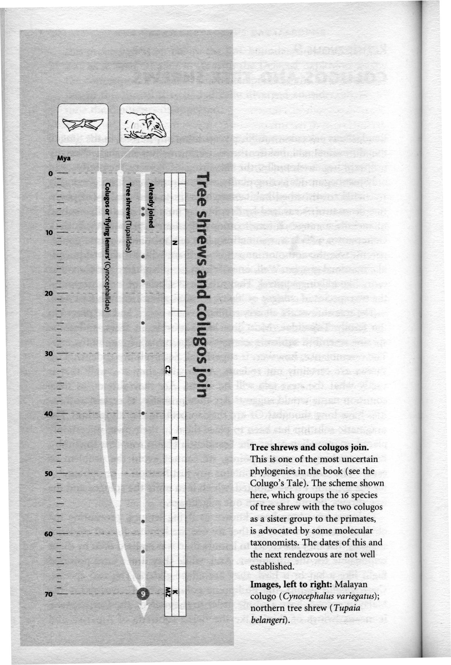
[1] From: Richard Dawkins, ''The
Ancestor's Tale'', (Boston, MA:
Houghton Mifflin Company, 2004),
p182. COPYRIGHTED
source: Richard Dawkins, "The
Ancestor's Tale", (Boston, MA: Houghton
Mifflin Company, 2004), p182.
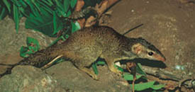
[2] Description English: Indian
Tree-shrew (Anathana ellioti) in
Yercaud, India. Date Taken on
film in the 1990s - scanned on
2005-09-26 (according to EXIF
data) Source Photographed by S.
Karthikeyan ( palmfly at gmail . com )
Please contact author for usage of any
higher resolution images. Author
S. Karthikeyan CC
source: http://upload.wikimedia.org/wiki
pedia/commons/7/78/Anathana_ellioti.jpg
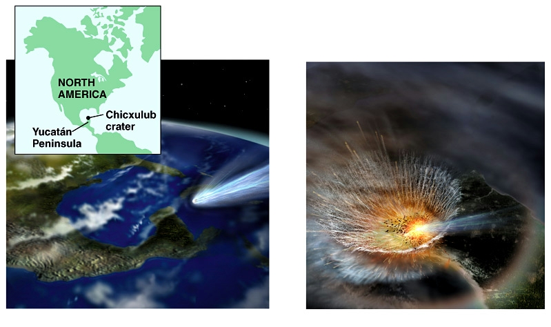
[1] Cretaceous meteor impact. Benjamin
Cummings. COPYRIGHTED
source: http://io.uwinnipeg.ca/~simmons/
16cm05/1116/16macro.htm
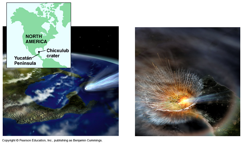
[2] Cretaceous meteor impact. Benjamin
Cummings. COPYRIGHTED
source: http://io.uwinnipeg.ca/~simmons/
16cm05/1116/16macro.htm
(cranes and rails).

[1] Fig. 4. Our phylogeny differs from
and agrees with previous
classifications. We merged
well-supported (>70% bootstrap values)
monophyletic clades at the tips with
the same ordinal designation across all
three classifications (e.g., 24 species
called Passerines). Only higher
relationships supported by bootstrap
values >50% are shown. Colors are as in
Fig. 2. Color bars to the right of the
tree show membership in three different
classifications: Peters' (25) (left),
Sibley and Monroe's (30) (middle), and
Livezey and Zusi's (13) (right). Black
text within the bars indicates
monophyletic orders in our phylogeny,
whereas white text within the bars
indicates nonmonophyletic orders.
Ordinal name codes: ANS (Anseriformes),
APO (Apodiformes), APT
(Apterygiformes), ARD (Ardeiformes),
BAL (Balaenicipitiformes), BUC
(Bucerotiformes), CAP
(Caprimulgiformes), CAS
(Casuariiformes), CHA
(Charadriiformes), CIC (Ciconiiformes),
CLM (Columbiformes), COL (Coliiformes),
COR (Coraciiformes), CRA (Craciformes),
CUC (Cuculiformes), FAL
(Falconiformes), GAL (Galliformes), GAV
(Gaviiformes), GLB (Galbuliformes), GRU
(Gruiformes), MUS (Musophagiformes),
OPI (Opisthocomiformes), PAS
(Passeriformes), PEL (Pelecaniformes),
PIC (Piciformes), POD
(Podicipediformes), PRO
(Procellariiformes), PSI
(Psittaciformes), RAL (Ralliformes),
RHE (Rheiformes), SPH
(Sphenisciformes), STH
(Struthioniformes), STR (Strigiformes),
TIN (Tinamiformes), TRC
(Trochiliformes), TRO (Trogoniformes),
TUR (Turniciformes), and UPU
(Upupiformes). Figure 4
from: Hackett, Shannon J. et al. “A
Phylogenomic Study of Birds Reveals
Their Evolutionary History.” Science
320.5884 (2008) : 1763 -1768.
Print. http://www.sciencemag.org/conten
t/320/5884/1763 COPYRIGHTED
source: http://www.sciencemag.org/conten
t/320/5884/1763/F4.large.jpg
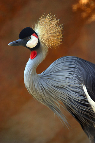
[2] By Aaron Logan, from
http://www.lightmatter.net/gallery/album
s.php w:en:Creative
Commons attribution CC
source: http://upload.wikimedia.org/wiki
pedia/commons/8/8d/Grey_Crowned_Crane.jp
g
[1] From: Richard Dawkins, ''The
Ancestor's Tale'', (Boston, MA:
Houghton Mifflin Company, 2004),
p231. COPYRIGHTED
source: Richard Dawkins, "The
Ancestor's Tale", (Boston, MA: Houghton
Mifflin Company, 2004), p231.
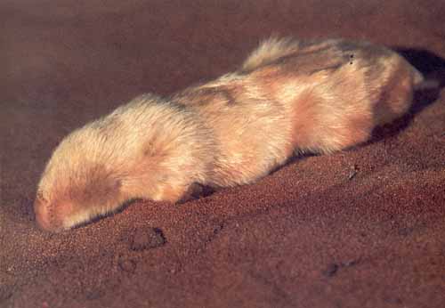
[2] English: The southern marsupial
mole (Notoryctes typhlops). Date
Originally uploaded to
pl.wikipedia on 10 May 2006. Source
Own work; originally from
pl.wikipedia; description page is/was
here. Author Bartus.malec at
pl.wikipedia. GNU
source: http://upload.wikimedia.org/wiki
pedia/commons/4/4b/Notoryctes_typhlops.j
pg
Numbat {nuMBaT}.
[1] From: Richard Dawkins, ''The
Ancestor's Tale'', (Boston, MA:
Houghton Mifflin Company, 2004),
p231. COPYRIGHTED
source: Richard Dawkins, "The
Ancestor's Tale", (Boston, MA: Houghton
Mifflin Company, 2004), p231.

[2] Description English: Quoll
imaged at a rescue park, Tasmania,
Austrailia, probably Tiger Quoll
(Dasyurus maculatus), indicated by
spots on tail Photographer's note.
This is a lucky through-the-fence shot
using an old Sony camera as the animal
was quite active. The small size of the
lens is a distinct advantage in this
case (my Canon xTi would not have been
able to get the
shot). Category:Dasyurus
maculatus Date Taken November 18,
2008, uploaded December 28, 2008 (28
December 2008 (original upload
date)) Source Transferred from
en.wikipedia; transferred to Commons by
User:Berichard using CommonsHelper. PD
source: http://upload.wikimedia.org/wiki
pedia/commons/f/f6/Dasyurus_maculatus.jp
g
{DIPrOTODoNsEu} evolve (Wombats,
Kangeroos, Possums, Koalas).
[1] From: Richard Dawkins, ''The
Ancestor's Tale'', (Boston, MA:
Houghton Mifflin Company, 2004),
p231. COPYRIGHTED
source: Richard Dawkins, "The
Ancestor's Tale", (Boston, MA: Houghton
Mifflin Company, 2004), p231.

[2] Eastern Grey Kangaroo with
joey PD
source: http://upload.wikimedia.org/wiki
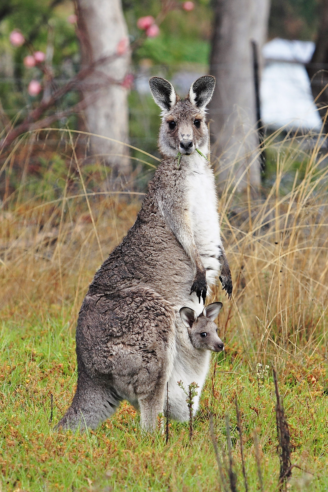
pedia/commons/0/0d/Kangaroo_and_joey03.j
pg
(rats, mice, gerbils, voles {VOLZ},
lemmings, hamsters).

[1] From: Richard Dawkins, ''The
Ancestor's Tale'', (Boston, MA:
Houghton Mifflin Company, 2004),
p187. COPYRIGHTED
source: Richard Dawkins, "The
Ancestor's Tale", (Boston, MA: Houghton
Mifflin Company, 2004), p187.
[2] From: Richard Dawkins, ''The
Ancestor's Tale'', (Boston, MA:
Houghton Mifflin Company, 2004),
p187. COPYRIGHTED
source: Richard Dawkins, "The
Ancestor's Tale", (Boston, MA: Houghton
Mifflin Company, 2004), p187.
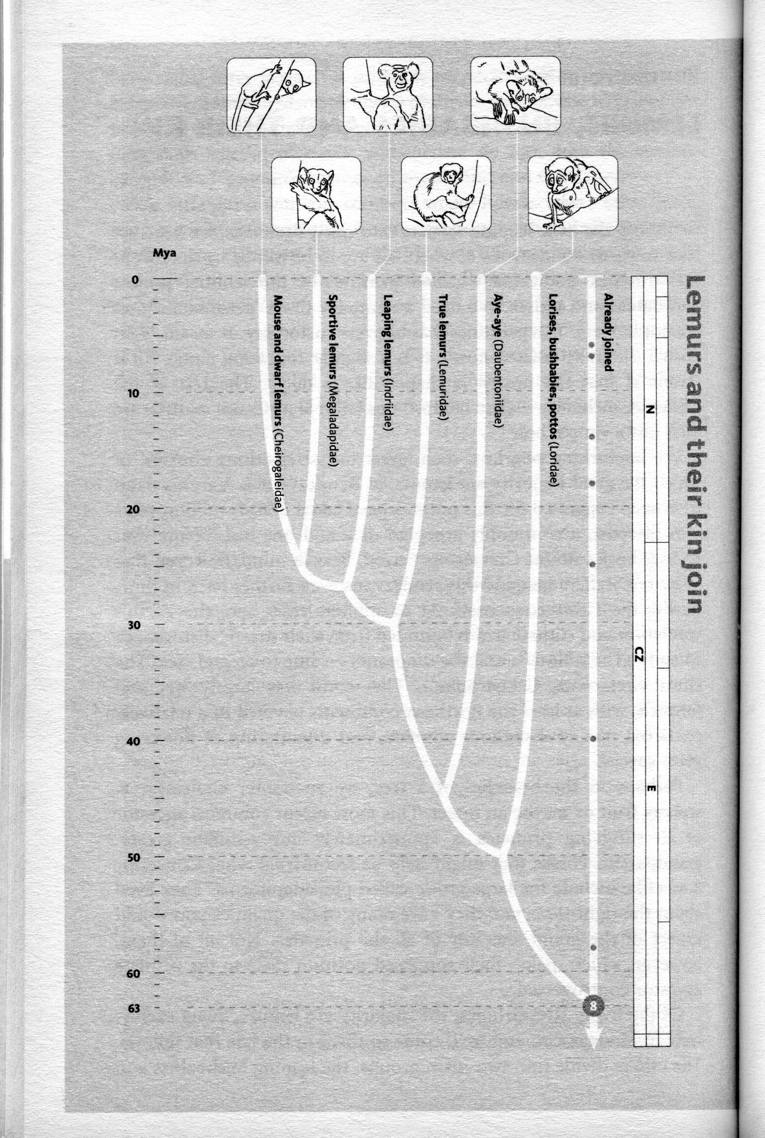
[1] From: Richard Dawkins, ''The
Ancestor's Tale'', (Boston, MA:
Houghton Mifflin Company, 2004),
p168. COPYRIGHTED
source: Richard Dawkins, "The
Ancestor's Tale", (Boston, MA: Houghton
Mifflin Company, 2004), p168.

[2] Description English: Gray
slender loris (Loris lydekkerianus)
photographed at Dindigal in Tamil
Nadu. Date 27 June 2008 Source
Own work Author Kalyan Varma
(Kalyanvarma) GNU
source: http://upload.wikimedia.org/wiki
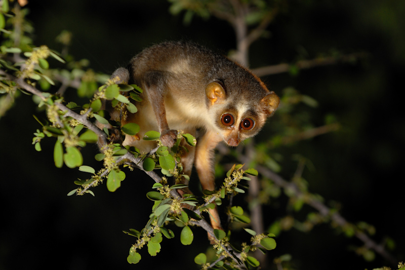
pedia/commons/8/8f/Slender_Loris.jpg
{STriJiFORmEZ} evolve (owls).
[1] Fig. 4. Our phylogeny differs from
and agrees with previous
classifications. We merged
well-supported (>70% bootstrap values)
monophyletic clades at the tips with
the same ordinal designation across all
three classifications (e.g., 24 species
called Passerines). Only higher
relationships supported by bootstrap
values >50% are shown. Colors are as in
Fig. 2. Color bars to the right of the
tree show membership in three different
classifications: Peters' (25) (left),
Sibley and Monroe's (30) (middle), and
Livezey and Zusi's (13) (right). Black
text within the bars indicates
monophyletic orders in our phylogeny,
whereas white text within the bars
indicates nonmonophyletic orders.
Ordinal name codes: ANS (Anseriformes),
APO (Apodiformes), APT
(Apterygiformes), ARD (Ardeiformes),
BAL (Balaenicipitiformes), BUC
(Bucerotiformes), CAP
(Caprimulgiformes), CAS
(Casuariiformes), CHA
(Charadriiformes), CIC (Ciconiiformes),
CLM (Columbiformes), COL (Coliiformes),
COR (Coraciiformes), CRA (Craciformes),
CUC (Cuculiformes), FAL
(Falconiformes), GAL (Galliformes), GAV
(Gaviiformes), GLB (Galbuliformes), GRU
(Gruiformes), MUS (Musophagiformes),
OPI (Opisthocomiformes), PAS
(Passeriformes), PEL (Pelecaniformes),
PIC (Piciformes), POD
(Podicipediformes), PRO
(Procellariiformes), PSI
(Psittaciformes), RAL (Ralliformes),
RHE (Rheiformes), SPH
(Sphenisciformes), STH
(Struthioniformes), STR (Strigiformes),
TIN (Tinamiformes), TRC
(Trochiliformes), TRO (Trogoniformes),
TUR (Turniciformes), and UPU
(Upupiformes). Figure 4
from: Hackett, Shannon J. et al. “A
Phylogenomic Study of Birds Reveals
Their Evolutionary History.” Science
320.5884 (2008) : 1763 -1768.
Print. http://www.sciencemag.org/conten
t/320/5884/1763 COPYRIGHTED
source: http://www.sciencemag.org/conten
t/320/5884/1763/F4.large.jpg

[2] Description Athene
noctua English: Little owl Español:
Mochuelo Date 2011-02-27 07:27
(UTC) Source
Athene_noctua_(portrait).jpg Author
Athene_noctua_(portrait).jpg:
Trebol-a derivative work:
Stemonitis (talk) CC
source: http://upload.wikimedia.org/wiki
pedia/commons/3/39/Athene_noctua_%28crop
ped%29.jpg
{KoRniVRu} (Cats, Dogs, Bears, Weasels,
Hyenas, Seals, Walruses).
[1] From: Richard Dawkins, ''The
Ancestor's Tale'', (Boston, MA:
Houghton Mifflin Company, 2004),
p200. COPYRIGHTED
source: Richard Dawkins, "The
Ancestor's Tale", (Boston, MA: Houghton
Mifflin Company, 2004), p200.

[2] Description English:
Two-spotted palm civet Nandinia
binotata mounted specimen in Manchester
Museum Date 2008-07-28 (original
upload date) (Original text : July
2008) GNU
source: http://upload.wikimedia.org/wiki
pedia/commons/5/5a/14-nandinia_binotata.
JPG
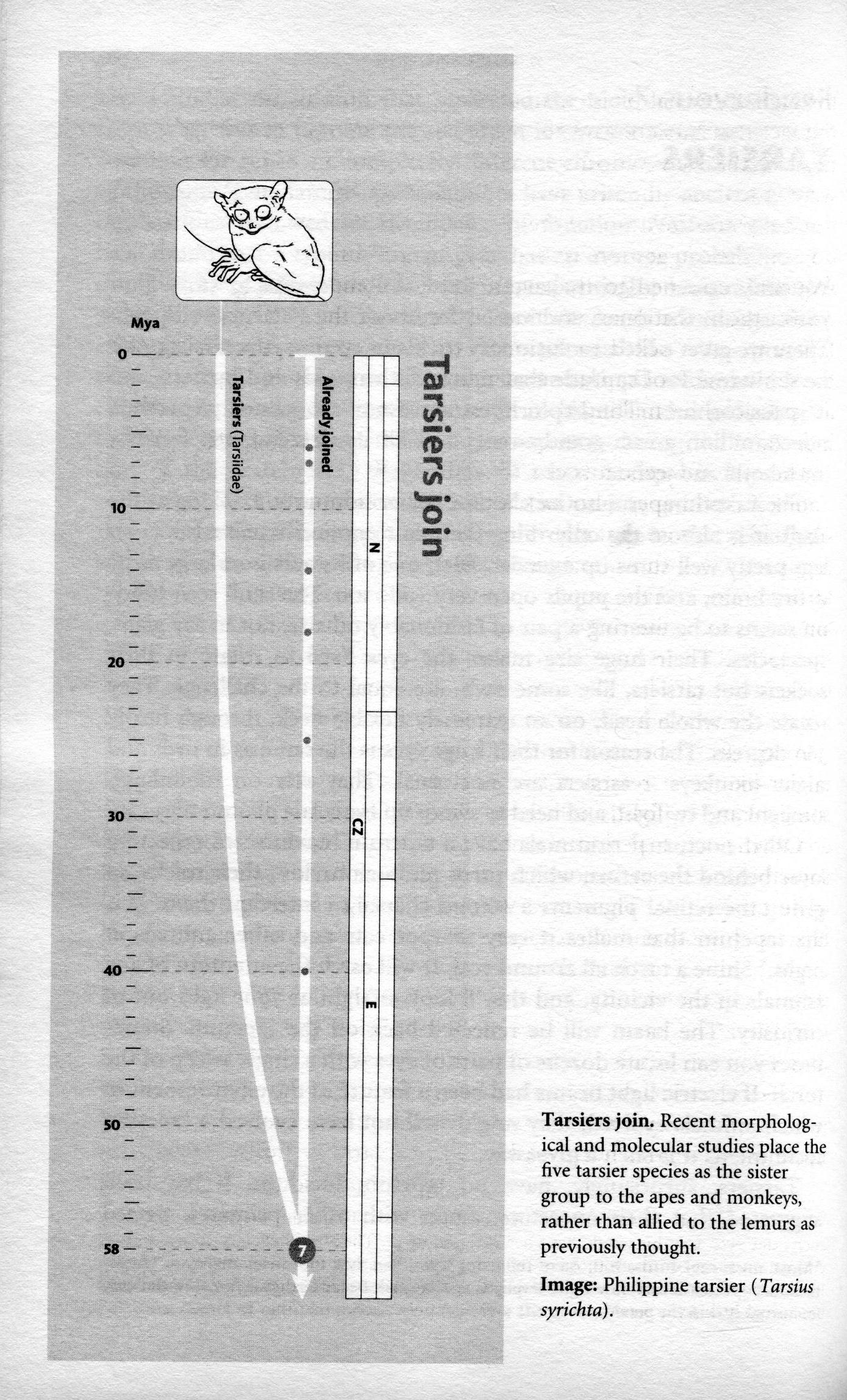
[1] From: Richard Dawkins, ''The
Ancestor's Tale'', (Boston, MA:
Houghton Mifflin Company, 2004),
p164. COPYRIGHTED
source: Richard Dawkins, "The
Ancestor's Tale", (Boston, MA: Houghton
Mifflin Company, 2004), p164.
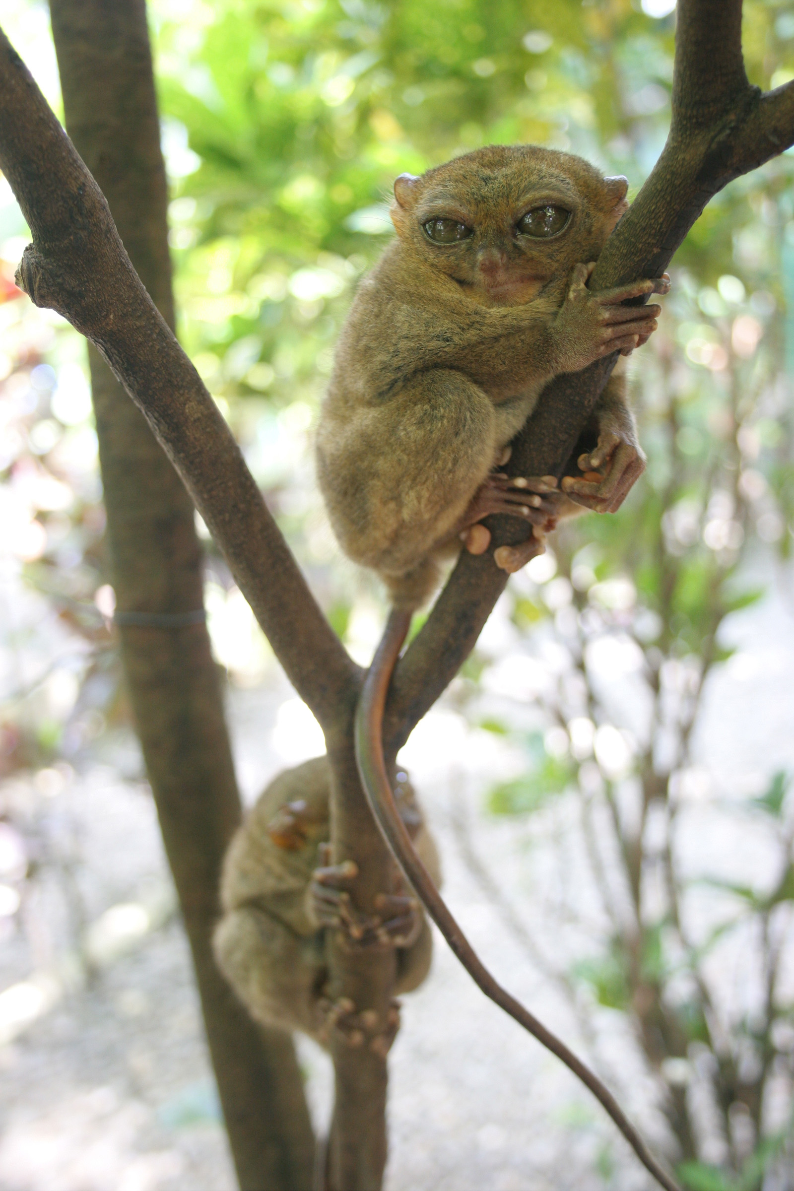
[2] Description Tarsius syrichta
(Philippine Tarsier) Date
- Source
http://www.sxc.hu/photo/490924 Aut
hor Jasper Greek Golangco PD
source: http://upload.wikimedia.org/wiki
pedia/commons/1/1d/Tarsius_Syrichta-GG.j
pg
{oPoD-i-FORmEZ} (hummingbirds, swifts).
[1] Fig. 4. Our phylogeny differs from
and agrees with previous
classifications. We merged
well-supported (>70% bootstrap values)
monophyletic clades at the tips with
the same ordinal designation across all
three classifications (e.g., 24 species
called Passerines). Only higher
relationships supported by bootstrap
values >50% are shown. Colors are as in
Fig. 2. Color bars to the right of the
tree show membership in three different
classifications: Peters' (25) (left),
Sibley and Monroe's (30) (middle), and
Livezey and Zusi's (13) (right). Black
text within the bars indicates
monophyletic orders in our phylogeny,
whereas white text within the bars
indicates nonmonophyletic orders.
Ordinal name codes: ANS (Anseriformes),
APO (Apodiformes), APT
(Apterygiformes), ARD (Ardeiformes),
BAL (Balaenicipitiformes), BUC
(Bucerotiformes), CAP
(Caprimulgiformes), CAS
(Casuariiformes), CHA
(Charadriiformes), CIC (Ciconiiformes),
CLM (Columbiformes), COL (Coliiformes),
COR (Coraciiformes), CRA (Craciformes),
CUC (Cuculiformes), FAL
(Falconiformes), GAL (Galliformes), GAV
(Gaviiformes), GLB (Galbuliformes), GRU
(Gruiformes), MUS (Musophagiformes),
OPI (Opisthocomiformes), PAS
(Passeriformes), PEL (Pelecaniformes),
PIC (Piciformes), POD
(Podicipediformes), PRO
(Procellariiformes), PSI
(Psittaciformes), RAL (Ralliformes),
RHE (Rheiformes), SPH
(Sphenisciformes), STH
(Struthioniformes), STR (Strigiformes),
TIN (Tinamiformes), TRC
(Trochiliformes), TRO (Trogoniformes),
TUR (Turniciformes), and UPU
(Upupiformes). Figure 4
from: Hackett, Shannon J. et al. “A
Phylogenomic Study of Birds Reveals
Their Evolutionary History.” Science
320.5884 (2008) : 1763 -1768.
Print. http://www.sciencemag.org/conten
t/320/5884/1763 COPYRIGHTED
source: http://www.sciencemag.org/conten
t/320/5884/1763/F4.large.jpg
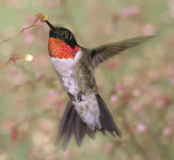
[2] Description Ruby-throated
hummingbird public domain USFWA Date
11 February 2003 Source
Cropped from U.S. Fish and
Wildlife Service Digital Library
System Author Steve Maslowski PD
source: http://upload.wikimedia.org/wiki
pedia/commons/8/87/Rubythroathummer65.jp
g
(woodpeckers, toucans).
[1] Fig. 4. Our phylogeny differs from
and agrees with previous
classifications. We merged
well-supported (>70% bootstrap values)
monophyletic clades at the tips with
the same ordinal designation across all
three classifications (e.g., 24 species
called Passerines). Only higher
relationships supported by bootstrap
values >50% are shown. Colors are as in
Fig. 2. Color bars to the right of the
tree show membership in three different
classifications: Peters' (25) (left),
Sibley and Monroe's (30) (middle), and
Livezey and Zusi's (13) (right). Black
text within the bars indicates
monophyletic orders in our phylogeny,
whereas white text within the bars
indicates nonmonophyletic orders.
Ordinal name codes: ANS (Anseriformes),
APO (Apodiformes), APT
(Apterygiformes), ARD (Ardeiformes),
BAL (Balaenicipitiformes), BUC
(Bucerotiformes), CAP
(Caprimulgiformes), CAS
(Casuariiformes), CHA
(Charadriiformes), CIC (Ciconiiformes),
CLM (Columbiformes), COL (Coliiformes),
COR (Coraciiformes), CRA (Craciformes),
CUC (Cuculiformes), FAL
(Falconiformes), GAL (Galliformes), GAV
(Gaviiformes), GLB (Galbuliformes), GRU
(Gruiformes), MUS (Musophagiformes),
OPI (Opisthocomiformes), PAS
(Passeriformes), PEL (Pelecaniformes),
PIC (Piciformes), POD
(Podicipediformes), PRO
(Procellariiformes), PSI
(Psittaciformes), RAL (Ralliformes),
RHE (Rheiformes), SPH
(Sphenisciformes), STH
(Struthioniformes), STR (Strigiformes),
TIN (Tinamiformes), TRC
(Trochiliformes), TRO (Trogoniformes),
TUR (Turniciformes), and UPU
(Upupiformes). Figure 4
from: Hackett, Shannon J. et al. “A
Phylogenomic Study of Birds Reveals
Their Evolutionary History.” Science
320.5884 (2008) : 1763 -1768.
Print. http://www.sciencemag.org/conten
t/320/5884/1763 COPYRIGHTED
source: http://www.sciencemag.org/conten
t/320/5884/1763/F4.large.jpg

[2] Description Hispaniolan
Woodpecker / Melanerpes striatus Date
20 January 2004 Source
http://www.pbase.com/wwcsig/image/4
1280575 Author Wolfgang
Wander GNU
source: http://upload.wikimedia.org/wiki
pedia/commons/1/1b/Melanerpes_striatus00
1.jpg
{PaSRiFORmEZ} (perching songbirds)
evolve. This order includes many common
birds: crows, jays, sparrows, warblers,
mockingbirds, robins, orioles,
bluebirds, vireos {VEREOZ}, larks,
finches.
Australia|Gondwana
[1] Fig. 4. Our phylogeny differs from
and agrees with previous
classifications. We merged
well-supported (>70% bootstrap values)
monophyletic clades at the tips with
the same ordinal designation across all
three classifications (e.g., 24 species
called Passerines). Only higher
relationships supported by bootstrap
values >50% are shown. Colors are as in
Fig. 2. Color bars to the right of the
tree show membership in three different
classifications: Peters' (25) (left),
Sibley and Monroe's (30) (middle), and
Livezey and Zusi's (13) (right). Black
text within the bars indicates
monophyletic orders in our phylogeny,
whereas white text within the bars
indicates nonmonophyletic orders.
Ordinal name codes: ANS (Anseriformes),
APO (Apodiformes), APT
(Apterygiformes), ARD (Ardeiformes),
BAL (Balaenicipitiformes), BUC
(Bucerotiformes), CAP
(Caprimulgiformes), CAS
(Casuariiformes), CHA
(Charadriiformes), CIC (Ciconiiformes),
CLM (Columbiformes), COL (Coliiformes),
COR (Coraciiformes), CRA (Craciformes),
CUC (Cuculiformes), FAL
(Falconiformes), GAL (Galliformes), GAV
(Gaviiformes), GLB (Galbuliformes), GRU
(Gruiformes), MUS (Musophagiformes),
OPI (Opisthocomiformes), PAS
(Passeriformes), PEL (Pelecaniformes),
PIC (Piciformes), POD
(Podicipediformes), PRO
(Procellariiformes), PSI
(Psittaciformes), RAL (Ralliformes),
RHE (Rheiformes), SPH
(Sphenisciformes), STH
(Struthioniformes), STR (Strigiformes),
TIN (Tinamiformes), TRC
(Trochiliformes), TRO (Trogoniformes),
TUR (Turniciformes), and UPU
(Upupiformes). Figure 4
from: Hackett, Shannon J. et al. “A
Phylogenomic Study of Birds Reveals
Their Evolutionary History.” Science
320.5884 (2008) : 1763 -1768.
Print. http://www.sciencemag.org/conten
t/320/5884/1763 COPYRIGHTED
source: http://www.sciencemag.org/conten
t/320/5884/1763/F4.large.jpg

[2] Western Bluebirds (female on
left) Irvine, CA PD
source: http://tedhuntington.com/bluebir
ds.jpg

[1] Description Moeritherium Date
1920 (probably) Source The Wonderful
Paleo Art of Heinrich Harder Author
Heinrich Harder (1858-1935) PD
source: http://upload.wikimedia.org/wiki
pedia/commons/9/97/Moeritherium.jpg
[2] From: Richard Dawkins, ''The
Ancestor's Tale'', (Boston, MA:
Houghton Mifflin Company, 2004),
p225. COPYRIGHTED
source: Richard Dawkins, "The
Ancestor's Tale", (Boston, MA: Houghton
Mifflin Company, 2004), p225.
{SiToRTEODaKTilu} evolve (ancestor of
all Artiodactyla {oRTEODaKTiLu}:
camels, pigs, ruminants, hippos, and
all Cetacea {SiTASEu or SiTAsEu}:
Whales, Dolphins).
[1] From: Richard Dawkins, ''The
Ancestor's Tale'', (Boston, MA:
Houghton Mifflin Company, 2004),
p200. COPYRIGHTED
source: Richard Dawkins, "The
Ancestor's Tale", (Boston, MA: Houghton
Mifflin Company, 2004), p200.

[2] [t may or may not be
accurate] Description Pakicetus
inachus, a whale ancestor from the
Early Eocene of Pakistan, after
Nummelai et al., (2006), pencil
drawing, digital coloring Date 29
November 2007 Source Own
work Author Nobu Tamura
email:nobu.tamura@yahoo.com
www.palaeocritti.com GNU
source: http://upload.wikimedia.org/wiki
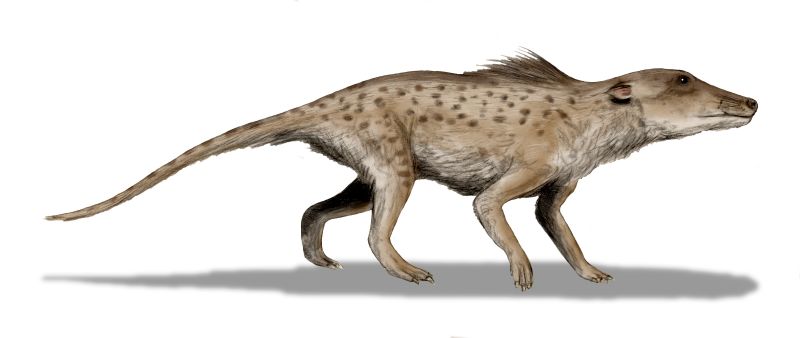
pedia/commons/3/34/Pakicetus_BW.jpg
{PeriSODaKTilu} (Horses, Tapirs {TAPRZ
}, Rhinos).
[1] From: Richard Dawkins, ''The
Ancestor's Tale'', (Boston, MA:
Houghton Mifflin Company, 2004),
p200. COPYRIGHTED
source: Richard Dawkins, "The
Ancestor's Tale", (Boston, MA: Houghton
Mifflin Company, 2004), p200.
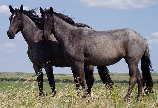
[2] Description Two young Nokota
mares Date 2010-02-11 22:34
(UTC) Source
Nokota_Horses.jpg Author
Nokota_Horses.jpg: François Marchal
derivative work: Dana boomer
(talk) CC
source: http://upload.wikimedia.org/wiki
pedia/commons/d/de/Nokota_Horses_cropped
.jpg
[1] From: Richard Dawkins, ''The
Ancestor's Tale'', (Boston, MA:
Houghton Mifflin Company, 2004),
p187. COPYRIGHTED
source: Richard Dawkins, "The
Ancestor's Tale", (Boston, MA: Houghton
Mifflin Company, 2004), p187.

[2] Description he was happily
sitting back and munching on something.
and munching, and munching... Date
4 July 2007, 12:55 Source
American Beaver Author Steve
from washington, dc,
usa Permission (Reusing this file)
See below. CC
source: http://upload.wikimedia.org/wiki
pedia/commons/6/6b/American_Beaver.jpg
[1] From: Richard Dawkins, ''The
Ancestor's Tale'', (Boston, MA:
Houghton Mifflin Company, 2004),
p187. COPYRIGHTED
source: Richard Dawkins, "The
Ancestor's Tale", (Boston, MA: Houghton
Mifflin Company, 2004), p187.
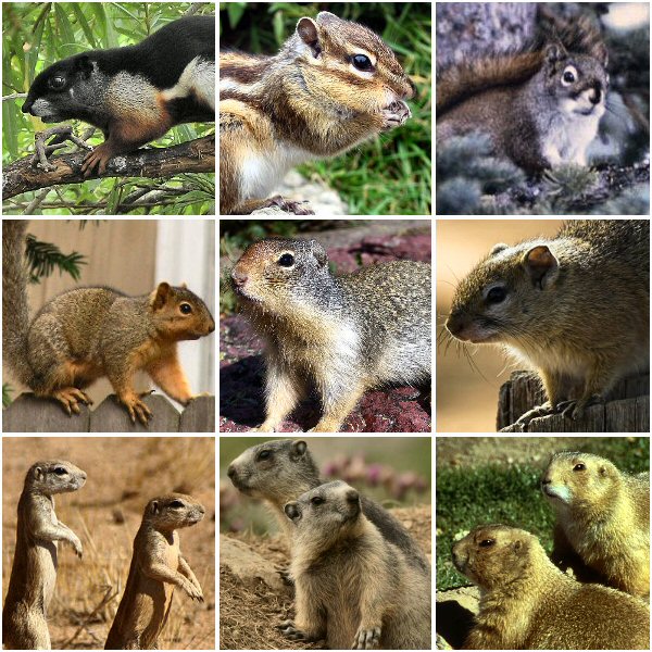
[2] Description Membres de la
famille des Suridés Date Source
Own work Author Chicoutimi
(montage) Montage 9 pictures.jpg
Karakal AndiW National Park
Service en:User:Markus Krötzsch
The Lilac Breasted Roller Nico
Conradie from Centurion, South Africa
Hans Hillewaert Sylvouille
National Park Service GNU
source: http://upload.wikimedia.org/wiki
pedia/commons/6/68/Sciuridae.jpg
hippos with dolphins and whales.
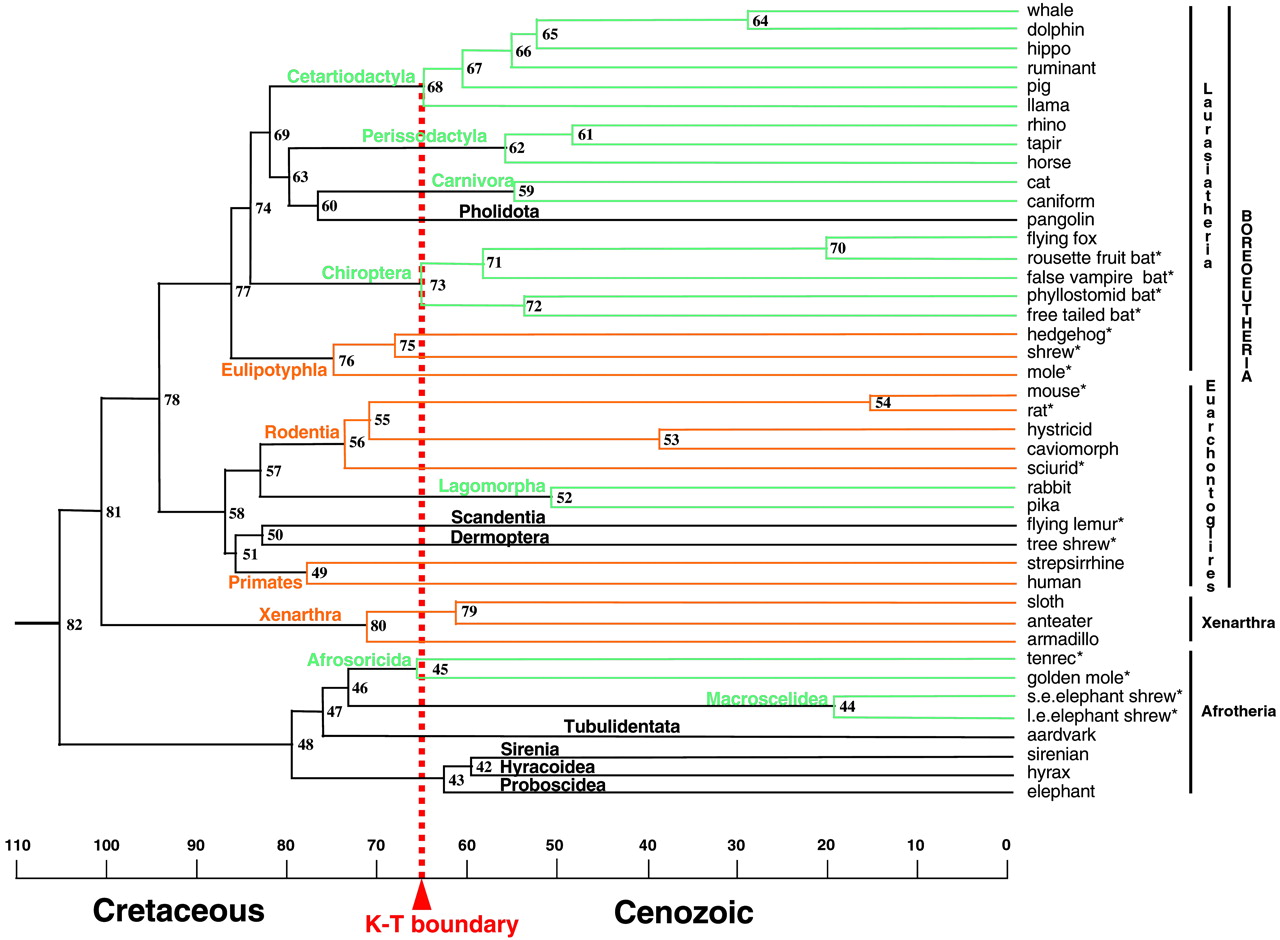
[1] Fig. 2. Molecular time scale for
the orders of placental mammals based
on the 16,397-bp data set and maximum
likelihood tree of ref. 14 with an
opossum outgroup (data not shown), 13
fossil constraints (Materials and
Methods), and a mean prior of 105 mya
for the placental root. Ordinal
designations are listed above the
branches. Orange and green lines denote
orders with basal diversification
before or after the K/T boundary,
respectively. Black lines depict orders
for which only one taxon was available.
Asterisks denote placental taxa
included in the ''K/T body size'' taxon
set. The composition of chimeric taxa,
including caniform, caviomorph,
strepsirrhine, and sirenian, is
indicated elsewhere (14). Numbers for
internal nodes are cross-referenced in
the supporting information.
COPYRIGHTED
source: http://www.pnas.org/content/vol1
00/issue3/images/large/pq0334222002.jpeg
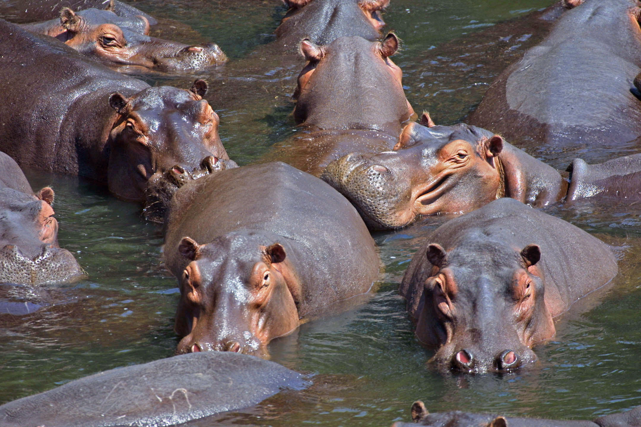
[2] Description Deutsch: Eine
Gruppe Flußpferde im Luangwa-Tal,
Sambia. English: Pod of Hippos
(Hippopotamus amphibius) in Luangwa
Valley, Zambia Français : Groupe
d'hippopotames (Hippopotamus amphibius)
dans la vallée du Luangua, en
Zambie Date 2005 Source Own
work Author Paul Maritz GNU
source: http://upload.wikimedia.org/wiki
pedia/commons/a/a3/Hippo_pod_edit.jpg
{KIroPTRu} (fruit bats, echolocating
bats).

[1] From: Richard Dawkins, ''The
Ancestor's Tale'', (Boston, MA:
Houghton Mifflin Company, 2004),
p200. COPYRIGHTED
source: Richard Dawkins, "The
Ancestor's Tale", (Boston, MA: Houghton
Mifflin Company, 2004), p200.
[2] From: Richard Dawkins, ''The
Ancestor's Tale'', (Boston, MA:
Houghton Mifflin Company, 2004),
p200. COPYRIGHTED
source: Richard Dawkins, "The
Ancestor's Tale", (Boston, MA: Houghton
Mifflin Company, 2004), p200.
[1] From: Richard Dawkins, ''The
Ancestor's Tale'', (Boston, MA:
Houghton Mifflin Company, 2004),
p187. COPYRIGHTED
source: Richard Dawkins, "The
Ancestor's Tale", (Boston, MA: Houghton
Mifflin Company, 2004), p187.

[2] Photograph of a brush-tailed
porcupine in Berlin Zoologischer
Garten. Taken by Eloquence in July 2005
and released into the public
domain. Public domain PD
source: http://upload.wikimedia.org/wiki
pedia/commons/2/21/Brush_tailed_porcupin
e_Berlin_Zoo.jpg
{FaLKoNiFORmEZ} (falcons, hawks,
eagles, Old World vultures).
[1] Fig. 4. Our phylogeny differs from
and agrees with previous
classifications. We merged
well-supported (>70% bootstrap values)
monophyletic clades at the tips with
the same ordinal designation across all
three classifications (e.g., 24 species
called Passerines). Only higher
relationships supported by bootstrap
values >50% are shown. Colors are as in
Fig. 2. Color bars to the right of the
tree show membership in three different
classifications: Peters' (25) (left),
Sibley and Monroe's (30) (middle), and
Livezey and Zusi's (13) (right). Black
text within the bars indicates
monophyletic orders in our phylogeny,
whereas white text within the bars
indicates nonmonophyletic orders.
Ordinal name codes: ANS (Anseriformes),
APO (Apodiformes), APT
(Apterygiformes), ARD (Ardeiformes),
BAL (Balaenicipitiformes), BUC
(Bucerotiformes), CAP
(Caprimulgiformes), CAS
(Casuariiformes), CHA
(Charadriiformes), CIC (Ciconiiformes),
CLM (Columbiformes), COL (Coliiformes),
COR (Coraciiformes), CRA (Craciformes),
CUC (Cuculiformes), FAL
(Falconiformes), GAL (Galliformes), GAV
(Gaviiformes), GLB (Galbuliformes), GRU
(Gruiformes), MUS (Musophagiformes),
OPI (Opisthocomiformes), PAS
(Passeriformes), PEL (Pelecaniformes),
PIC (Piciformes), POD
(Podicipediformes), PRO
(Procellariiformes), PSI
(Psittaciformes), RAL (Ralliformes),
RHE (Rheiformes), SPH
(Sphenisciformes), STH
(Struthioniformes), STR (Strigiformes),
TIN (Tinamiformes), TRC
(Trochiliformes), TRO (Trogoniformes),
TUR (Turniciformes), and UPU
(Upupiformes). Figure 4
from: Hackett, Shannon J. et al. “A
Phylogenomic Study of Birds Reveals
Their Evolutionary History.” Science
320.5884 (2008) : 1763 -1768.
Print. http://www.sciencemag.org/conten
t/320/5884/1763 COPYRIGHTED
source: http://www.sciencemag.org/conten
t/320/5884/1763/F4.large.jpg

[2] Description English: Bald Eagle
(Haliaeetus leucocephalus) in
Tree Date July 2005 Source
U.S. Fish and Wildlife
Service Author Hillebrand,
Steve PD
source: http://upload.wikimedia.org/wiki
pedia/commons/6/69/Haliaeetus_leucocepha
lus-tree-USFWS.jpg
guinea pigs, capybaras {KaPuBoRoZ}.
[1] From: Richard Dawkins, ''The
Ancestor's Tale'', (Boston, MA:
Houghton Mifflin Company, 2004),
p187. COPYRIGHTED
source: Richard Dawkins, "The
Ancestor's Tale", (Boston, MA: Houghton
Mifflin Company, 2004), p187.
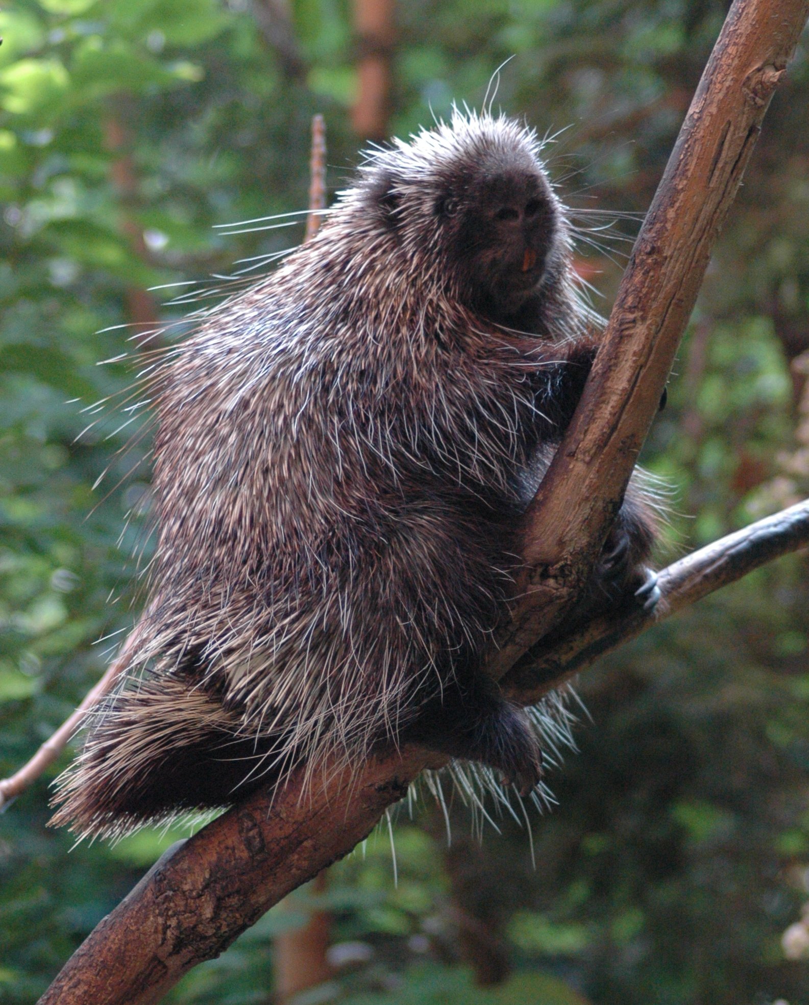
[2] Description English: A North
American porcupine (Erethizon dorsatum)
rests in a tree in Montreal's
BioDome. Date 20 July
2004 Source self-made with a
Nikon D70 Author J. Glover CC
source: http://upload.wikimedia.org/wiki
pedia/commons/8/83/Porcupine-BioDome.jpg
(Sakis, Spider, Howler and Squirrel
monkeys, Capuchins {KaP YU CiNZ},
Tamarins).
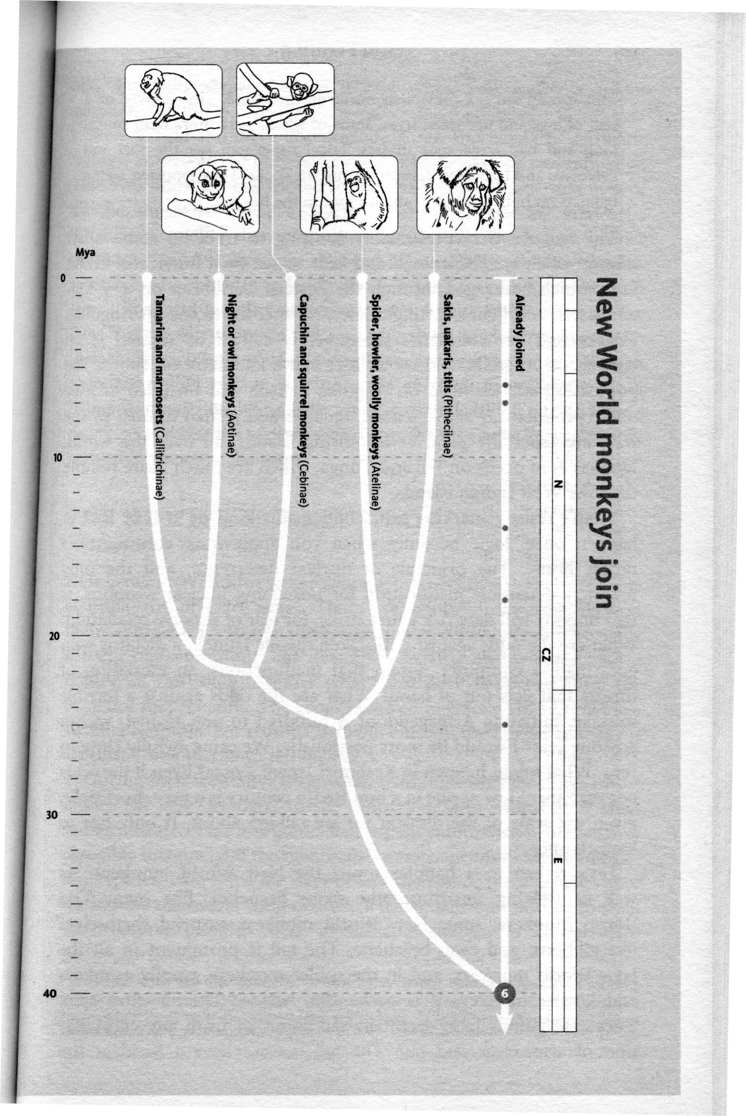
[1] From: Richard Dawkins, ''The
Ancestor's Tale'', (Boston, MA:
Houghton Mifflin Company, 2004),
p149. COPYRIGHTED
source: Richard Dawkins, "The
Ancestor's Tale", (Boston, MA: Houghton
Mifflin Company, 2004), p149.

[2] Description English: A
critically endangered Brown Spider
Monkey, Ateles hybridus, with uncommon
blue eyes. Shot in captivity in
Barquisimeto,
Venezuela Русский:
Паукообразная
обезьяна Ateles hybridus с
редко встречающимися
голубыми глазами.
Сфотографирована в
неволе в
Венесуэле. Date
September 2008 Source
Image:BrownSpiderMonkey.jpg Author
http://www.birdphotos.com edit by
Fir0002 Permission (Reusing this
file) See below. Attribution must
appear on same page as photo. CC
source: http://upload.wikimedia.org/wiki
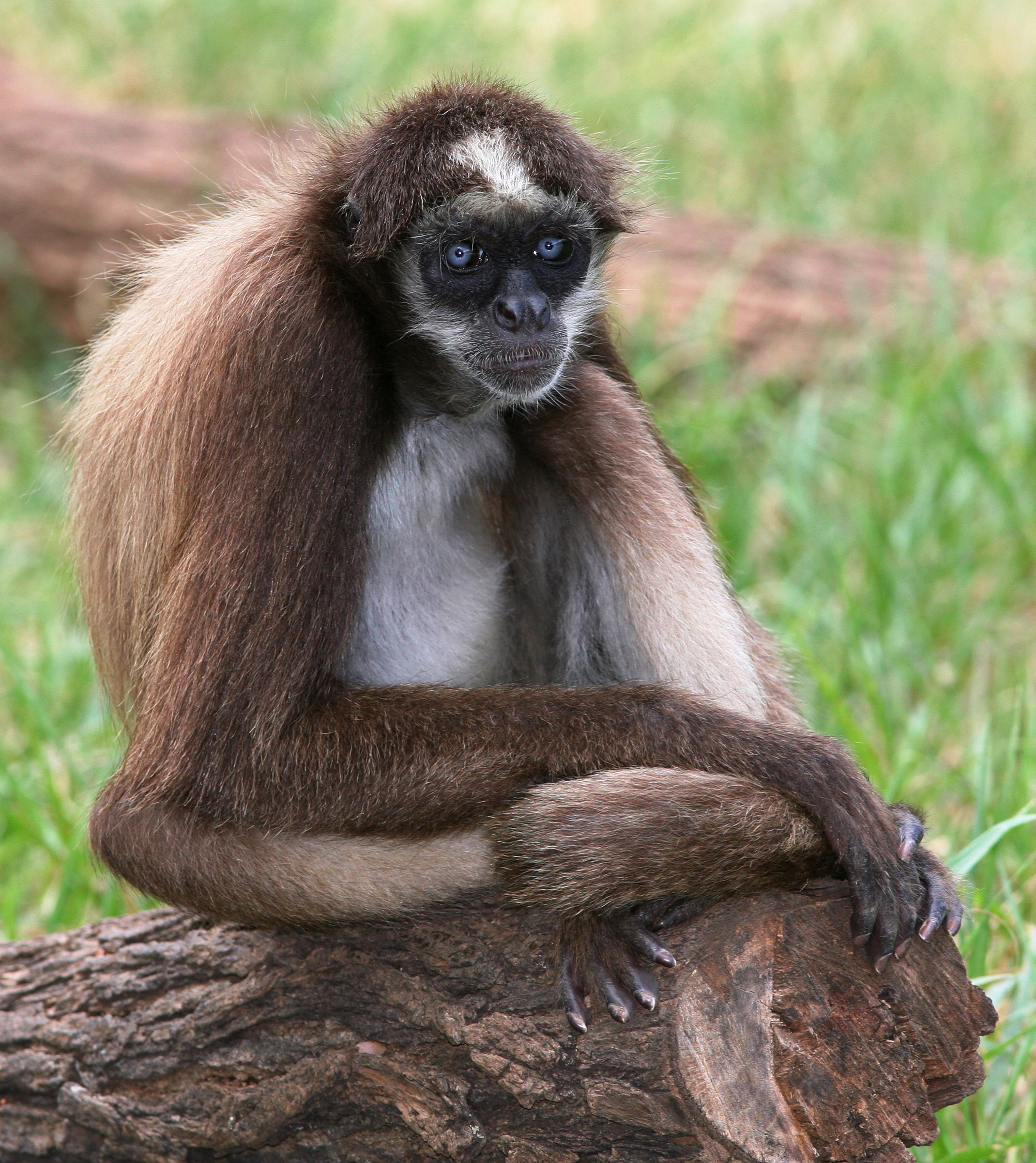
pedia/commons/d/dc/BrownSpiderMonkey_%28
edit2%29.jpg
evolve (cuckoos, roadrunners).
[1] Fig. 4. Our phylogeny differs from
and agrees with previous
classifications. We merged
well-supported (>70% bootstrap values)
monophyletic clades at the tips with
the same ordinal designation across all
three classifications (e.g., 24 species
called Passerines). Only higher
relationships supported by bootstrap
values >50% are shown. Colors are as in
Fig. 2. Color bars to the right of the
tree show membership in three different
classifications: Peters' (25) (left),
Sibley and Monroe's (30) (middle), and
Livezey and Zusi's (13) (right). Black
text within the bars indicates
monophyletic orders in our phylogeny,
whereas white text within the bars
indicates nonmonophyletic orders.
Ordinal name codes: ANS (Anseriformes),
APO (Apodiformes), APT
(Apterygiformes), ARD (Ardeiformes),
BAL (Balaenicipitiformes), BUC
(Bucerotiformes), CAP
(Caprimulgiformes), CAS
(Casuariiformes), CHA
(Charadriiformes), CIC (Ciconiiformes),
CLM (Columbiformes), COL (Coliiformes),
COR (Coraciiformes), CRA (Craciformes),
CUC (Cuculiformes), FAL
(Falconiformes), GAL (Galliformes), GAV
(Gaviiformes), GLB (Galbuliformes), GRU
(Gruiformes), MUS (Musophagiformes),
OPI (Opisthocomiformes), PAS
(Passeriformes), PEL (Pelecaniformes),
PIC (Piciformes), POD
(Podicipediformes), PRO
(Procellariiformes), PSI
(Psittaciformes), RAL (Ralliformes),
RHE (Rheiformes), SPH
(Sphenisciformes), STH
(Struthioniformes), STR (Strigiformes),
TIN (Tinamiformes), TRC
(Trochiliformes), TRO (Trogoniformes),
TUR (Turniciformes), and UPU
(Upupiformes). Figure 4
from: Hackett, Shannon J. et al. “A
Phylogenomic Study of Birds Reveals
Their Evolutionary History.” Science
320.5884 (2008) : 1763 -1768.
Print. http://www.sciencemag.org/conten
t/320/5884/1763 COPYRIGHTED
source: http://www.sciencemag.org/conten
t/320/5884/1763/F4.large.jpg
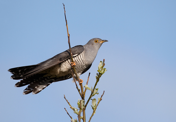
[2] Description English: Common
cuckoo Deutsch: Kuckuck Date
Source Own work Author
Vogelartinfo GNU
source: http://upload.wikimedia.org/wiki
pedia/commons/b/b0/Cuculus_canorus_vogel
artinfo_chris_romeiks_CHR0791.jpg
[1] From: Richard Dawkins, ''The
Ancestor's Tale'', (Boston, MA:
Houghton Mifflin Company, 2004),
p168. COPYRIGHTED
source: Richard Dawkins, "The
Ancestor's Tale", (Boston, MA: Houghton
Mifflin Company, 2004), p168.
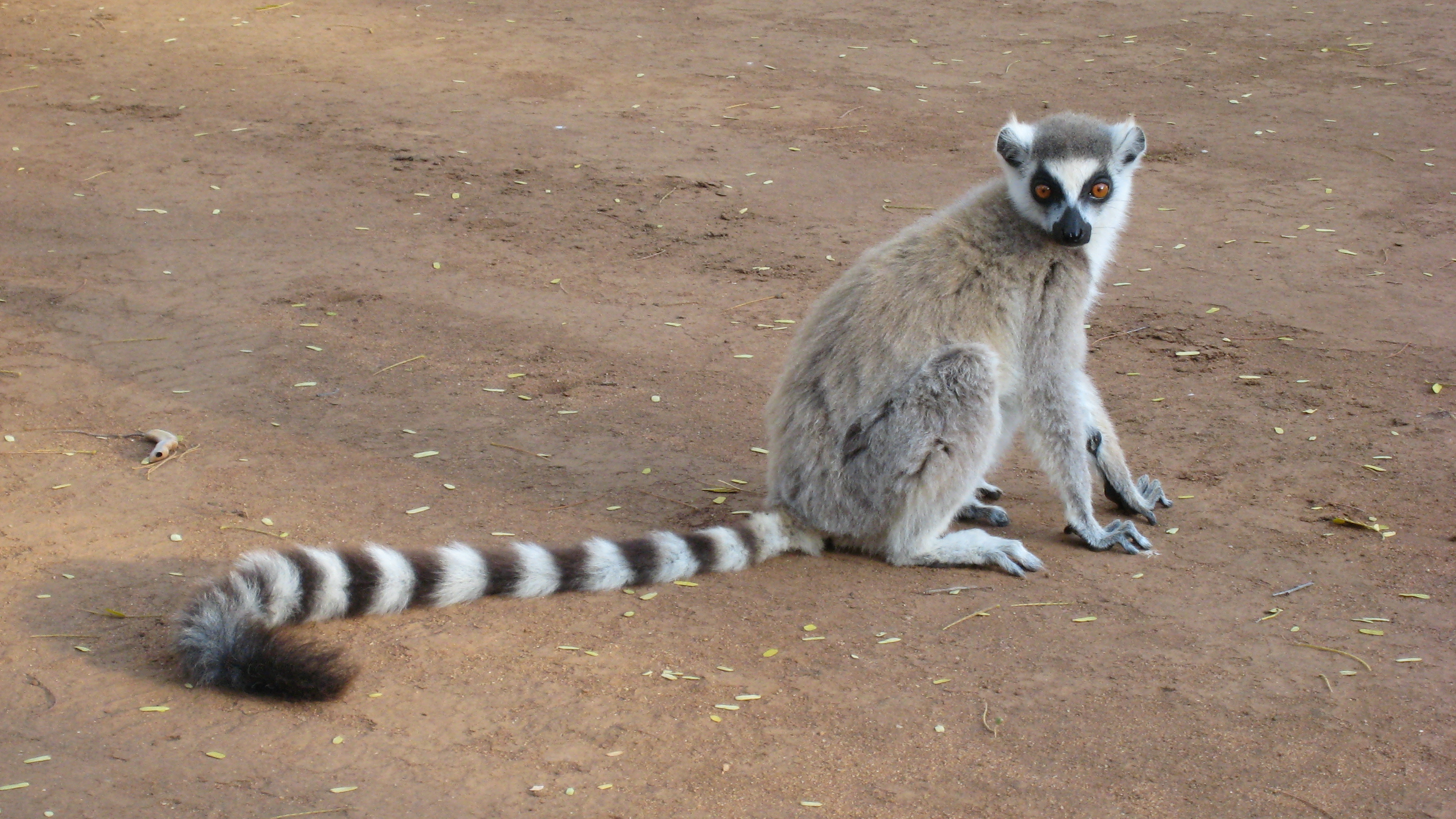
[2] Description English:
Ring-tailed Lemur (Lemur catta) at
Berenty Private Reserve in
Madagascar Date 4 October
2009 Source Own work Author
Alex Dunkel
(Visionholder) Permission (Reusing
this file) See below. CC
source: http://upload.wikimedia.org/wiki
pedia/commons/f/f5/Lemur_catta_001.jpg
(Macaques, Baboons, Mandrills,
Proboscis and Colobus {KoLiBeS}
monkeys).
Africa

[1] From: Stewart, Caro-Beth, and Todd
R Disotell. “Primate evolution - in
and out of Africa.” Current Biology
8.16 (1998) :
R582-R588. http://www.sciencedirect.com
/science/article/pii/S0960982207003673
Figure 2. A synthetic hypothesis of
catarrhine primate evolution. The
branching order shown for the living
species is well-supported by numerous
molecular phylogenetic studies (for
example [6, 7, 8, 9, 10, 11, 12, 18, 24
and 25]). We present the dates of
divergence calculated by Goodman and
colleagues [11], on the understanding
that these are still rough estimates
and more precise measurements are
needed, especially for the Old World
monkeys. The fossil species (genus
names in italics) were placed on this
tree by parsimony analyses of
relatively large morphological datasets
[4, 11, 14 and 15]. Known dates for
fossils [1, 2 and 21] are indicated by
the thicker lines; these lines are
attached to the tree as determined by
the parsimony analyses, although the
dates of the attachment points are our
best guesses. Species found in Africa
are in red and species found in Eurasia
are in black. The continental locations
of the ancestral lineages were inferred
by parsimony using the computer program
MacClade [30]. The intercontinental
dispersal events required, at a
minimum, to explain the distribution of
the living and fossil species are
indicated by the arrows. COPYRIGHTED
source: http://www.sciencedirect.com/sci
ence?_ob=ArticleURL&_udi=B6VRT-4C4DVM4-D
&_user=4422&_handle=V-WA-A-W-WC-MsSAYVW-
UUW-U-AAVECYCCBC-AAVDAZZBBC-YCACYAZCV-WC
-U&_fmt=full&_coverDate=07%2F30%2F1998&_
rdoc=12&_orig=browse&_srch=%23toc%236243
%231998%23999919983%23494082!&_cdi=6243&
view=c&_acct=C000059600&_version=1&_urlV
ersion=0&_userid=4422&md5=5558415c4ccd34
6c64e2e6be03c3865e
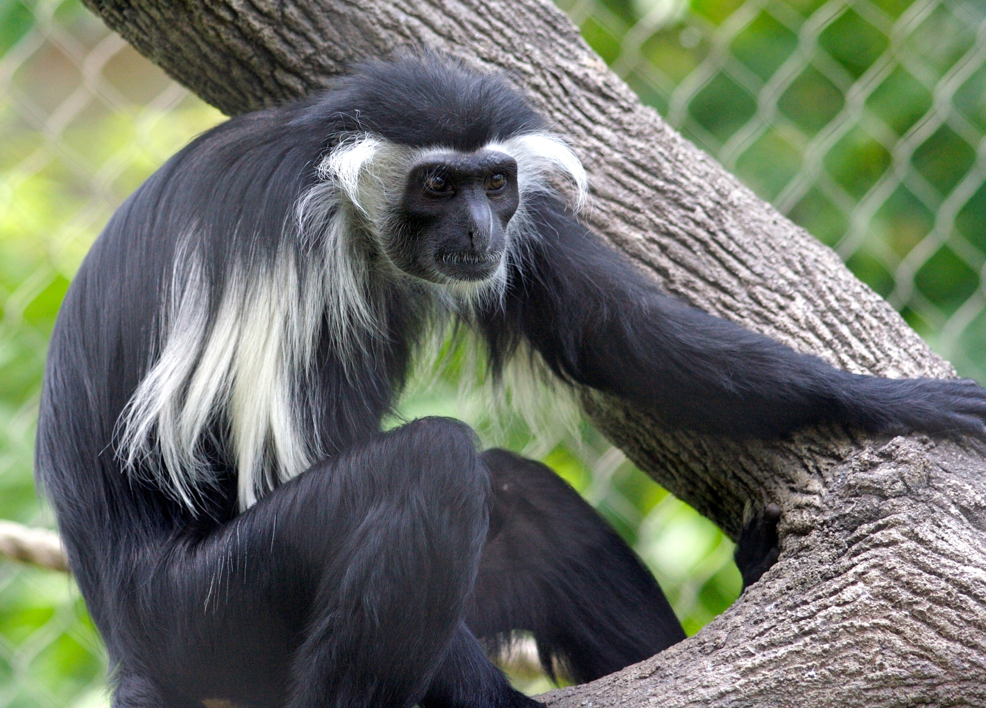
[2] Description Colobus
angolensis monkey Date 13 June
2007, 13:13 Source Angola Colobus
Monkey #6 Author Ryan E.
Poplin CC
source: http://upload.wikimedia.org/wiki
pedia/commons/5/5a/Colobus_angolensis.jp
g
(Gibbons and Hominids) loses its tail.
[1] From: Stewart, Caro-Beth, and Todd
R Disotell. “Primate evolution - in
and out of Africa.” Current Biology
8.16 (1998) :
R582-R588. http://www.sciencedirect.com
/science/article/pii/S0960982207003673
Figure 2. A synthetic hypothesis of
catarrhine primate evolution. The
branching order shown for the living
species is well-supported by numerous
molecular phylogenetic studies (for
example [6, 7, 8, 9, 10, 11, 12, 18, 24
and 25]). We present the dates of
divergence calculated by Goodman and
colleagues [11], on the understanding
that these are still rough estimates
and more precise measurements are
needed, especially for the Old World
monkeys. The fossil species (genus
names in italics) were placed on this
tree by parsimony analyses of
relatively large morphological datasets
[4, 11, 14 and 15]. Known dates for
fossils [1, 2 and 21] are indicated by
the thicker lines; these lines are
attached to the tree as determined by
the parsimony analyses, although the
dates of the attachment points are our
best guesses. Species found in Africa
are in red and species found in Eurasia
are in black. The continental locations
of the ancestral lineages were inferred
by parsimony using the computer program
MacClade [30]. The intercontinental
dispersal events required, at a
minimum, to explain the distribution of
the living and fossil species are
indicated by the arrows. COPYRIGHTED
source: http://www.sciencedirect.com/sci
ence?_ob=ArticleURL&_udi=B6VRT-4C4DVM4-D
&_user=4422&_handle=V-WA-A-W-WC-MsSAYVW-
UUW-U-AAVECYCCBC-AAVDAZZBBC-YCACYAZCV-WC
-U&_fmt=full&_coverDate=07%2F30%2F1998&_
rdoc=12&_orig=browse&_srch=%23toc%236243
%231998%23999919983%23494082!&_cdi=6243&
view=c&_acct=C000059600&_version=1&_urlV
ersion=0&_userid=4422&md5=5558415c4ccd34
6c64e2e6be03c3865e

[2] Gregoire: 62-year-old
chimpanzee Description English:
Chimpanzee named ''Gregoire'' born in
1944 (Jane Goodall sanctuary of
Tchimpounga in Congo Brazzaville) -
Picture taken the 9th of December
2006 Français : Chimpanzé nommé
''Grégoire'' né en 1944 (sanctuaire
Jane Goodall de Tchimpounga au Congo
Brazzaville) - Photo prise le 9
décembre 2006 Date 9 December
2006 Source Own work Author
Delphine
Bruyère Permission (Reusing this
file) Attribution : Delphine
Bruyere GNU
source: http://upload.wikimedia.org/wiki
pedia/commons/b/ba/2006-12-09_Chimpanzee
_Gregoire_D_Bruyere.JPG
Guinea
[1] From: Richard Dawkins, ''The
Ancestor's Tale'', (Boston, MA:
Houghton Mifflin Company, 2004),
239. COPYRIGHTED
source: Richard Dawkins, "The
Ancestor's Tale", (Boston, MA: Houghton
Mifflin Company, 2004), 239.
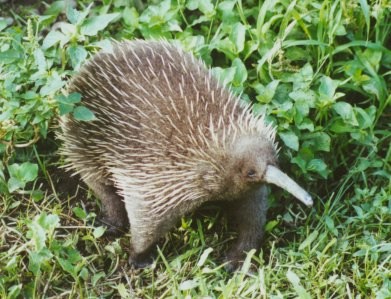
[2] The echidna is one of a handful of
mammals to give birth to its offspring
by laying eggs. GNU
source: http://upload.wikimedia.org/wiki
pedia/commons/3/3b/Long-beakedEchidna.jp
g
[1] From: Richard Dawkins, ''The
Ancestor's Tale'', (Boston, MA:
Houghton Mifflin Company, 2004),
239. COPYRIGHTED
source: Richard Dawkins, "The
Ancestor's Tale", (Boston, MA: Houghton
Mifflin Company, 2004), 239.
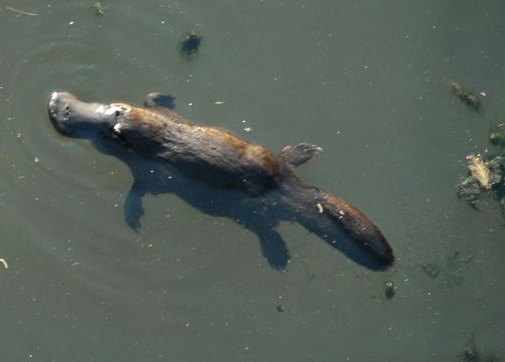
[2] Description Description
Swiming Platypus * Photographer Peter
Scheunis * Source self-made Date
September 2004 Location Broken
River-Queensland-Australia Date
2010-01-18 03:46 (UTC) Source
Platypus_BrokenRiver_QLD_Australia.jpg
Author
Platypus_BrokenRiver_QLD_Australia.jpg:
Peterdvv derivative work: Bobisbob
(talk) CC
source: http://upload.wikimedia.org/wiki
pedia/commons/1/12/Platypus_BrokenRiver_
QLD_Australia2.png
[1] Figure 2. A synthetic hypothesis of
catarrhine primate evolution. The
branching order shown for the living
species is well-supported by numerous
molecular phylogenetic studies (for
example [6, 7, 8, 9, 10, 11, 12, 18, 24
and 25]). We present the dates of
divergence calculated by Goodman and
colleagues [11], on the understanding
that these are still rough estimates
and more precise measurements are
needed, especially for the Old World
monkeys. The fossil species (genus
names in italics) were placed on this
tree by parsimony analyses of
relatively large morphological datasets
[4, 11, 14 and 15]. Known dates for
fossils [1, 2 and 21] are indicated by
the thicker lines; these lines are
attached to the tree as determined by
the parsimony analyses, although the
dates of the attachment points are our
best guesses. Species found in Africa
are in red and species found in Eurasia
are in black. The continental locations
of the ancestral lineages were inferred
by parsimony using the computer program
MacClade [30]. The intercontinental
dispersal events required, at a
minimum, to explain the distribution of
the living and fossil species are
indicated by the arrows. COPYRIGHTED
source: http://www.sciencedirect.com/sci
ence?_ob=ArticleURL&_udi=B6VRT-4C4DVM4-D
&_user=4422&_handle=V-WA-A-W-WC-MsSAYVW-
UUW-U-AAVECYCCBC-AAVDAZZBBC-YCACYAZCV-WC
-U&_fmt=full&_coverDate=07%2F30%2F1998&_
rdoc=12&_orig=browse&_srch=%23toc%236243
%231998%23999919983%23494082!&_cdi=6243&
view=c&_acct=C000059600&_version=1&_urlV
ersion=0&_userid=4422&md5=5558415c4ccd34
6c64e2e6be03c3865e
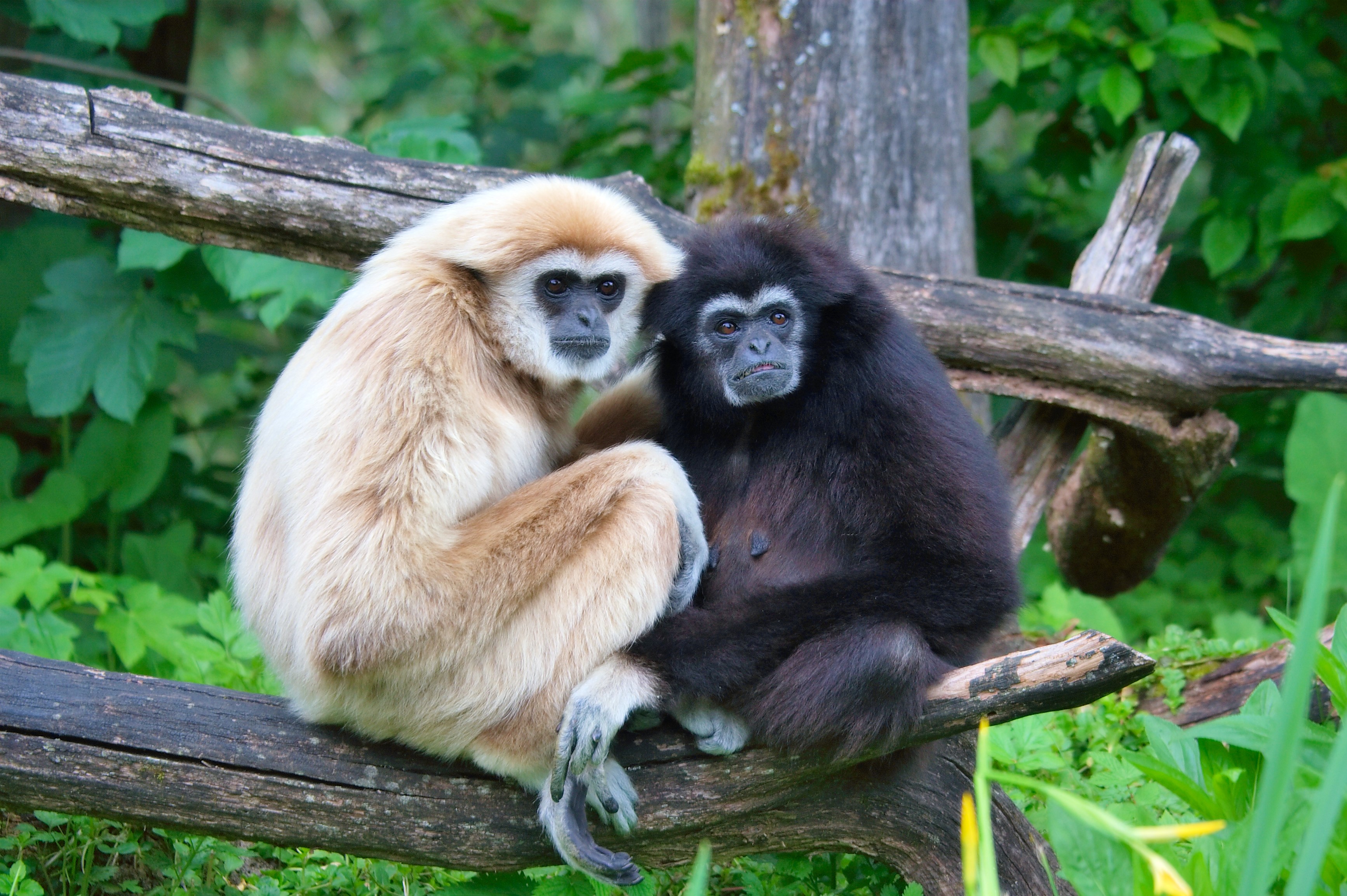
[2] Description Deutsch:
Weißhandgibbons Date 25 May
2006 Source Own work Author
User:MatthiasKabel GNU
source: http://upload.wikimedia.org/wiki
pedia/commons/3/38/Hylobates_lar_pair_of
_white_and_black_01.jpg
Orangutans.
[1] From: Stewart, Caro-Beth, and Todd
R Disotell. “Primate evolution - in
and out of Africa.” Current Biology
8.16 (1998) :
R582-R588. http://www.sciencedirect.com
/science/article/pii/S0960982207003673
Figure 2. A synthetic hypothesis of
catarrhine primate evolution. The
branching order shown for the living
species is well-supported by numerous
molecular phylogenetic studies (for
example [6, 7, 8, 9, 10, 11, 12, 18, 24
and 25]). We present the dates of
divergence calculated by Goodman and
colleagues [11], on the understanding
that these are still rough estimates
and more precise measurements are
needed, especially for the Old World
monkeys. The fossil species (genus
names in italics) were placed on this
tree by parsimony analyses of
relatively large morphological datasets
[4, 11, 14 and 15]. Known dates for
fossils [1, 2 and 21] are indicated by
the thicker lines; these lines are
attached to the tree as determined by
the parsimony analyses, although the
dates of the attachment points are our
best guesses. Species found in Africa
are in red and species found in Eurasia
are in black. The continental locations
of the ancestral lineages were inferred
by parsimony using the computer program
MacClade [30]. The intercontinental
dispersal events required, at a
minimum, to explain the distribution of
the living and fossil species are
indicated by the arrows. COPYRIGHTED
source: http://www.sciencedirect.com/sci
ence?_ob=ArticleURL&_udi=B6VRT-4C4DVM4-D
&_user=4422&_handle=V-WA-A-W-WC-MsSAYVW-
UUW-U-AAVECYCCBC-AAVDAZZBBC-YCACYAZCV-WC
-U&_fmt=full&_coverDate=07%2F30%2F1998&_
rdoc=12&_orig=browse&_srch=%23toc%236243
%231998%23999919983%23494082!&_cdi=6243&
view=c&_acct=C000059600&_version=1&_urlV
ersion=0&_userid=4422&md5=5558415c4ccd34
6c64e2e6be03c3865e
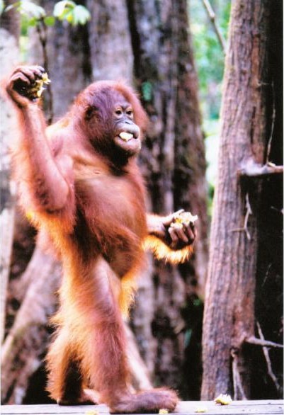
[2] Taken from Wikipedia. Same
name. ''Orangutan image taken by Tom
Low at Camp Leakey, Tanjung Puting,
Kalimantan, Indonesia (2003).'' PD
source: http://upload.wikimedia.org/wiki
pedia/commons/0/0b/Orangutan.jpg
[1] From: Stewart, Caro-Beth, and Todd
R Disotell. “Primate evolution - in
and out of Africa.” Current Biology
8.16 (1998) :
R582-R588. http://www.sciencedirect.com
/science/article/pii/S0960982207003673
Figure 2. A synthetic hypothesis of
catarrhine primate evolution. The
branching order shown for the living
species is well-supported by numerous
molecular phylogenetic studies (for
example [6, 7, 8, 9, 10, 11, 12, 18, 24
and 25]). We present the dates of
divergence calculated by Goodman and
colleagues [11], on the understanding
that these are still rough estimates
and more precise measurements are
needed, especially for the Old World
monkeys. The fossil species (genus
names in italics) were placed on this
tree by parsimony analyses of
relatively large morphological datasets
[4, 11, 14 and 15]. Known dates for
fossils [1, 2 and 21] are indicated by
the thicker lines; these lines are
attached to the tree as determined by
the parsimony analyses, although the
dates of the attachment points are our
best guesses. Species found in Africa
are in red and species found in Eurasia
are in black. The continental locations
of the ancestral lineages were inferred
by parsimony using the computer program
MacClade [30]. The intercontinental
dispersal events required, at a
minimum, to explain the distribution of
the living and fossil species are
indicated by the arrows. COPYRIGHTED
source: http://www.sciencedirect.com/sci
ence?_ob=ArticleURL&_udi=B6VRT-4C4DVM4-D
&_user=4422&_handle=V-WA-A-W-WC-MsSAYVW-
UUW-U-AAVECYCCBC-AAVDAZZBBC-YCACYAZCV-WC
-U&_fmt=full&_coverDate=07%2F30%2F1998&_
rdoc=12&_orig=browse&_srch=%23toc%236243
%231998%23999919983%23494082!&_cdi=6243&
view=c&_acct=C000059600&_version=1&_urlV
ersion=0&_userid=4422&md5=5558415c4ccd34
6c64e2e6be03c3865e
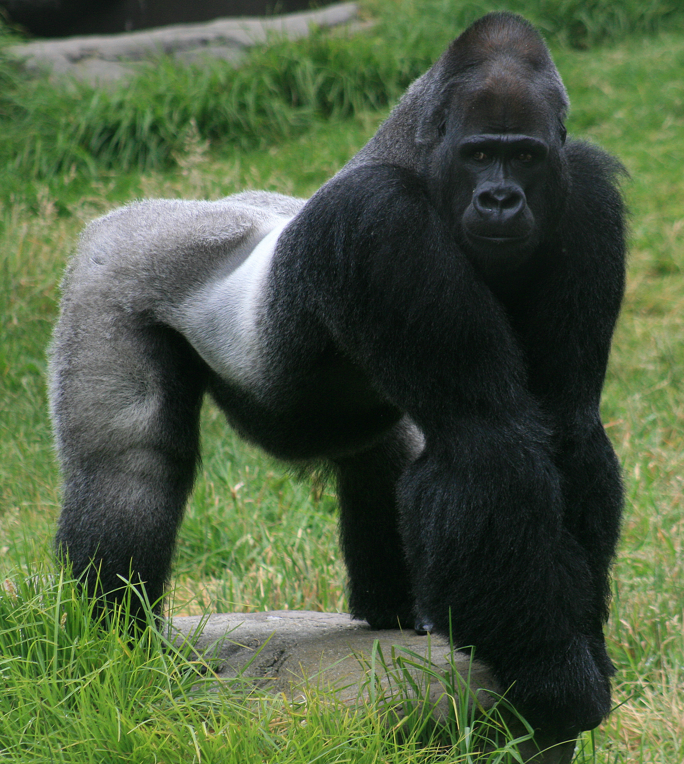
[2] Description English: Male
silverback w:Gorilla, Gorilla gorilla
in SF zoo Date Source Own
work Author Mila
Zinkova Permission (Reusing this
file) See below. CC
source: http://upload.wikimedia.org/wiki
pedia/commons/5/50/Male_gorilla_in_SF_zo
o.jpg
ancestor of chimpanzees and humans.

[1] From: Richard Dawkins, ''The
Ancestor's Tale'', (Boston, MA:
Houghton Mifflin Company, 2004),
p106. COPYRIGHTED
source: Richard Dawkins, "The
Ancestor's Tale", (Boston, MA: Houghton
Mifflin Company, 2004), p106.
[2] Gregoire: 62-year-old
chimpanzee Description English:
Chimpanzee named ''Gregoire'' born in
1944 (Jane Goodall sanctuary of
Tchimpounga in Congo Brazzaville) -
Picture taken the 9th of December
2006 Français : Chimpanzé nommé
''Grégoire'' né en 1944 (sanctuaire
Jane Goodall de Tchimpounga au Congo
Brazzaville) - Photo prise le 9
décembre 2006 Date 9 December
2006 Source Own work Author
Delphine
Bruyère Permission (Reusing this
file) Attribution : Delphine
Bruyere GNU
source: http://upload.wikimedia.org/wiki
pedia/commons/b/ba/2006-12-09_Chimpanzee
_Gregoire_D_Bruyere.JPG
bipedal primate.
Africa
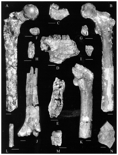
[1] Fig. 1. Orrorin tugenensis nov.
gen. nov. sp. A: BAR 1002′00, left
femur, posterior view; B: BAR
1002′00, left femur, anterior view;
C: BAR 1000′00, right mandibular
fragment with M3, buccal view; D: BAR
1000′00, left mandibular fragment
with M2–3, lingual view; E: BAR
1000′00, left mandibular fragment
with M2–3, occlusal view; F: BAR
1900′00, right M3, occlusal view; G:
BAR 1390′00, right P4, distal view;
H: BAR 1001′00, upper I1, labial
view; I: BAR 1425′00, right
Image , lingual view; J: BAR
1004′00, right distal humerus,
posterior view; K: BAR 1003′00,
proximal left femur, anterior view; L:
BAR 349′00, manual proximal phalanx,
superior view; M: BAR 1426′00, left
M3, distal view; N: BAR 1215′00,
fragmentary right proximal femur,
posterior view. Scale bars = 1
cm.Orrorin tugenensis nov. gen. nov.
sp. A : BAR 1002′00, fémur gauche,
vue postérieure ; B : BAR 1002′00,
fémur gauche, vue antérieure ; C :
BAR 1000′00, fragment mandibulaire
droit avec M3, vue buccale ; D : BAR
1000′00, fragment mandibulaire gauche
avec M2–3, vue linguale ; E : BAR
1000′00, fragment mandibulaire gauche
avec M2–3, vue occlusale ; F : BAR
1900′00, M3 droite, vue occlusale ; G
: BAR 1390′00, P4 droite, vue distale
; H : BAR 1001′00, I1, vue labiale ;
I : BAR 1425′00, Image droite, vue
linguale ; J : BAR 1004′00, humérus
distal droit, vue postérieure ; K :
BAR 1003′00, fémur proximal gauche,
vue antérieure ; L : BAR 349′00,
phalange proximale de la main, vue
supérieure ; M : BAR 1426′00, M3
gauche, vue distale ; N : BAR
1215′00, fémur proximal
fragmentaire, vue postérieure. Chaque
barre équivaut à 1 cm. COPYRIGHTED
source: http://www.sciencedirect.com/cac
he/MiamiImageURL/B6VJ3-42FS9XV-9-1/0?wch
p=dGLzVlz-zSkzS

[2] Description Ardipithecus
ramidus specimen, nicknamed
?Ardi?. After Gen Suwa, Berhane
Asfaw, Reiko T. Kono, Daisuke Kubo, C.
Owen Lovejoy, Tim D. White (2009):
''The Ardipithecus ramidus Skull and
Its Implications for Hominid Origins.''
Science, 2 October 2009: Vol. 326. no.
5949, pp. 68e1-68e7, Fig. 2 Date
14 November 2009, 16:50 Source
Zanclean skull Uploaded by
FunkMonk Author T. Michael
Keesey Permission (Reusing this file)
CC
source: http://upload.wikimedia.org/wiki
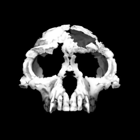
pedia/commons/e/e1/Ardi.jpg
(x-STrA-lO-PitiKuS}.
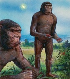
[1] Australopithecus squinted at the
blue African sky. He had never seen a
star in broad daylight before, but he
could see one today. White. Piercing.
Not as bright as the Sun, yet much more
than a full moon. Was it dangerous? He
stared for a long time, puzzled, but
nothing happened, and after a while he
strode across the savanna
unconcerned. Millions of years
later, we know better. ''That star
was a supernova, one of many that
exploded in our part of the galaxy
during the past 10 million years,''
says astronomer Mark Hurwitz of the
University of
California-Berkeley. Right: Human
ancestors, unconcerned by odd lights in
the daytime sky. This image is based on
a painting featured in The
Economist. PD
source: http://science.nasa.gov/headline
s/y2003/06jan_bubble.htm?list847478

[2] Image Source *
http://www.familie-rebmann.de/photo11.ht
m COPYRIGHTED CLAIMED FAIR USE
source: http://en.wikipedia.org/wiki/Ima
ge:Laetoliafar.jpg.jpg
SCIENCE

[1] a, The exterior surface of
DIK-55-2, and the location of each of
the surface marks. The rib is oriented
such that the rib head (broken off)
would be to the left. Dashed rule,
4 cm. b, Marks A1 and A2
(high-confidence stone-tool cut marks)
under low-power optical magnification;
the yellow rectangle demarcates c.
Scale bar, 5 mm. c, ESEM image
showing microstriations indicative of
cutting with a stone tool. Scale bar,
100 μm. d, Mark B (high-confidence
stone-tool-inflicted mark) under
low-power optical magnification,
indicative of a cutting and scraping
action or percussion; the yellow
rectangle demarcates e. Scale bar,
5 mm. e, ESEM image showing
microstriations indicative of stone
tool action. Scale bar, 500 μm.
b–e, The direction of the rib head is
indicated by the black arrows. See
Supplementary Information for the
details of mark C. COPYRIGHTED
source: http://www.nature.com/nature/jou
rnal/v466/n7308/images/nature09248-f2.2.
jpg
PuS} evolves.
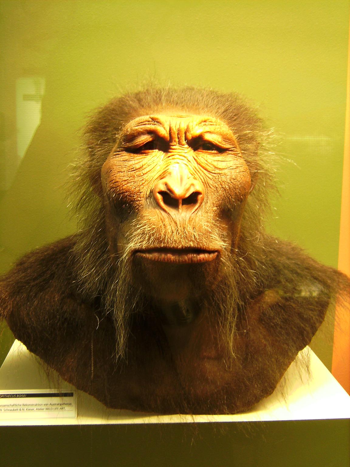
[1] Description Deutsch: plastische
wissenschaftliche Rekonstruktion eines
Paranthropus boisei English:
scientiffic reconstruction of a
Paranthropus boisei Date 25 March
2007 Source Photographed at
Westfälisches Museum für
Archäologie, Herne Author
Photographed by
User:Lillyundfreya Permission (Reusing
this file) own work GNU
source: http://upload.wikimedia.org/wiki
pedia/commons/6/6c/Paranthropus_boisei.J
PG

[2] Skull of Paranthropus
boisei. From Smithsonian Institute
website. COPYRIGHTED CLAIMED FAIR USE
source: http://en.wikipedia.org/wiki/Ima
ge:Zinj3.jpg

[1] Figure 3 from: Semaw, S. et al.
2.5-million-year-old stone tools from
Gona, Ethiopia. Nature 385, 333–336
(1997)
http://www.nature.com/nature/journal/v
385/n6614/abs/385333a0.html COPYRIGHTED
source: http://www.nature.com/nature/jou
rnal/v385/n6614/abs/385333a0.html

[2] Early man lived on elephant meat,
so much they died out in the Middle
East 400,000 years ago Submitted by
Anonymous on Wed Dec 14 2011 17:23:00
GMT-0800 (Pacific Standard Time) -
Source: dailymail.co.uk Docile,
lumbering elephants were so perfect for
Homo erectus, that they provided up to
60 per cent of their diet - until
constant hunting wiped out elephants in
the Middle East. The disappearance
of elephants helped kill off Homo
erectus, and paved the way for Homo
sapiens - modern humans - to take
over. Findings from the University
of Tel Aviv reveal how important the
huge animals were to the diet of early
humans - researchers that elephants
provided 60 per cent of the meat eaten
by Homo erectus. UNKNOWN
source: http://i4.asntown.net/Mastodon-t
vfm.jpg
evolve (earliest member of the genus
"Homo").
This is when the human brain begins to
get bigger.

[1] KNM ER 1813 Homo habilis This
image is from the website of the
Smithsonian Institution [1] and may be
copyrighted. The Smithsonian
Institution explicitly considers the
use of its content for non-commercial
educational purposes to qualify as fair
use under United States copyright law,
if: 1. The author and source of the
content is clearly cited. 2. Any
additional copyright information about
the photograph from the Smithsonian
Institution website is included. 3.
None of the content is modified or
altered.
source: http://en.wikipedia.org/wiki/Ima
ge:KNM_ER_1813.jpg
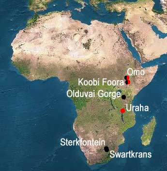
[2] red= Homo rudolfensis black=Homo
habilis COPYRIGHTED
source: http://sesha.net/eden/Eerste_men
sen.asp
[1] From: Richard Dawkins, ''The
Ancestor's Tale'', (Boston, MA:
Houghton Mifflin Company, 2004),
p106. COPYRIGHTED
source: Richard Dawkins, "The
Ancestor's Tale", (Boston, MA: Houghton
Mifflin Company, 2004), p106.
[2] Gregoire: 62-year-old
chimpanzee Description English:
Chimpanzee named ''Gregoire'' born in
1944 (Jane Goodall sanctuary of
Tchimpounga in Congo Brazzaville) -
Picture taken the 9th of December
2006 Français : Chimpanzé nommé
''Grégoire'' né en 1944 (sanctuaire
Jane Goodall de Tchimpounga au Congo
Brazzaville) - Photo prise le 9
décembre 2006 Date 9 December
2006 Source Own work Author
Delphine
Bruyère Permission (Reusing this
file) Attribution : Delphine
Bruyere GNU
source: http://upload.wikimedia.org/wiki
pedia/commons/b/ba/2006-12-09_Chimpanzee
_Gregoire_D_Bruyere.JPG
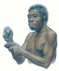
[1] Homo ergaster. Capacité
crânienne de 800 à 950
cm3 COPYRIGHTED
source: http://ma.prehistoire.free.fr/er
gaster.htm

[2] Turkana Boy COPYRIGHTED
source: http://www.anthropology.at/virta
nth/evo_links/turkana%20boy.jpg
from Africa.
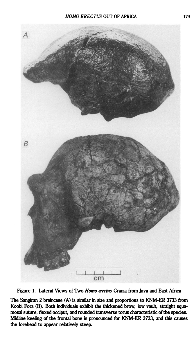
[1] G. Philip Rightmire, ''The
Dispersal of Homo erectus from Africa
and the Emergence of More Modern
Humans'', Journal of Anthropological
Research, Vol. 47, No. 2, A Quarter
Century of Paleoanthropology: Views
from the U.S.A. (Summer, 1991), pp.
177-191 Published by: University of
New Mexico Article Stable URL:
http://www.jstor.org/stable/3630324
COPYRIGHTED
source: http://www.jstor.org/stable/3630
324
[2] All statistically significant
inferences in Tables 1 and 2 are
incorporated into this single model.
Major expansions of human populations
are indicated by red arrows. Genetic
descent is indicated by vertical lines,
and gene flow by diagonal lines. The
timing of inferences lacking resolution
at the 5% level and/or not validated by
more than one locus are indicated by
question marks. COPYRIGHTED
source: http://www.nature.com/nature/jou
rnal/v416/n6876/images/416045a-f1.2.jpg
Africa
[1] Description English: A fire lit
using twigs and pine cones. Date
2008-03-27 (original upload
date) (Original text : 10:58, 27 March
2008 (UTC)) Source Transferred
from en.wikipedia (Original text :
http://waxingnonsensical.blogspot.com)
Author Original uploader was
Emeldil at en.wikipedia (Original text
: Pavan Srinath) Permission (Reusing
this file) CC-BY-SA-3.0. CC
source: http://upload.wikimedia.org/wiki
pedia/commons/thumb/0/0f/Campfire_Pineco
ne.png/450px-Campfire_Pinecone.png

[2] Swartkrans Caves For any
picture requests, please email:
marketing@maropeng.co.za All photos
should be credited (© Maropeng),
unless otherwise stated in the caption.
UNKNOWN
source: http://maropeng.flowcommunicatio
.netdna-cdn.com/images/sized/images/medi
agallery/IMG_7223-600x450.JPG
hair.

[1] escription English: A diorama in
National Museum of Indonesia, Jakarta,
depicting the life size model of stone
equipped hunter, a Homo erectus family
living in Sangiran about 900,000 years
ago. Bahasa Indonesia: Sebuah diorama
di Museum Nasional Indonesia di Jakarta
menampilkan adegan pemburu dengan
alat-alat batu, sebuah keluarga Homo
erectus yang hidup di Sangiran sekitar
900.000 tahun yang lalu. Date 24
August 2010 Source Own
work Author Gunkarta Gunawan
Kartapranata CC
source: http://upload.wikimedia.org/wiki
pedia/commons/thumb/1/13/Sangiran_Homo_e
rectus_Diorama.jpg/1280px-Sangiran_Homo_
erectus_Diorama.jpg
[1] Homo erectus, artwork C010/4389
Rights Managed Credit: JOSE ANTONIO
PEÑAS/SCIENCE PHOTO
LIBRARY Caption: Homo erectus.
Computer artwork of a Homo erectus man
standing in a prehistoric landscape.
Homo erectus is the most widespread and
longest-surviving of all the fossil
hominids. Its geographical spread
included north and east Africa, Europe,
Indonesia and China, where it lived
between 1 and 2 million years
ago. Release details: Model and
property releases are not available
UNKNOWN
source: http://www.sciencephoto.com/imag
e/417426/large/C0104389-Homo_erectus,_ar
twork-SPL.jpg

[2] Flint artefacts include
hard-hammer flakes, notches, retouched
flakes and cores (a–c, hard-hammer
flake; d, e, multiple notch; f,
hard-hammer flake; g, h, hard-hammer
flake, showing pronounced point of
percussion on plain butt).
Supplementary Information includes
micro-CT volume rendering of artefacts
(still example shown as a) with
three-dimensional animations (see
Supplementary Movies 1–10). i, Cone
of Pinus cf. sylvestris. j, Upper
second molar of Mammuthus cf.
meridionalis. COPYRIGHTED
source: http://nature.com/nature/journal
/v466/n7303/images/nature09117-f2.2.jpg
Germany.)

[1] (Photo : Jayne Wilkins) Replicas of
the 500,000-year-old stone points from
Kathu Pan 1 were hafted onto wooden
dowels with acacia resin and sinew, and
plunged into antelope carcasses Read
more at
http://www.latinospost.com/articles/6938
/20121115/oldest-stone-spear-tips-found-
came-200.htm#irhScS4kokLTYQJp.99 UNKNOW
N
source: http://images.latinospost.com/da
ta/images/full/8174/early-hafted-spears.
jpg?w=600
[2] Fig. 2 (A and B) Distal
step-terminating bending fractures on
ventral surfaces of complete
nonretouched convergent blades, banded
ironstone. (C) Distal impact burination
on ventral surface of a complete
nonretouched convergent flake, banded
ironstone. (D) Distal impact burination
on ventral surface of complete
nonretouched convergent blade, banded
ironstone. (E) Comparison of DIF
frequencies (95% confidence intervals)
at Holocene kill and habitation sites
with well-established weapon tips (11,
27, 28, 36), experimental weapon tip
studies (10, 27, 37–40), trampling
experiments (32, 41), and KP1.
Wilkins, Jayne et al. “Evidence for
Early Hafted Hunting Technology.”
Science 338.6109 (2012):
942–946. http://www.sciencemag.org/co
ntent/338/6109/942 COPYRIGHTED
source: http://www.sciencemag.org/conten
t/338/6109/942
[1] Figure from: Day, M. H. ''Omo
human skeletal remains.'' Nature 222,
1135–1138 (1969)
http://www.nature.com/nature/journal/v
222/n5199/pdf/2221135a0.pdf COPYRIGHTED
source: http://www.nature.com/nature/jou
rnal/v222/n5199/pdf/2221135a0.pdf
[2] Figure 1 from: Tim D. White,
Berhane Asfaw, David DeGusta, Henry
Gilbert, Gary D. Richards, Gen Suwa &
F. Clark Howell, ''Pleistocene Homo
sapiens from Middle Awash, Ethiopia'',
Nature 423, 742-747 (12 June
2003) http://www.nature.com/nature/jour
nal/v423/n6941/full/nature01669.html CO
PYRIGHTED
source: http://www.nature.com/nature/jou
rnal/v423/n6941/images/nature01669-f1.2.
jpg
sounds begins to develop. All words are
single syllable.
[1] EARLY HUMANS SETTLED IN BRITAIN
800,000 YEARS AGO July 7, 2010 --
During the harsh winters, early humans
almost certainly relied on hunting
animals, as edible plants would have
been in very short supply, the study
says. UNKNOWN
source: http://news.discovery.com/archae
ology/2010/07/07/early-humans-zoom.jpg
[2] Phonetic Alphabet Symbols used by
Ted Huntington PD
source: http://tedhuntington.com/fonikal
f.jpg
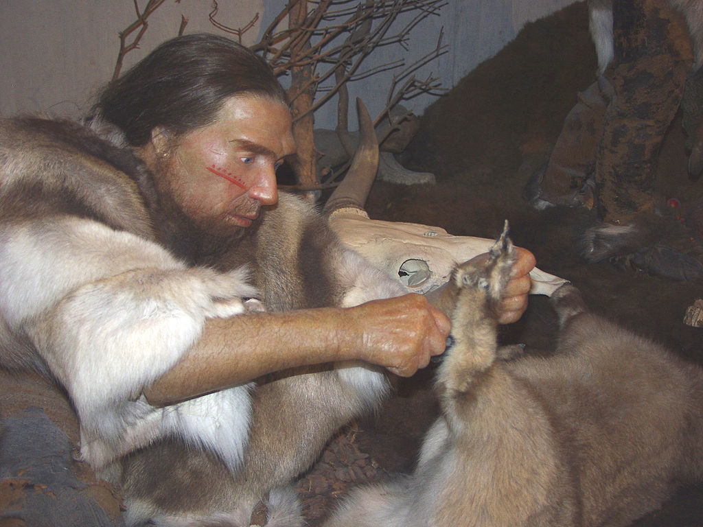
[1] Description Deutsch:
Rekonstruierter Neandertaler im
Neanderthal-Museum Date 2007 Source
Own
work Author Ökologix Permission
(Reusing this file) See below. PD
source: http://upload.wikimedia.org/wiki
pedia/commons/thumb/9/95/Neandertaler-im
-Museum.jpg/1024px-Neandertaler-im-Museu
m.jpg
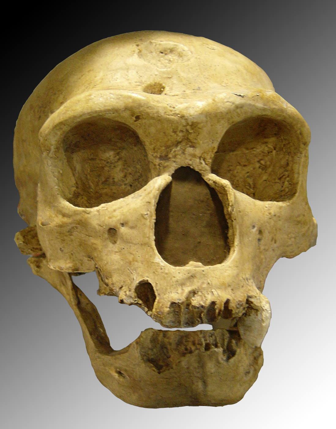
[2] Description English: Homo
neanderthalensis. Skull discovered in
1908 at La Chapelle-aux-Saints
(France). Date October
2005 Source Own
work Author Luna04 GNU
source: http://upload.wikimedia.org/wiki
pedia/commons/e/e0/Homo_sapiens_neandert
halensis.jpg
[99000 BC]
into Eurasia. Beginning of differences
in race.
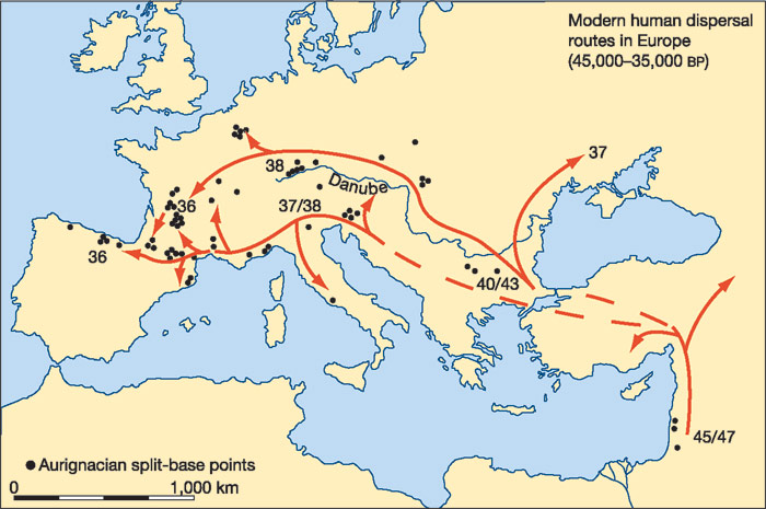
[1] The northern route (along the
Danube) is represented by the 'classic'
Aurignacian technologies, while the
southern (Mediterranean) route is
represented by the 'proto-Aurignacian'
bladelet technologies (Fig. 3)-with
their inferred origins in the preceding
early Upper Palaeolithic technologies
in the Near East and southeastern
Europe. Dates (in thousands of years
bp) indicate the earliest radiocarbon
dates for these technologies in
different areas, expressed in thousands
of radiocarbon years before present
(bp). (These are likely to
underestimate the true (calendar) ages
of the sites by between 2,000 and 4,000
yr; see ref. 32). Dashed lines indicate
uncertain routes. COPYRIGHTED
source: http://www.nature.com/nature/jou
rnal/v432/n7016/fig_tab/nature03103_F1.h
tml
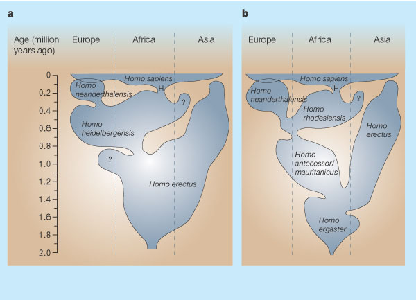
[2] The figure shows the geographical
and temporal distribution of hominid
populations, based on fossil finds,
using different taxonomic schemes. The
new finds from Herto4, 5 (H) represent
early Homo sapiens. a, This reflects
the view that both Neanderthals and
modern humans derived from a widespread
ancestral species called H.
heidelbergensis2. b, However, evidence
is growing that Neanderthal features
have deep roots in Europe2, 8, so H.
neanderthalensis might extend back over
400,000 years. The roots of H. sapiens
might be similarly deep in Africa, but
this figure represents the alternative
view that the ancestor was a separate
African species called H. rhodesiensis.
Different views of early human
evolution are also shown. Some workers
prefer to lump the earlier records
together and recognize only one
widespread species, H. erectus2 (shown
in a). Others recognize several
species, with H. ergaster and H.
antecessor (or H. mauritanicus) in the
West, and H. erectus only in the Far
East8 (shown in b). Adapted with
permission from refs 8, 11. 8.
Hublin, J.-J in Human Roots: Africa
and Asia in the Middle Pleistocene (eds
Barham, L. & Robson-Brown, K.) 99-121
(Western Academic & Specialist Press,
Bristol, 2001). 11. Rightmire, G. P.
in Human Roots: Africa and Asia in the
Middle Pleistocene (eds Barham, L. &
Robson-Brown, K.) 123-133 (Western
Academic & Specialist Press, Bristol,
2001). COPYRIGHTED
source: http://www.nature.com/nature/jou
rnal/v423/n6941/fig_tab/423692a_F1.html
[98000 BC]

[1] {ULSF: Skhul IV from Mugharet
Es-Skhul, corresponds to images in
pages between p112 and p113 Garrod,
''The Stone Age of Mount Carmel
excavations at the
Wady-el-Mughara'',1937} Reconstructio
n of the burial of Skhul 4 , a
strongly-built, ''early modern''
human. (photograph from Stringer C.S.
et al., 1994) UNKNOWN
source: http://indiaeng.com/Tsunami-2004
--Andaman%20fault/Toba%20Volcano,%20ch_5
%20-%20Human%20evolution_files/mht1055(1
).TMP
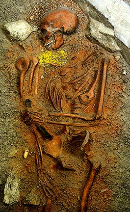
[2] The following is taken from James
Shreeve's book The Neandertal Enigma:
solving the mystery of modern human
origins (William Morrow and Company,
New York, 1995.) UNKNOWN
source: http://www.mesacc.edu/dept/d10/a
sb/origins/hominid_journey/pictures/buri
al.jpeg
[59000 BC]
[1] Lucinda Backwella, Francesco
d'Erricob and Lyn Wadley (june 2008).
''Middle Stone Age bone tools from the
Howiesons Poort layers, Sibudu Cave,
South Africa''. ScienceDirect.
http://www.sciencedirect.com/science/a
rticle/pii/S0305440307002142
COPYRIGHTED
source: http://www.sciencedirect.com/sci
ence/article/pii/S0305440307002142

[2] Objects found in the
archaeological site called Border Cave
include a) a wooden digging stick; b) a
wooden poison applicator; c) a bone
arrow point decorated with a spiral
incision filled with red pigment; d) a
bone object with four sets of notches;
e) a lump of beeswax; and f) ostrich
eggshell beads and marine shell beads
used as personal ornaments. (Francesco
d'Errico and Lucinda Backwell / July
30, 2012) COPYRIGHTED
source: http://www.trbimg.com/img-5016e6
83/turbine/la-sci-sn-modern-culture-afri
ca-20120730-001/600
[44000 BC]
Australia.
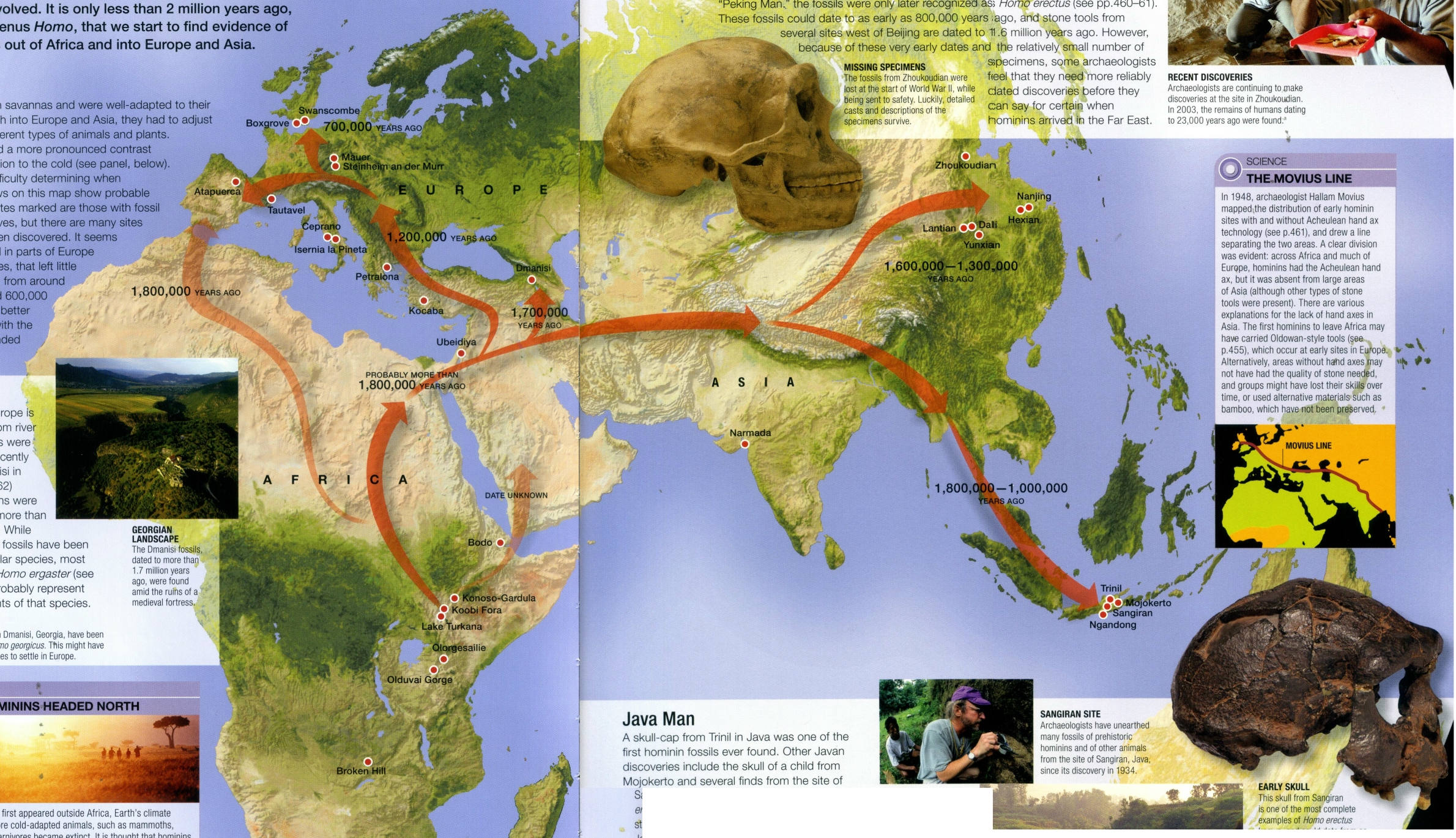
[1] Palmer, et al, ''Prehistoric
Life'', 2009, p470-471. COPYRIGHTED
source: Palmer, et al, "Prehistoric
Life", 2009, p470-471.

[2] World map of human migrations,
with the North Pole at center. Africa,
harboring the start of the migration,
is at the top left and South America at
the far right. Migration patterns are
based on studies of mitochondrial
(matrilinear) DNA. Numbers represent
thousand years before present. The
blue line represents area covered in
ice or tundra during the last great ice
age. The letters are the mitochondrial
DNA haplogroups (pure motherly
lineages); Haplogroups can be used to
define genetic populations and are
often geographically oriented. For
example, the following are common
divisions for mtDNA
haplogroups: African: L, L1, L2,
L3 Near Eastern: J, N Southern
European: J, K General European: H,
V Northern European: T, U, X Asian:
A, B, C, D, E, F, G (note: M is
composed of C, D, E, and G) Native
American: A, B, C, D, and sometimes
X [edit]Data
derivation Image:Northern icesheet
hg.png shows the region that was
covered by ice or tundra in the last
ice age All migration data based on
mitomap Geographic data from
http://en.wikipedia.org/wiki/Image:Last_
glacial_vegetation_map.png and adding
the following data
http://en.wikipedia.org/wiki/Image:Ice_A
ge_Temperature.png we get this
interesting result
http://en.wikipedia.org/wiki/Image:Human
-migration-temperature.jpg GNU
source: http://upload.wikimedia.org/wiki
pedia/commons/3/37/Map-of-human-migratio
ns.jpg
[38000 BC]
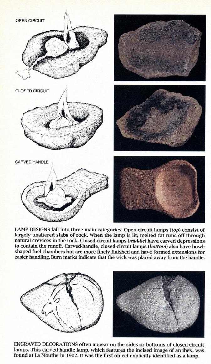
[1] Figure from: Sophie A. de Beaune
and Randall White, ''Ice Age Lamps'',
Scientific American, March
1993. http://halshs.archives-ouvertes.f
r/docs/00/42/17/69/PDF/Sc.Amer.1993.pdf
source: http://halshs.archives-ouvertes.
fr/docs/00/42/17/69/PDF/Sc.Amer.1993.pdf
[38000 BC]
Cave, Spain|Southern France

[1] Drawings of horses from Chauvet
Cave GNU
source: http://www.sciencemag.org/conten
t/336/6087/F5.large.jpg
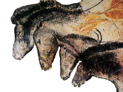
[2] Fig 3 from: Pike, A. W. G. et al.
“U-Series Dating of Paleolithic Art
in 11 Caves in Spain.” Science
336.6087 (2012): 1409 –1413.
Print. http://www.sciencemag.org/conten
t/336/6087/1409.abstract A time line
of the cave art dated. A single arrow
represents a minimum age, but, where
two dates are indicated, both maximum
and minimum ages have been obtained.
The error bars for O-21 reflect the
variation resulting from the two
different methods of detrital
correction (11). Larger versions of
these images showing sample locations
are available in the supplementary
materials, figs. S2 to
S12. COPYRIGHTED
source: http://en.wikipedia.org/wiki/Ima
ge:Chauvethorses.jpg
[38000 BC]
flute.
[1] Prehistorian historian Nicholas
Conard presents the bone flute from
Hohle Fels to journalists COPYRIGHTED
source: http://www.google.com/hostednews
/afp/media/ALeqM5hlF6Vh9FxCmW4OYCeiBOJqR
J3VgA?size=l

[2] Conard et al.1 have discovered the
oldest known flute, at Hohle Fels Cave
in Germany. The flute is made from bird
bone, and dates from the early
Aurignacian, 40,000 years ago. H.
JENSEN/UNIV. TÜBINGEN COPYRIGHTED
source: http://www.nature.com/nature/jou
rnal/v460/n7256/images/460695a-f1.2.jpg
[30000 BC]
[1] Fig. 1 (1 to 7) Fibers from
Dzudzuana, Georgia, unit D. 1, twisted
flax fibers; 2 to 4, flax fibers; and 5
to 7, unraveled flax fibers. (8 to 12)
Fibers from Dzudzuana, unit C. 8 and 9,
twisted flax fibers; 10 and 12, flax
fibers; and 11, dyed flax fibers.
COPYRIGHTED
source: http://www.sciencemag.org/conten
t/325/5946/-CSCO-3h--1359/-CSCO-3h--F1.l
arge.jpg

[2] On a lump of fired clay from the
Dolní Věstonice / Pavlov area were
found the impressions of substances
from plant fibres. The whole process of
picking nettles, crushing the dried
stem, preparation of tow, spinning the
thread and then weaving was tested and
shown to be possible using tools of the
time by M. Bunatova. Urbanová (ca
1999) http://www.donsmaps.com/dolnivpot
tery.html Dexterity of the First
Weavers A decade ago, experts did
not dare to think about people living
in the last ice age making
fabric. However, on a lump of fired
clay from the Dolní Věstonice /
Pavlov area were found the impressions
of substances from plant fibres. The
whole process of picking nettles,
crushing the dried stem, preparation of
tow, spinning the thread and then
weaving was tested and shown to be
possible using tools of the time by M.
Bunatova. Urbanová (ca
1999) Source: Display, Dolní
Věstonice Museum From Buňatová
(1999) and Sosna (2000): Buňatová,
M., 1999: Textilní produkce v mladém
paleolitu, experiment pro
dokumentární film ''Úsvit géniů'',
in: AR LI, Praha, 104 - 111. Sosna,
D., 2000: Počátky textilnictví. PhD.
Dissertation, Department of
Anthropology, Masaryk University, Brno.
UNKNOWN
source: http://www.ancient-wisdom.co.uk/
Images/countries/Czech%20pics/dolnifabri
c.jpg
[29700 BC]
[1] Description Deutsch:
Europäischer Grauwolf (Canis
lupus) English: grey wolf Date
February 2009 Source Own
work (own photo) Author Gunnar
Ries Amphibol Permission (Reusing
this file) You must give the
original author credit. If you use my
pictures outside the wiki projects,
please let me know. CC
source: http://upload.wikimedia.org/wiki
pedia/commons/thumb/9/9d/Grauwolf_P11302
75.jpg/1024px-Grauwolf_P1130275.jpg
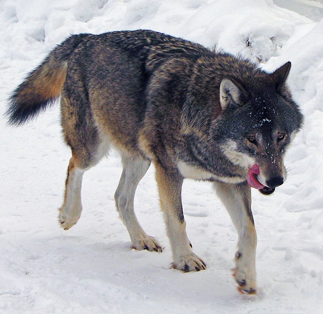
[2] Description Español: Lobo en
el zoo de Kolmården (Suecia). Date
2010-12-23 18:10 (UTC) Source
Wolf_Kolmården.jpg Author
Wolf_Kolmården.jpg: Daniel
Mott from Stockholm, Sweden
derivative work:
Mariomassone Permission (Reusing
this file) See below. CC
source: http://upload.wikimedia.org/wiki
pedia/commons/5/5f/Kolm%C3%A5rden_Wolf.j
pg
[27000 BC]

[1] Description Věstonická
venuše na výstavě Lovci mamutů v
Národním muzeu v Praze Date 2.
9. 2007 Source che Author
che (Please credit as ''Petr
Novák, Wikipedia'' in case you use
this outside WMF projects.) guidance:
Danny B. Permission (Reusing this
file) As they reached the Summit,
he said: “Thou shall take this
Snapshot and use it according to the
Code of License, and let your people
flourish all around the world.” They
brought the Snapshot to their homes and
there was much rejoicing. CC
source: http://upload.wikimedia.org/wiki
pedia/commons/b/b8/Vestonicka_venuse_edi
t.jpg

[2] Description Deutsch: Venus von
Willendorf Date 1 January
2007 Source Own work Author
User:MatthiasKabel Own work,
attribution required (Multi-license
with GFDL and Creative Commons CC-BY
2.5) GNU Figure 2 from: O. Soffer,
J. M. Adovasio, D. C. Hyland, ''The
“Venus” Figurines: Textiles,
Basketry, Gender, and Status in the
Upper Paleolithic'', Current
Anthropology, Vol. 41, No. 4
(August/October 2000), pp.
511-537 URL:
http://www.jstor.org/stable/10.1086/3173
81 COPYRIGHTED
source: http://upload.wikimedia.org/wiki
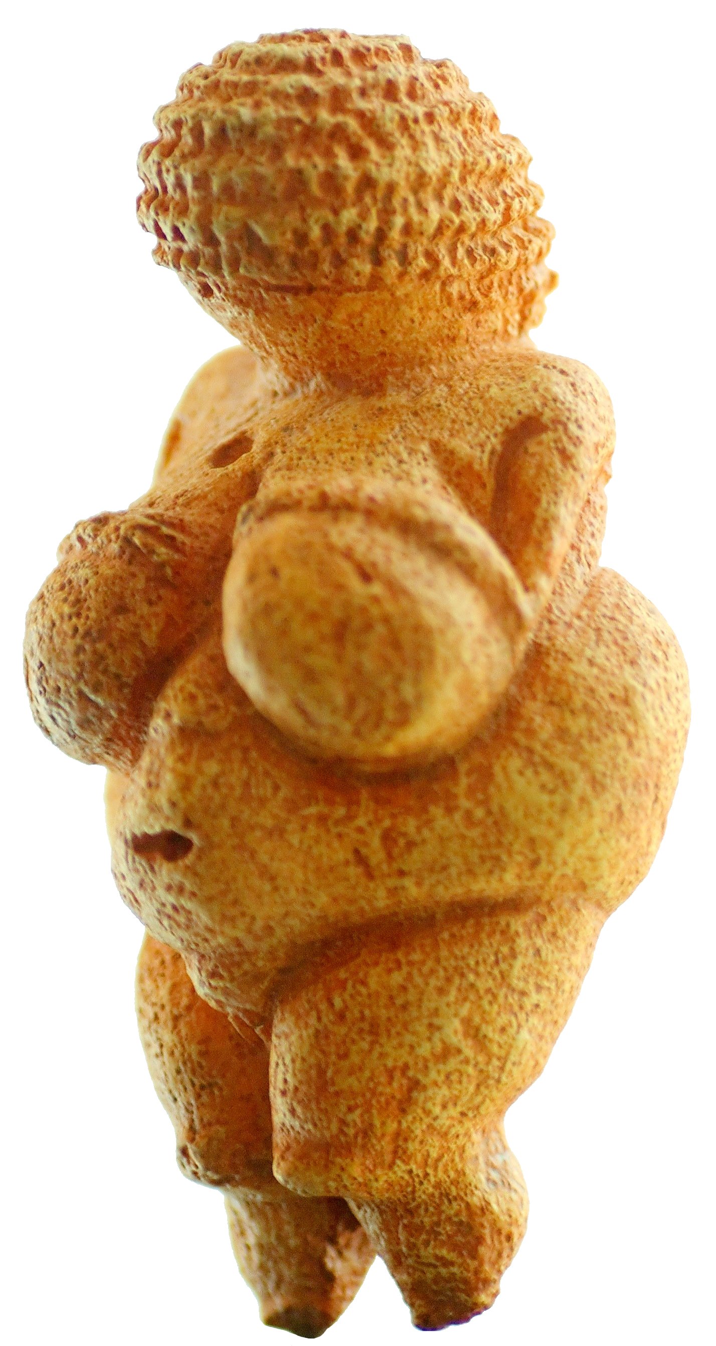
pedia/commons/5/50/Venus_von_Willendorf_
01.jpg
[21000 BC]
Greece
[1] Picture: Remains of the stone wall.
From the Greek Ministry of Culture.
UNKNOWN
source: http://blogs.discovery.com/files
/wall.jpg
[17000 BC]
Lebanon, Israel, Iraq, Jordan, Saudi
Arabia)
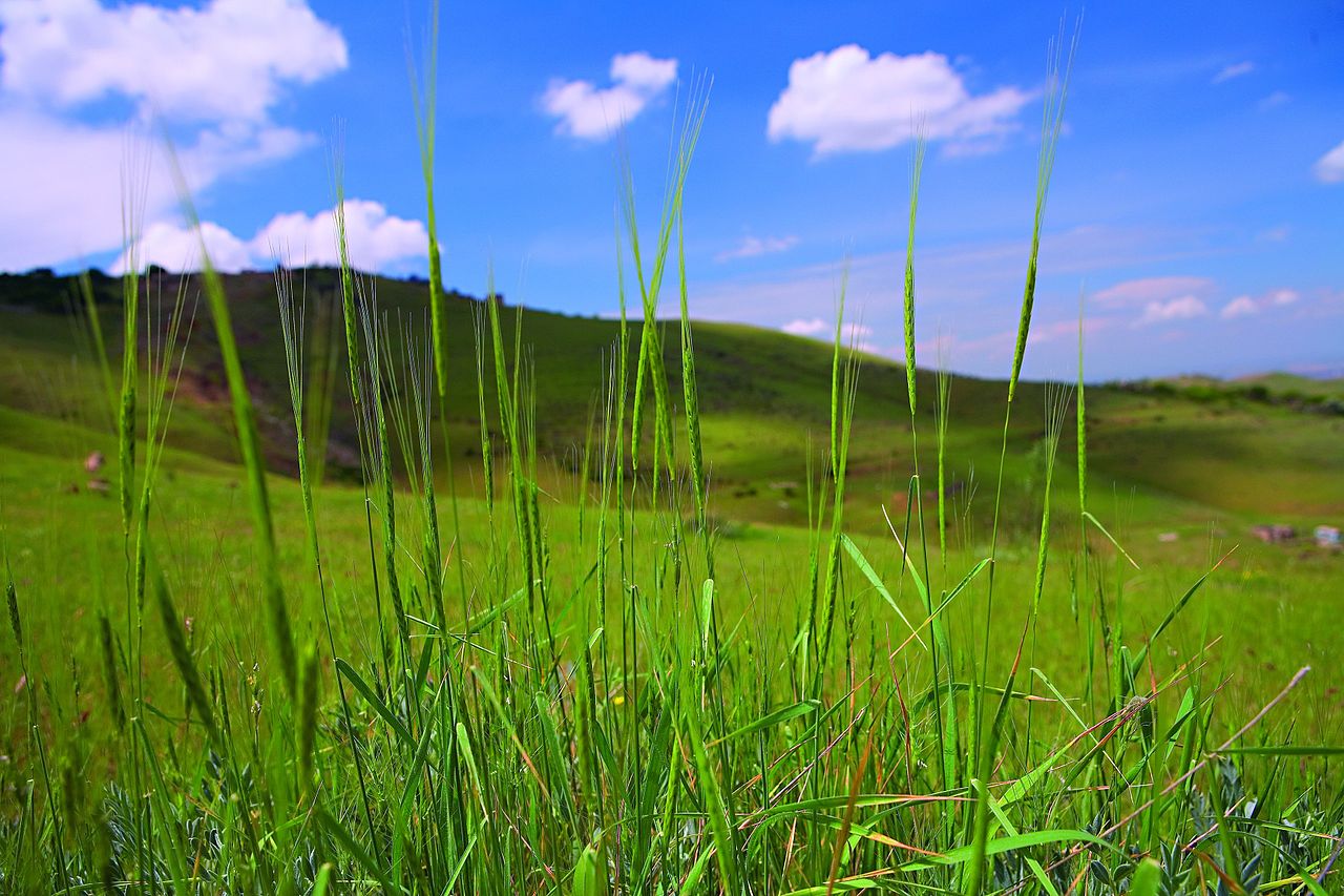
[1] escription Русский:
Дикая пшеница
Эребунийского
заповедника -
Однозернянка
араратская (Triticum
araraticum) English: The Wild Wheat of
Erebuni Reserve (Triticum
araraticum) Date 5 June 2007 Source
for-wikimedia.wowarmenia.ru Author
uncredited Permission (Reusing this
file) Released by WOWARMENIA for
Wikimedia under Creative Commons
Attribution-Share Alike license
(Multi-license with GFDL and Creative
Commons CC-BY-SA-3.0 and older versions
(2.5, 2.0 and 1.0)). If you wish to
reuse the photos elsewhere, please read
the instructions at COM:REUSE. GNU
source: http://upload.wikimedia.org/wiki
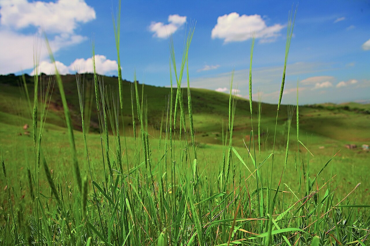
pedia/commons/thumb/d/d3/WildWheat_Erebu
ni_Reserve.jpg/1280px-WildWheat_Erebuni_
Reserve.jpg

[2] Description Česky:
Pšenice. Deutsch: Weizen. English:
Wheat. Español: Trigo. Français :
Blé. Magyar: Búza. Tiếng Việt:
Lúa mì. Date August
2005 Source Own work Author
User:Bluemoose GNU
source: http://upload.wikimedia.org/wiki
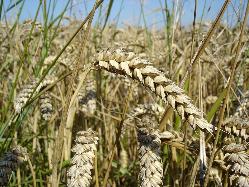
pedia/commons/thumb/b/b4/Wheat_close-up.
JPG/800px-Wheat_close-up.JPG
[16000 BC]
Province, China

[1] Pottery Fu (Cooking Vessel)-Shaped
Vessel Paleolithic Age to Neolithic
Age 12000 years ago Diameter at mouth
32.5cm height 29.8cm Restored on the
basis of unearthed pottery pieces at
Yuchanyan, Dao County in 1995 It is by
far the earliest pottery discovered, a
cooking vessel. [t Note that there
are apparently fragments of 2 or more
pottery vessels, and they are redated
in the article to 18000ybn: Elisabetta
Boaretto, Xiaohong Wu, Jiarong Yuan,
Ofer Bar-Yosef, Vikki Chu, Yan Pan,
Kexin Liu, David Cohen, Tianlong Jiao,
Shuicheng Li, Haibin Gu, Paul Goldberg,
and Steve Weiner, ''Radiocarbon dating
of charcoal and bone collagen
associated with early pottery at
Yuchanyan Cave, Hunan Province, China
PNAS 2009 106 (24) 9595-9600;''
published ahead of print June 1, 2009,
doi:10.1073/pnas.0900539106
http://www.pnas.org/content/106/24/959
5.full?sid=4a6f1743-94c2-4be8-b046-575b4
f27ab46]
source: http://www.hnmuseum.com/hnmuseum
/eng/whatson/exhibition/images/kg/2.jpg
[15000 BC]
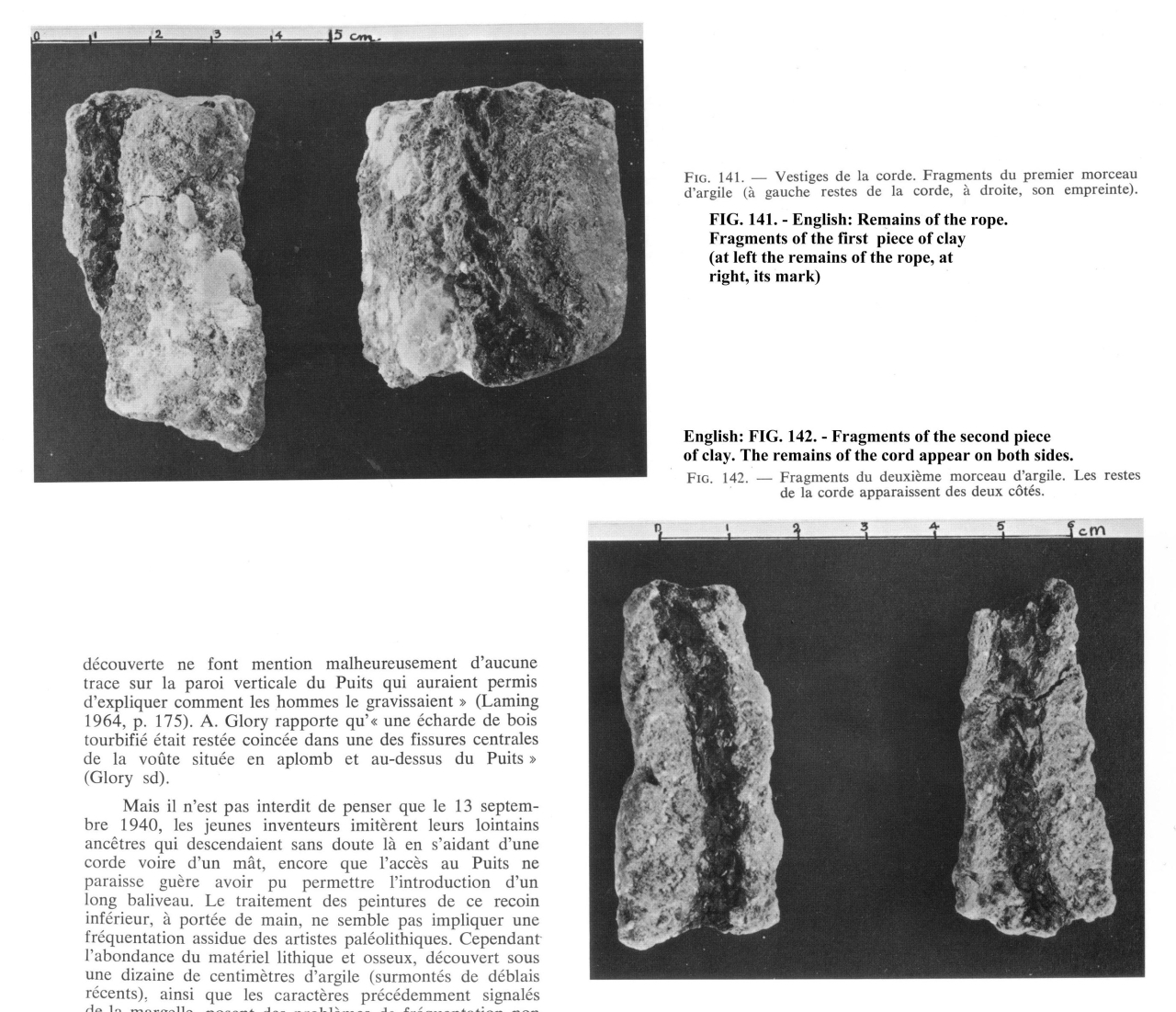
[1] Remains of the rope. Fragments of
the first piece of clay (at left the
remains of the rope, at right, its
mark). Images from: LEROI-GOURHAN,
A., Lascaux Inconnu (A. LEROIGOURHAN &
J. ALLAIN, eds.), Xlle Suppl. à Gallia
Préhistoire, CNRS: Paris, 1979,
p183. COPYRIGHTED
source: LEROI-GOURHAN, A., Lascaux
Inconnu (A. LEROIGOURHAN & J. ALLAIN,
eds.), Xlle Suppl. à Gallia
Préhistoire, CNRS: Paris, 1979, p183.
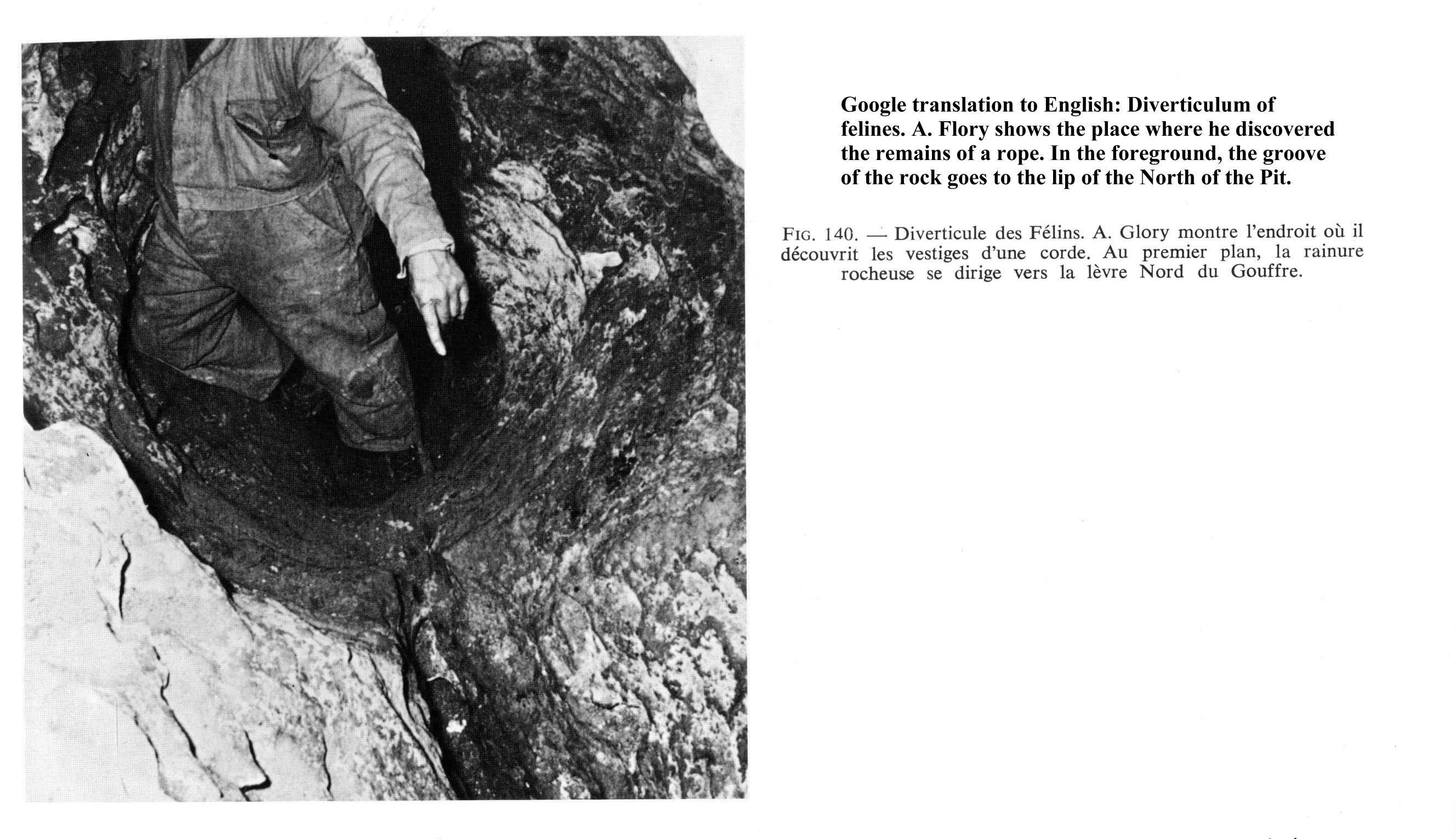
[2] Figure 142. - Fragments of the
second piece of clay. The remains of
the cord appear on both sides. Images
from: LEROI-GOURHAN, A., Lascaux
Inconnu (A. LEROIGOURHAN & J. ALLAIN,
eds.), Xlle Suppl. à Gallia
Préhistoire, CNRS: Paris, 1979,
p183. COPYRIGHTED
source: LEROI-GOURHAN, A., Lascaux
Inconnu (A. LEROIGOURHAN & J. ALLAIN,
eds.), Xlle Suppl. à Gallia
Préhistoire, CNRS: Paris, 1979, p183.
[11000 BC]
Santa Rosa Island, California,
USA
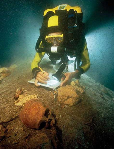
[1] A member of Arturo González's
underwater archaeological team studies
a skull in an underwater cave on the
coast of the Yucatán Peninsula near
Tulum, Mexico. Skeletons found in
similar caves may be among the oldest
human remains ever found in North or
South America. Photograph courtesy
Arturo Gonzáles COPYRIGHTED
source: http://news.nationalgeographic.c
om/news/bigphotos/images/080903-oldest-s
keletons_big.jpg
[2] Tulum, Mexico UNKNOWN
source: maps.google.com
[9000 BC]
Palestine

[1] An aerial view of Jericho showing
the ruins of Tell
es-Sultan Description Italiano:
veduta aerea dell'area archeologica di
Gerico Date 2008-03-05 (original
upload date) Source Transferred
from it.wikipedia Author Original
uploader was Fullo88 at
it.wikipedia PD
source: http://upload.wikimedia.org/wiki
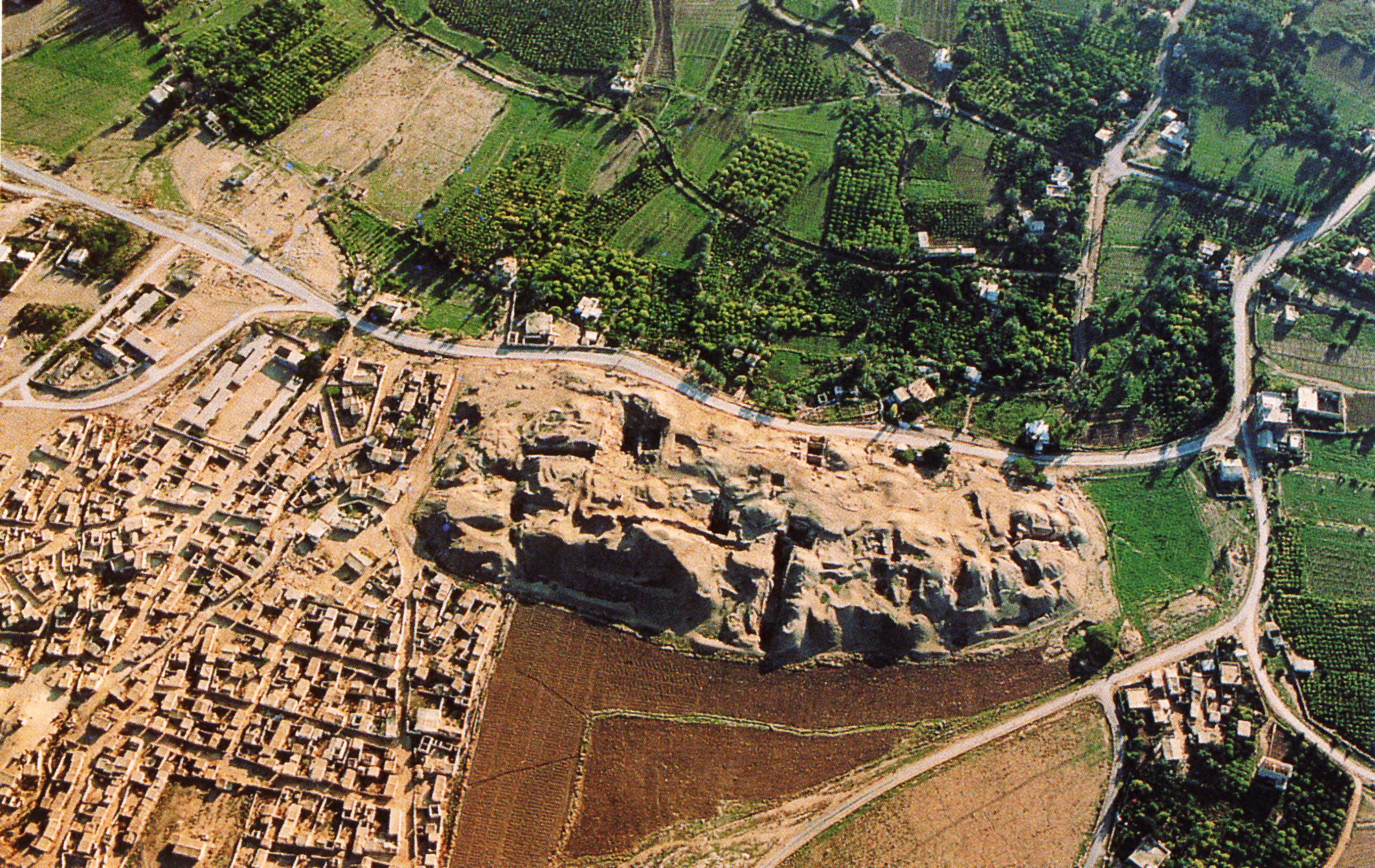
pedia/commons/f/f4/Tell_es-sultan.jpg

[2] Plastered skulls figures
from: Kathleen Kenyon, ''Excavations
at Jericho'', 1981,
vol5. {Kenyon_Excavations_At_Jericho_19
81.pdf} COPYRIGHTED
source: Kenyon_Excavations_At_Jericho_19
81.pdf
[7500 BC]
Turkey and northern Syria (Nevali Cori,
Turkey)
[1] Description Česky:
Pšenice. Deutsch: Weizen. English:
Wheat. Español: Trigo. Français :
Blé. Magyar: Búza. Tiếng Việt:
Lúa mì. Date August
2005 Source Own work Author
User:Bluemoose GNU
source: http://upload.wikimedia.org/wiki
pedia/commons/thumb/b/b4/Wheat_close-up.
JPG/800px-Wheat_close-up.JPG
[2] Nevali Cori, Turkey from Google
Maps UNKNOWN
source: https://maps.google.com/maps?ll=
37.518333,38.605556&spn=0.01,0.01&t=m&q=
37.518333,38.605556 http://localhost/ul
sf/images/Nevali_Cori_Turkey.jpg
[7240 BC]
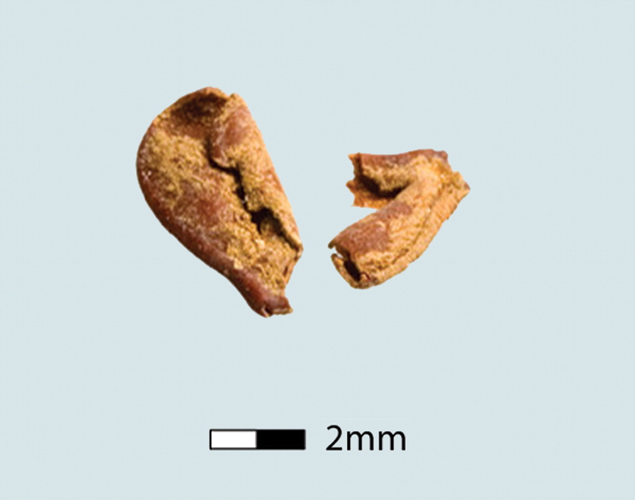
[1] Fig. 3. Close-up of two dark
brown squash seed (C. moschata)
fragments recovered from a buried house
floor at CA-09-27. from: Tom D.
Dillehay, Jack Rossen, Thomas C.
Andres, and David E. Williams,
''Preceramic Adoption of Peanut,
Squash, and Cotton in Northern Peru'',
Science 29 June 2007: 316 (5833),
1890-1893. http://www.sciencemag.org/co
ntent/316/5833/1890.abstract COPYRIGHTE
D
source: http://www.sciencemag.org/conten
t/316/5833/1890/F3.large.jpg
[6000 BC]
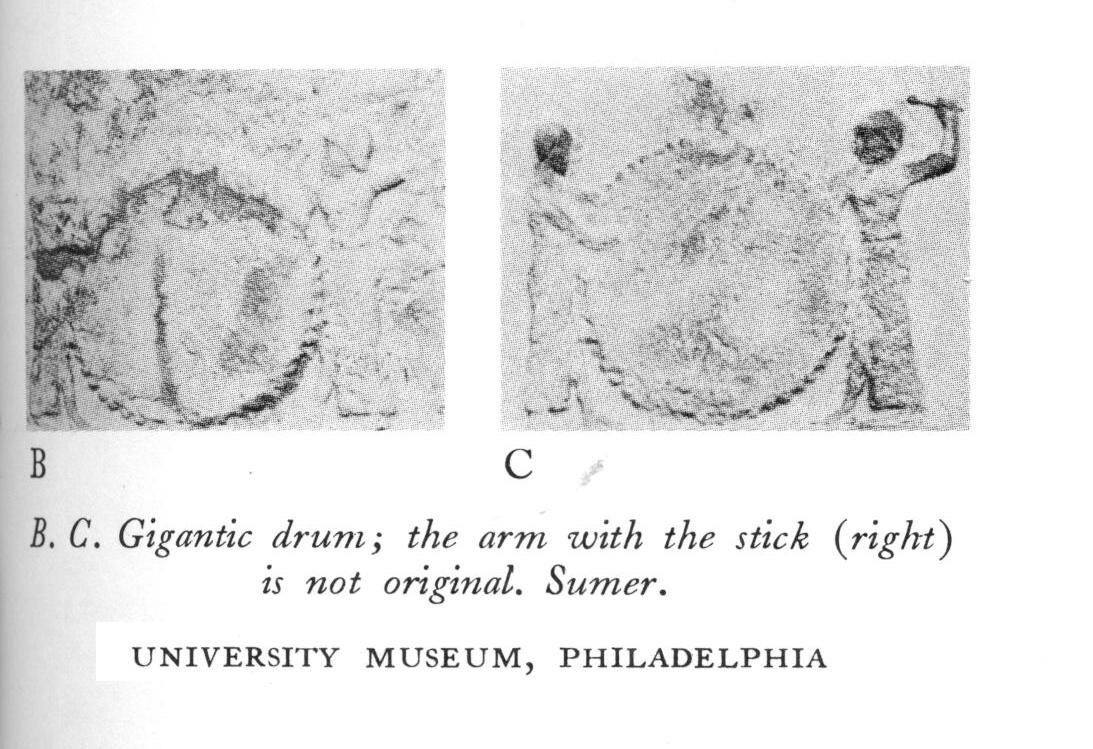
[1] Curt Sachs, ''The History of
Musical Instruments'', 1940, p81. PD
source: Curt Sachs, "The History of
Musical Instruments", 1940, p81.
[5700 BC]
Province|Yangtze (in Hubei and Hunan
provinces), China
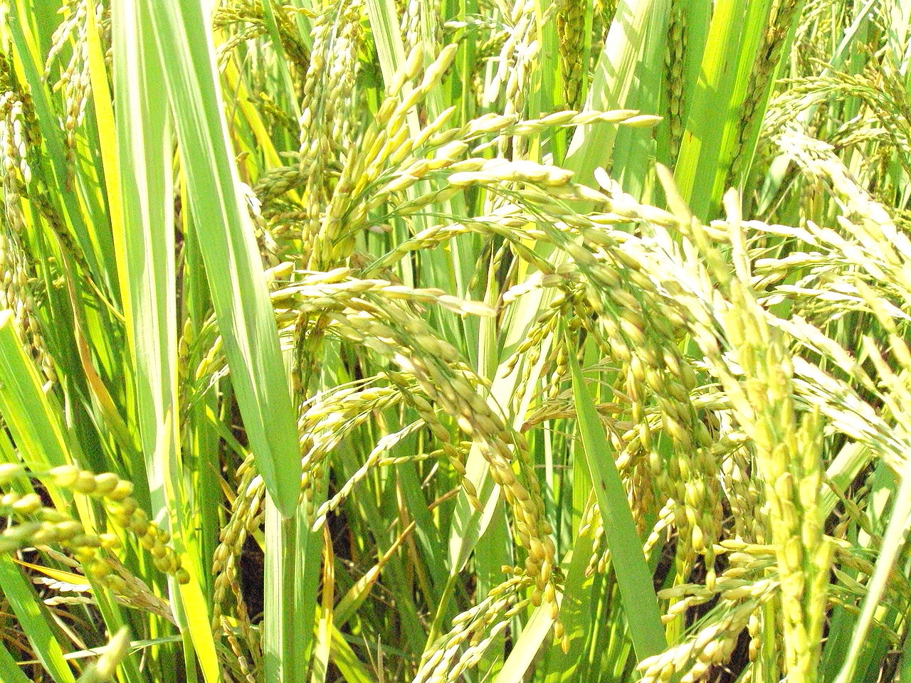
[1] Description English: Paddy in
West Bengal, India Date 18 October
2009 Source Own
work Author Amartyabag CC
source: http://upload.wikimedia.org/wiki
pedia/commons/thumb/9/98/Paddy_West_Beng
al.jpg/1280px-Paddy_West_Bengal.jpg

[2] Description: Cambodia, Kratie: A
worker is removing the rice
seedlings. Capture date: August
2002 Photographer: Oliver Spalt
Published under CC
source: http://upload.wikimedia.org/wiki
pedia/commons/0/07/Rice_02.jpg
[5100 BC]
Mexico)
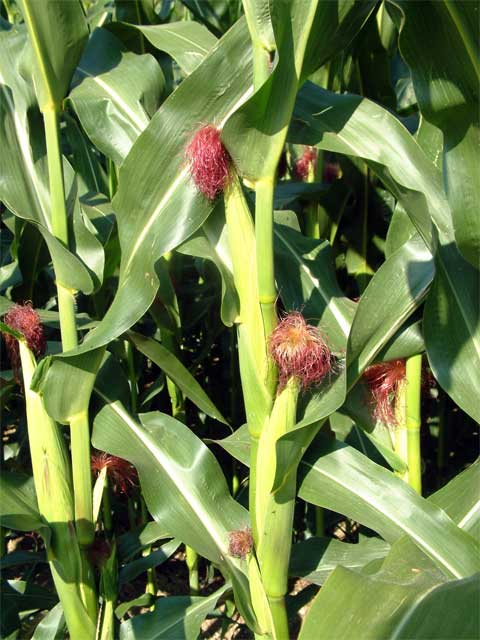
[1] Description Deutsch:
Maispflanzen (Zea mays) English: Maize
(Zea mays) plant with ears, the baby
corn growing level தமிழ்:
இளங்கதிர்கள்,
நன்கு
வளர்நிலையில்
இருக்கிறது. Date
2004 Source Own work Author
burgkirsch CC
source: http://upload.wikimedia.org/wiki
pedia/commons/3/32/Maispflanze.jpg
[5000 BC]
(copper).
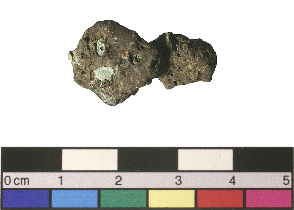
[1] Copper slag from Belovode (sample
No. 21). Figure 3 from: Miljana
Radivojević, Thilo Rehren, Ernst
Pernicka, Dušan Šljivar, Michael
Brauns, Dušan Borić, On the origins
of extractive metallurgy: new evidence
from Europe, Journal of Archaeological
Science, Volume 37, Issue 11, November
2010, Pages 2775-2787, ISSN 0305-4403,
10.1016/j.jas.2010.06.012. (http://www.
sciencedirect.com/science/article/pii/S0
305440310001986) COPYRIGHTED
source: http://www.sciencedirect.com/sci
ence/article/pii/S0305440310001986
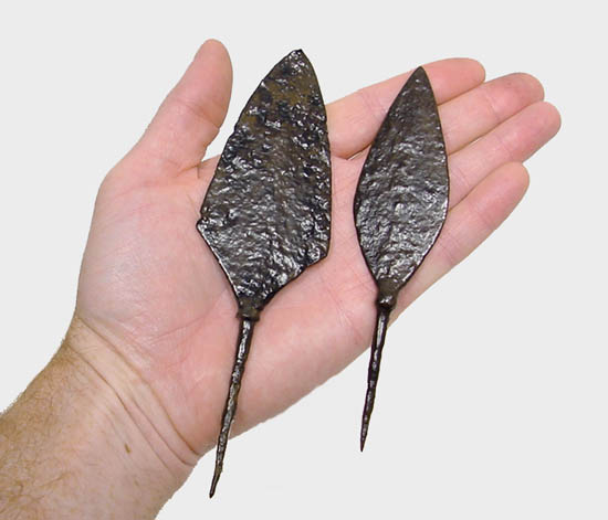
[2] Byzantine Roman
Collection tanged arrowhead Inventory
#: 308-316 Type:
Arrowhead Material: Iron Period:
Byzantine (Eastern Roman) 6th - 14th
Cent. A.D. Provenance: Balkan
Region Measurements: (lengths in cm,
left to right): 14.5, 17 cm UNKNOWN
source: http://www.worldmuseumofman.org/
img1000/308316b.jpg
[4000 BC]
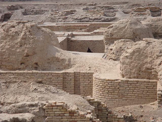
[1] The Royal Tombs (Cemetery) of Ur.
Courtesy Nathanm, Creative Commons. CC
source: http://popular-archaeology.com/u
pload/2697/urroyaltombs.jpg

[2] Pre-Historic Tell Uqair UNKNOWN
source: http://ancientneareast.tripod.co
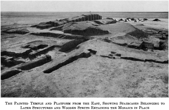
m/IMAGES/Uqair.jpg
[3500 BC]
numbers. First stamp (or seal).
Sumer, Highland Iran)

[1] Fig 109 from: Nissen, Archaic
Bookkeeping, 1993, p127. COPYRIGHTED
source: Archaic Bookkeeping, Nissen,
1993, p127.
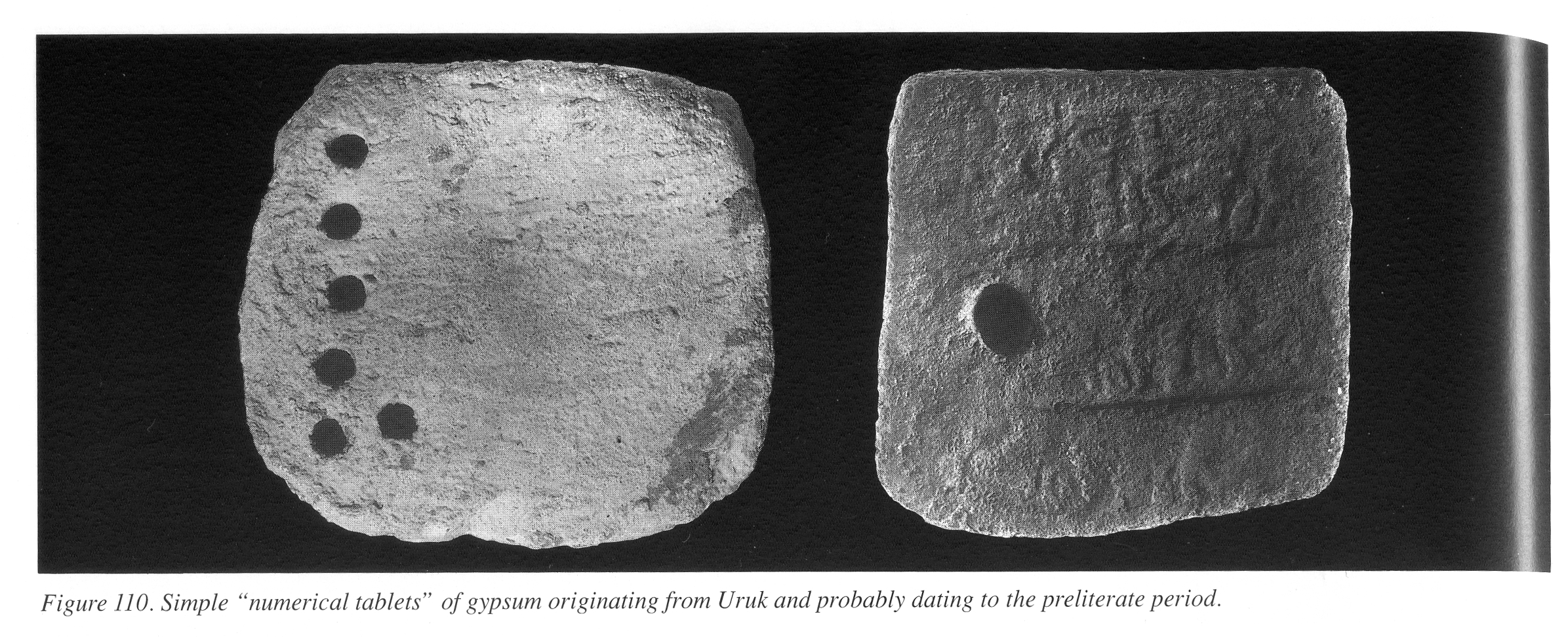
[2] Fig 110 from: Nissen, Archaic
Bookkeeping, 1993, p128. COPYRIGHTED
source: Archaic Bookkeeping, Nissen,
1993, p128.
[3500 BC]

[1] Stick in sand with shadow UNKNOWN
source: http://farm1.static.flickr.com/1
77/484077420_e01337d101.jpg
[2] Description English: Ancient
sundial from Marcianopolis, Museum of
Mosaicas, Devnya,
Bulgaria Български:
Слънчев часовник от
Марцианополис, Музей
на мозайките,
Девня Date 21 September
2010 Source Own work Author
Edal Anton Lefterov CC
source: http://upload.wikimedia.org/wiki
pedia/commons/f/ff/Sundial-from-Marciano
polis.jpg
[3500 BC]
wheel from Choga Mish, Iran)

[1] These pots, found at al`Ubaid type
site itself are typical of last phase
of Ubaid pottery found throughout much
of Mesopotamia, including Uruk. London:
British Museum. [t Note that the
first and tihrd match figures in
Woolley's 1982 book.] PD
source: http://www.hartford-hwp.com/imag
e_archive/ue/pottery03.jpg
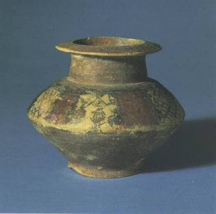
[2] 14. Pottery jar of Jemdat Nasr
type. It was found in the al`Ain region
of the United Arab Emirates, which
attests to contacts between Mesopotamia
and Oman peninsula—an important
source of copper. Ca. 3000 BC. London:
British Museum. UNKNOWN
source: http://www.hartford-hwp.com/imag
e_archive/ue/pottery02.jpg
[3490 BC]
[1] English: cotton plant, Texas, 1996,
after chemical haulm (topkilling
Chemical ; usually by the Monosodium
methyl arsenate used to quickly kill
the leaves that would interfere with
harvesting machines). This chemical is
a growing source of residual
contamination of soils by arsenic,
which is not degradable; Photo courtesy
of USDA Natural Resources Conservation
Service. http://photogallery.nrcs.usda.
gov/Index.asp This came from the
website PD
source: http://upload.wikimedia.org/wiki
pedia/commons/thumb/6/68/CottonPlant.JPG
/1024px-CottonPlant.JPG
[3350 BC]
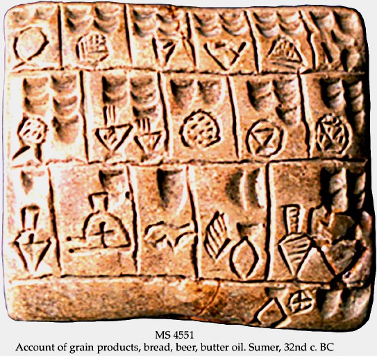
[1] MS 4551 Account of grain products,
bread, beer, butter oil. Sumer 32nd
century COPYRIGHTED?
source: http://www.earth-history.com/_im
ages/ms4551.jpg
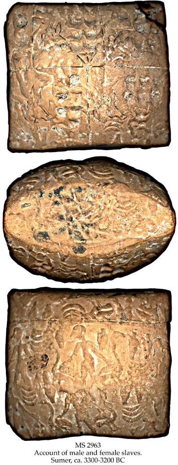
[2] MS2963 Account of male and female
slaves Sumer
c3300-3200BCE COPYRIGHTED?
source: http://www.earth-history.com/_im
ages/ms2963.jpg
[3310 BC]
Bronocice, Krakow, Poland

[1] Stuart and Piggott, ''The Earliest
Wheeled Transport'', 1983,
p40,62-63. COPYRIGHTED
source: Stuart and Piggott, "The
Earliest Wheeled Transport", 1983,
p40,62-63.
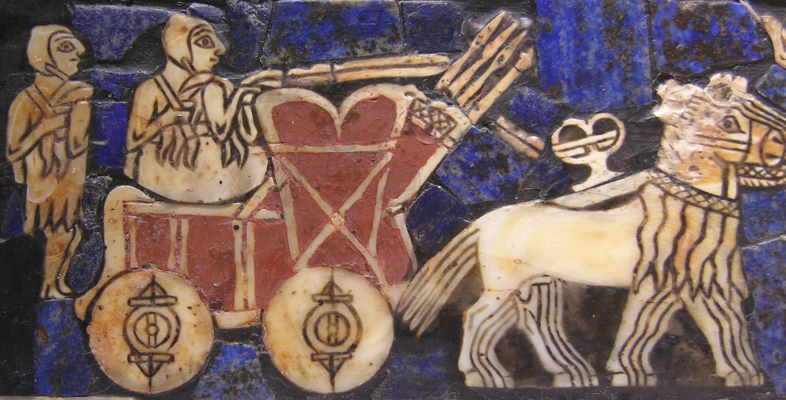
[2] According
to: http://www.britishmuseum.org/explor
e/highlights/highlight_objects/me/t/the_
standard_of_ur.aspx 2600-2400
BC According to:
http://sumerianshakespeare.com/687045.ht
ml this image is 4500 years old -
putting it at 2500bce - get more
evidence of age [1] Description
English: detail of the ''Standard of
Ur'', ca. 2500 BC. Date 2500
BC Source
http://www.alexandriaarchive.org/op
encontext/iraq_ghf/ur_standard/ur_standa
rd_8.jpg Author
Anonymous Permission (Reusing
this file) See below. PD
source: http://upload.wikimedia.org/wiki
pedia/commons/7/7d/Ur_chariot.jpg
[3000 BC]
and harp).

[1] Bearded Harpists, detail from
Sumerian tablet in the Temple of Sin in
Khafage, Mesopotamia (presently Iraq) c
3000 BC. Reprinted by permission
from The Harp by Rajka
Dobronic-Mazzoni. Published by Graficki
Zavrod Hrvatske, OOUR, Izdavcka
djelatnost, Preobrazenska 4, Zagreb,
Croatia, 1989 PD
source: http://www.harpspectrum.org/time
line/images/mesopotamia_1.jpg
[2] Harp-player of Sumer, from a
plaque of Khafaje (After Heras, 1953,
p. 182). PD
source: http://www.hindunet.org/hindu_hi
story/sarasvati/html/HARPPL-1.jpg
[3000 BC]
[1] Fischer, S.R. A History of Writing.
Reaktion, 2001. Globalities Series.
p47-48. COPYRIGHTED
source: Fischer, S.R. A History of
Writing. Reaktion, 2001. Globalities
Series. p47-48.
[2] Source:
http://pandora.cii.wwu.edu/vajda/ling201
/writingsystems/sumeriancuneiform.htm U
NKNOWN
source: http://www.omniglot.com/images/w
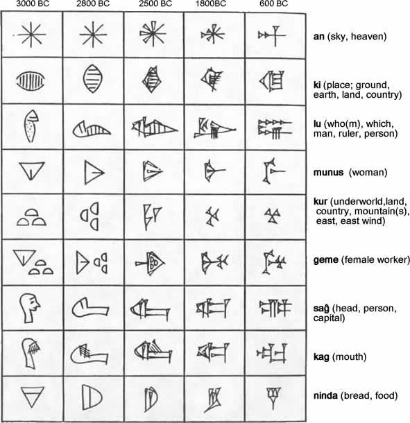
riting/sumerian_glyphs.jpg
[3000 BC]

[1] Tell Judaidah bronze
figurines These figurines of men and
women from Tell Judaidah, Turkey, are
the oldest examples of true bronze
(combination of copper and tin) known.
They date to about 3000 B.C. The male
figures were originally equipped as
warriors, and the women were dressed
with accessories of precious metal.
They are the forerunners of later
figurines of gods who were ''dressed''
in gold and silver. Recently, the ore
content of the figurines was tested at
the Advanced Photon Source at Argonne
National Laboratory. UNKNOWN
source: http://www-news.uchicago.edu/rel
eases/05/050112.oi-3.jpg

[2] Female Figurine Amuq Valley Tell
Judaidah Turkey Amuq G Early Bronze Age
I (3400-2750 BCE)
Bronze Photographed at the Oriental
Institute of the University of Chicago,
Chicago, Illinois. UNKNOWN
source: http://farm3.staticflickr.com/26
18/3859375883_ccc6b90ec4_b.jpg
[3000 BC]

[1] Description A free body
diagram of a mass on an inclined
plane Date 27 May 2007 Source
Own work Author Mets501 CC
source: http://upload.wikimedia.org/wiki
pedia/commons/thumb/8/85/Free_body.svg/1
000px-Free_body.svg.png
[2500 BC]
(modern Turkey)|Palestine|Tell Hammeh
(az-Zarqa), Jordan|Central Europe and
north Assyria

[1] Description Dagger with iron blade
and golden hilt from Alaca Höyük.
Early evidence for the use of iron in
Anatolia. Date 2005 Source Own
work Author Stipich Béla GNU
source: http://upload.wikimedia.org/wiki
pedia/commons/b/b9/Alaca_H%C3%BCy%C3%BCk
_dagger.jpg
[2] Xander Veldhuijzen and Eveline van
der Steen, ''Iron Production Center
Found in the Jordan Valley'', Near
Eastern Archaeology, Vol. 62, No. 3
(Sep., 1999), pp. 195-199 Published
by: The American Schools of Oriental
Research Article Stable URL:
http://www.jstor.org/stable/3210714 COP
YRIGHTED
source: http://www.jstor.org/stable/3210
714
[2300 BC]
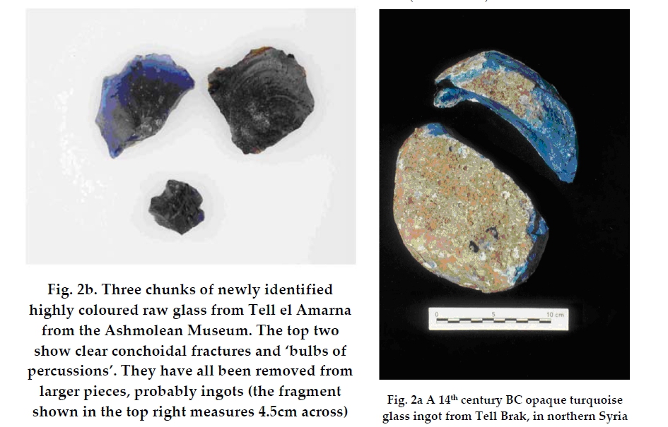
[1] Figures 2b and 2a from: J.
Henderson, J. Evans and K. Nikita,
''ISOTOPIC EVIDENCE FOR THE PRIMARY
PRODUCTION, PROVENANCE AND TRADE OF
LATE BRONZE AGE GLASS IN THE
MEDITERRANEAN'', Mediterranean
Archaeology and Archaeometry, Vol. 10,
No. 1, pp. 1‐24.
2010. http://www.rhodes.aegean.gr/maa_j
ournal/Henderson%2010_1.pdf COPYRIGHTED
source: http://www.rhodes.aegean.gr/maa_
journal/Henderson%2010_1.pdf
[2] Glass ingots (inset) from a Bronze
Age shipwreck near Turkey fit Egyptian
molds. COPYRIGHTED
source: http://www.toutankharton.com/IMG
/jpg/a6260_1449.jpg
[2300 BC]
[1] Photo of Creation and deluge tablet
- I did verify that this is the
earliest tablet of the earliest written
story with Kramer's Sumerian
Mythology[t] Arno Poebel, ''Historical
and grammatical texts'', vols 1-5,
1914. vol 1:
http://books.google.com/books?id=tg0TAAA
AYAAJ vol 4:
http://books.google.com/books?id=mxwYAAA
AYAAJ vol 5:
http://books.google.com/books?id=_A0TAAA
AYAAJ PD
source: http://books.google.com/books?id
=_A0TAAAAYAAJ
[2] Photo of Creation and deluge
tablet - I did verify that this is the
earliest tablet of the earliest written
story with Kramer's Sumerian
Mythology[t] Arno Poebel, ''Historical
and grammatical texts'', vols 1-5,
1914. vol 1:
http://books.google.com/books?id=tg0TAAA
AYAAJ vol 4:
http://books.google.com/books?id=mxwYAAA
AYAAJ vol 5:
http://books.google.com/books?id=_A0TAAA
AYAAJ PD
source: http://books.google.com/books?id
=_A0TAAAAYAAJ
[2130 BC]

[1] [t Note that this is not evidence
of the earliest horn, but is from
around 1250BC or 700 BCE] Hittites:
Musical scene, Carchemish Height:
100 cm, 700 BC. Museum of Anatolian
Civilizations, Ankara Three men are
playing a drum, while on the left a man
is holding a horn-shaped instrument to
his mouth with both hands. PD
source: http://farm1.staticflickr.com/6/
10156251_017f473153_b.jpg
[2000 BC]
River

[1] Ancient wooden lock and key from
Khorsabad (Much reduced) COPYRIGHTED
source: http://www.usgennet.org/usa/topi
c/preservation/science/inventions/chpt8.
htm
[1531 BC]

[1] Description English: Venus Tablet
of Ammisaduqa. Neo-Assyrian
period. Date 15 July 2010 Current
location [show]British
Museum Source/Photographer Fæ (Own
work) Permission (Reusing this
file) See below. British Museum
reference K.160 Detailed
description Upper part of a clay
tablet, 3 pieces, beginning of obverse
and the end of reverse are wanting,
astrological forecasts, a copy of the
so-called Venus Tablet of Ammisaduqa,
Neo-Assyrian. ~ Description extract
from BM record. Size Length: 17.14 cm
(6.75 in) Width: 9.2 cm (3.6 in)
Thickness: 2.22 cm (0.87
in) Location Room 55 CC
source: http://upload.wikimedia.org/wiki
pedia/commons/b/bb/Venus_Tablet_of_Ammis
aduqa.jpg
[1500 BC]

[1] Part of a Bas-relief showing a
Pulley, and a Warrior originally in
the most ancient palace of Nimroud. PD
source: http://www.ctesiphon.com/auction
s/Nineve-Remains-NY-1854-s-g.jpg
[2] Description Ilustración de una
polea simple fija. Date 7 agugust
2004 Source Own work Author César
Rincón GNU
source: http://upload.wikimedia.org/wiki
pedia/commons/a/a3/Polea-simple-fija.jpg
[785 BC]
[1] by Ted Huntington PD
source: my own based on info from
http://www.britannica.com/eb/art-3466?ar
ticleTypeId=1 and
http://nssdc.gsfc.nasa.gov/planetary/fac
tsheet/sunfact.html
[529 BC]
[1] Description: Phytagoras, coin made
under emperor Decius Source:
Baumeister, Denkmäler des klassischen
Altertums. 1888. Band III., Seite
1429 s Roman Emperor from 249 to
251. PD
source: http://www-groups.dcs.st-and.ac.
uk/~history/BigPictures/Pythagoras_4.jpe
g
[2] Bust of Pythagoras UNKNOWN
source: http://www-groups.dcs.st-and.ac.
uk/~history/BigPictures/Pythagoras.jpeg
[467 BC]
known.
Miletus)|Athens|Did not move to Athens
until around 462 bce

[1] Description English: Detail of
the right-hand facade fresco, showing
Anaxagoras. National and Kapodistrian
University of Athens. Date c.
1888 Source http://nibiryukov.narod.r
u/nb_pinacoteca/nbe_pinacoteca_artists_l
.htm Author Eduard Lebiedzki,
after a design by Carl Rahl PD
source: http://upload.wikimedia.org/wiki
pedia/commons/2/2c/Anaxagoras_Lebiedzki_
Rahl.jpg
[467 BC]
communication.

[1] This image was moved from
Image:Image62.gif Description A
drawing of the lighthouse by German
archaeologist Prof. H. Thiersch
(1909). Date 2007-01-16 (original
upload date) Source Originally from
en.wikipedia; description page is/was
here. Author Original uploader
was Ragemanchoo at
en.wikipedia Permission (Reusing this
file) PD
source: http://upload.wikimedia.org/wiki
pedia/commons/2/2e/Lighthouse_-_Thiersch
.gif

[2] English: Mosaic Lighthouse of
Alexandria: was found in the Qasr Libya
in Libya, which was known by several
names including history and Olbia
Theodorias, This is a painting that was
left over to show the form of
lighthouse after the quake, which
destroyed the lighthouse. Qasr Libya
Museum PD
source: http://freespace.virgin.net/ric.
martin/vectis/hookeweb/roberthooke.htm
[460 BC]
atoms.

[1] Coin with the head of Leukippos on
it from around 330-320
BC.[t] Greece,Metapont
330-320BC,Leukkipos,1/3stater. Hammer
price 2002: CHF 12.000. UNKNOWN
source: http://numisbooks.dk/info/fotos/
romanphotos/leukippos330-320.jpg
[260 BC]

[1] Description Español: Esta
imagen ilustra la ventaja mecánica de
la palanca. Deutsch: Illustration des
Hebelgesetzes. Copyright © 2004
César Rincón. Imagen creada para la
Wikipedia en Español. Date
2004-08-05 (first version);
2004-08-07 (last version) Source
Originally from es.wikipedia;
description page is/was here. Author
Original uploader was CR at
es.wikipedia Permission (Reusing this
file) Released under the GNU Free
Documentation License. GNU
source: http://upload.wikimedia.org/wiki
pedia/commons/f/f2/Palanca-ejemplo.jpg

[2] Publishing, DK. Science: The
Definitive Visual Guide. DK Publishing,
2009, p40. COPYRIGHTED
source: Publishing, DK. Science: The
Definitive Visual Guide. DK Publishing,
2009, p40.
[260 BC]

[1] Description Archimedes' screw.
Public domain, from Chambers's
Encyclopedia (Philadelphia: J. B.
Lippincott Company, 1875). Added to
illustrate article en:Archimedes. Date
2007-06-18 (original upload
date) Source Originally from
en.wikipedia; description page is/was
here. Author Original uploader
was Ianmacm at en.wikipedia PD
source: http://upload.wikimedia.org/wiki
pedia/commons/8/82/Archimedes_screw.JPG

[2] Description Deutsch: animierte
Prinzip einer Foerderschnecke oder auch
Archimedesche Spirale genannt, mit
einer Kugel zur Demonstration der
Foerderbewegung. Date published
06.Mai 2007 Source
File:Archimedes-screw_one-screw-thr
eads_with-ball_3D-view_animated.gif
created by Silberwolf Author
Silberwolf (size changed by:
Jahobr) Permission (Reusing this
file) Own work, share alike,
attribution required (Creative Commons
CC-BY-SA-2.5) CC
source: https://upload.wikimedia.org/wik
ipedia/commons/a/a1/Archimedes-screw_one
-screw-threads_with-ball_3D-view_animate
d.gif
[260 BC]
its own axis once a day and around the
Sun once a year is understood.
Egpyt

[1] Aristarchus's 3rd century BC
calculations on the relative sizes of
from left the Sun, Earth and Moon, from
a 10th century CE Greek copy PD
source: http://www.thelivingmoon.com/43a
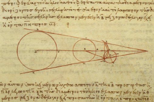
ncients/04images/Artifacts/Aristarchus_w
orking.jpg

[2] Statue of Aristarchus at Aristotle
University in Thessalonica,
Greece UNKNOWN
source: http://www.thelivingmoon.com/43a
ncients/04images/People/Aristarchos_Samo
s.png
[246 BC]
calculated.
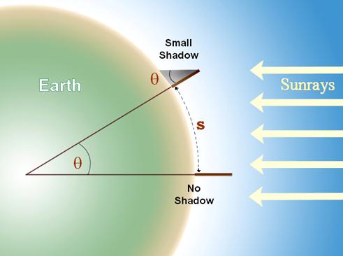
[1] Eratosthenes experiment UNKNOWN
source: http://www.iucaa.ernet.in/~scipo
p/Obsetion/eratos/image008.jpg

[2] Eratosthenes (portrait) Copied
from w:es
Imagen:Eratostenes-retrato.png
(originally from Enciclopedia
Libre) PD
source: http://upload.wikimedia.org/wiki
pedia/commons/a/a2/Portrait_of_Eratosthe
nes.png
[231 BC]
[1] Description Archimedes' screw.
Public domain, from Chambers's
Encyclopedia (Philadelphia: J. B.
Lippincott Company, 1875). Added to
illustrate article en:Archimedes. Date
2007-06-18 (original upload
date) Source Originally from
en.wikipedia; description page is/was
here. Author Original uploader
was Ianmacm at en.wikipedia PD
source: http://upload.wikimedia.org/wiki
pedia/commons/8/82/Archimedes_screw.JPG
[2] Description Deutsch: animierte
Prinzip einer Foerderschnecke oder auch
Archimedesche Spirale genannt, mit
einer Kugel zur Demonstration der
Foerderbewegung. Date published
06.Mai 2007 Source
File:Archimedes-screw_one-screw-thr
eads_with-ball_3D-view_animated.gif
created by Silberwolf Author
Silberwolf (size changed by:
Jahobr) Permission (Reusing this
file) Own work, share alike,
attribution required (Creative Commons
CC-BY-SA-2.5) CC
source: https://upload.wikimedia.org/wik
ipedia/commons/a/a1/Archimedes-screw_one
-screw-threads_with-ball_3D-view_animate
d.gif
[160 BC]
its motion).
Nicaea)|(observatory on) Island of
Rhodes, Greece

[1] Hipparchus (196 BC – 120 BC) was
born in Nicaea (now Iznik, Turkey), and
probably died on the island of Rhodes.
He is known to have been a working
astronomer at least from 147 BC to 127
BC. UNKNOWN
source: http://web.jccc.edu/gallery/astr
otext/Bills%20Files/Astronomy%20Textbook
/Chapter%203_files/img12qwe.jpg
[2] image of Hipparchos from coin?
http://www-history.mcs.st-and.ac.uk/hist
ory/Mathematicians/Hipparchus.html PD
source: http://en.wikipedia.org/wiki/Ima
ge:Hipparchos_1.jpeg
[140 BC]
province of China|Xian, China

[1] Description Early Chinese hemp
fiber paper, used for wrapping not
writing, on display at the Shaanxi
history museum in Xi'An, China.
Excavated from the Han Tomb of Wu Di
(140-87 BC) at Baqiao, Xi'An. Photo by
Yannick Trottier, 2007 Date 22
June 2007 Source Own work Author
Ytrottier GNU
source: http://upload.wikimedia.org/wiki
pedia/commons/7/7f/Chinese_hemp_paper_we
stern_han.jpg

[2] It's the earliest Paper in the
world : Western Han (140-87
BC) UNKNOWN
source: http://www.amateras.com/trip/chi
na/12Sha-Paper360x240.jpg
[56 BC]
atoms that move very fast.

[1] Text copied from: Titus Carus
Lucretius, ''T. Lucreti Cari De rerum
natura libri sex, Volume 1'', 1866,
lines 176-229,
p530 http://books.google.com/books?id=o
iUTAAAAQAAJ PD
source: http://books.google.com/books?id
=oiUTAAAAQAAJ

[2] Text copied from: Titus Carus
Lucretius, ''T. Lucreti Cari De rerum
natura libri sex, Volume 1'', 1866,
lines 176-229,
p530 http://books.google.com/books?id=o
iUTAAAAQAAJ PD
source: http://books.google.com/books?id
=oiUTAAAAQAAJ
[50 AD]

[1] Name of Image: Hero's Engine MIX
#: 9513982 NIX #: MSFC-9513982 Date
of Image: 2004-04-15 Category: Early
Rockets Full Description:
Legendary characters used the power of
mythology to fly through the heavens.
About 200 BC, a Greek inventor known as
Hero of Alexandria came up with a new
invention that depended on the
mechanical interaction of heat and
water. He invented a rocket-like device
called an aeolipile. It used steam for
propulsion. Hero mounted a sphere on
top of a water kettle. A fire below the
kettle turned the water into steam, and
the gas traveled through the pipes to
the sphere. Two L-shaped tubes on
opposite sides of the sphere allowed
the gas to escape, and in doing so gave
a thrust to the sphere that caused it
to rotate. (MRPO) MRD/SPD
Discipline(s): n/a (MRPO) Subject
Type: n/a Keywords: Hero's Engine,
Aeolipile MSFC Negative Number:
9513982 Reference Number:
MSFC-75-SA-4105-2C n/a
n/a from: http://mix.msfc.nasa.gov/ab
stracts.php?p=1867 PD
source: http://mix.msfc.nasa.gov/IMAGES/
HIGH/9513982.jpg
[2] Hero's aeolipile From Knight's
American Mechanical Dictionary, 1876.
PD
source: http://en.wikipedia.org/wiki/Ima
ge:Aeolipile_illustration.JPG
[83 AD]

[1] Figure from: Joseph Needham,
''Science and Civilization in China'',
vol 4, part 1, 1962,
p230-268. {Needham_China_compass_1962.p
df} COPYRIGHTED
source: Joseph Needham, "Science and
Civilization in China", vol 4, part 1,
1962, p230-268.
[2] ''The south-pointing fish'' was
recorded in the documents of the
Northern Song Dynasty. Such
direction-pointing device is a thin
steel plate cut into the shape of a
fish magnetized in the geomagnetic
field. The tail of the fish is
magnetized in the geological direction
of the North Pole, thus the tail has
the south magnetic pole and the head of
the fish has the north magnetic pole.
When put into the water, the floating
fish has its head pointing to the
south. UNKNOWN
source: http://kaleidoscope.cultural-chi
na.com/chinaWH/images/exbig_images/3ee20
b9ad9430ca4fcd43b3165a315c5.jpg
[600 AD]

[1] (Images via: Ullesthorpe,
BluePlanet, DeutschesMuseum and
WorldofEnergy) UNKNOWN
source: http://cdn.webecoist.com/wp-cont
ent/uploads/2009/01/ancient-persian-wind
mills.jpg
[850 AD]

[1] Description The earliest known
written description of the formula for
gunpowder, from the Chinese Wujing
Zongyao military manuscript that was
compiled by 1044 during the Song
Dynasty of China. It was written and
compiled by the 11th century Song
scholars Zeng Gongliang (曾公亮),
Ding Du (丁度), and Yang Weide
(楊惟德). The entry for this
specific page is headed with the title
''method for making the fire-chemical''
(''huo yao fa''). This picture can
also be found on page 119 of Joseph
Needham's book Science and Civilization
in China: Volume 5, Part 7. Date
11 August 2007 Source Own
work (My book) Author
PericlesofAthens Permission (Reus
ing this file) See below. PD
source: http://upload.wikimedia.org/wiki
pedia/commons/c/c2/Chinese_Gunpowder_For
mula.JPG
[920 AD]
America.
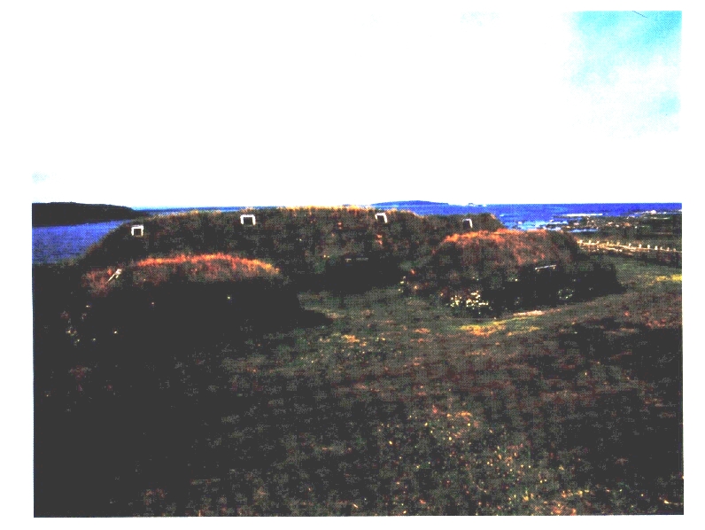
[1] Figure from: Helge Ingstad, ''The
Viking Discovery of America: The
Excavation of a Norse Settlement in
L'Anse aux Meadows, Newfoundland'',
2001. COPYRIGHTED
source: Helge Ingstad, "The Viking
Discovery of America: The Excavation of
a Norse Settlement in L'Anse aux
Meadows, Newfoundland", 2001.

[2] Figure 24 from: Helge Ingstad,
''The Viking Discovery of America: The
Excavation of a Norse Settlement in
L'Anse aux Meadows, Newfoundland'',
2001. COPYRIGHTED
source: Helge Ingstad, "The Viking
Discovery of America: The Excavation of
a Norse Settlement in L'Anse aux
Meadows, Newfoundland", 2001.
[960 AD]
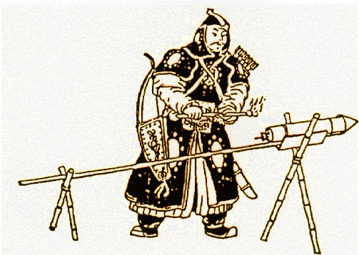
[1] Description Drawing of an
early Chinese soldier lighting a
rocket Date 2007 Source
http://history.msfc.nasa.gov/rocket
ry/03.html Author
NASA Permission (Reusing this
file) NASA still images, audio
files and video generally are not
copyrighted. You may use NASA imagery,
video and audio material for
educational or informational purposes,
including photo collections, textbooks,
public exhibits and Internet Web
pages. PD
source: http://upload.wikimedia.org/wiki
pedia/commons/6/63/Chinese_rocket.gif

[2] Widely reputed as the world's
first ''astronaut'', Wan Hu was a minor
Chinese official of the Ming Dynasty
(1368-1644). Early in the 16th century,
Wan Hu decided to take advantage of
China's advanced power and fireworks
technology to launch himself into outer
space. He had a chair built with 47
''rockets'' attached. On the day of
lift-off, Wan climbed into his rocket
chair and held one enormous kite in
each hand. The ignition of the 47 fuses
caused a huge explosion and sent him
into the sky. But unfortunately, he
failed to go into orbit and his body
smashed into pieces on the ground.
UNKNOWN
source: http://images.china.cn/images1/2
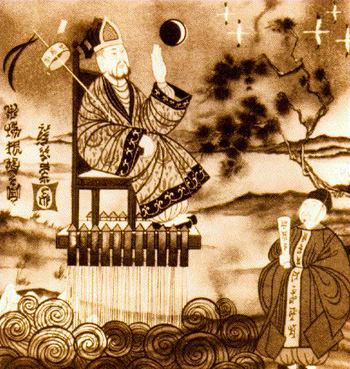
00710/410673.jpg
[1132 AD]
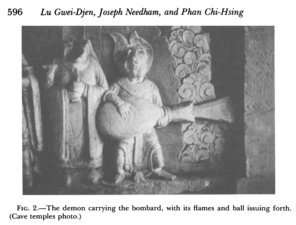
[1] Figure 2 from: Gwei-Djen, Lu,
Joseph Needham, and Phan Chi-Hsing.
“The Oldest Representation of a
Bombard.” Technology and Culture 29.3
(1988): 594–605.
Print. http://www.jstor.org/stable/3105
275 {Gwei-Djen_1988.pdf} COPYRIGHTED
source: Gwei-Djen, Lu, Joseph Needham,
and Phan Chi-Hsing. “The Oldest
Representation of a Bombard.”
Technology and Culture 29.3 (1988):
594–605.
Print. http://www.jstor.org/stable/3105
275

[2] Figure 3 from: Gwei-Djen, Lu,
Joseph Needham, and Phan Chi-Hsing.
“The Oldest Representation of a
Bombard.” Technology and Culture 29.3
(1988): 594–605.
Print. http://www.jstor.org/stable/3105
275 {Gwei-Djen_1988.pdf} COPYRIGHTED
source: Gwei-Djen, Lu, Joseph Needham,
and Phan Chi-Hsing. “The Oldest
Representation of a Bombard.”
Technology and Culture 29.3 (1988):
594–605.
Print. http://www.jstor.org/stable/3105
275
[1228 AD]
light.

[1] Record Number: 19885 Shelfmark:
Royal 6 E. V Page Folio Number:
f.6 Description: [Miniature only]
Initial 'A', portrait of Robert
Grosseteste, Bishop of Lincoln. The
beginning of one of the bishop's
sermons Title of Work: Works of
Robert Grosseteste, Bishop of
Lincoln Author: Grosseteste,
Robert Illustrator: - Production:
England; 15th
century Language/Script: Latin /
- PD
source: http://www.imagesonline.bl.uk/br
itishlibrary/controller/textsearch?text=
grosseteste&y=0&x=0&startid=31330&width=
4&height=2&idx=2

[2] Robert Grosseteste (1168-1253).
Scientist, Philosopher, Bishop of
Lincoln. PD
source: http://commons.wikimedia.org/wik
i/Image:Grosseteste-color.png
[10/12/1492 AD]

[1] Artist [show]Sebastiano del
Piombo (1485–1547) Link back to
Creator infobox template
wikidata:Q285423 Description
Christopher Columbus Date
1519 Medium painting Current
location [show]Metropolitan Museum
of ArtLink back to Institution infobox
template wikidata:Q160236 Inscriptions
Text top center Source/Photographer
http://www.metmuseum.org/works_of_art/c
ollection_database/european_paintings/po
rtrait_of_a_man_said_to_be_christopher_c
olumbus_sebastiano_del_piombo_sebastiano
_luciani/objectview.aspx?page=1&sort=6&s
ortdir=asc&keyword=Piombo&fp=1&dd1=11&dd
2=0&vw=0&collID=11&OID=110002098&vT=1&hi
=0&ov=0 PD
source: http://upload.wikimedia.org/wiki
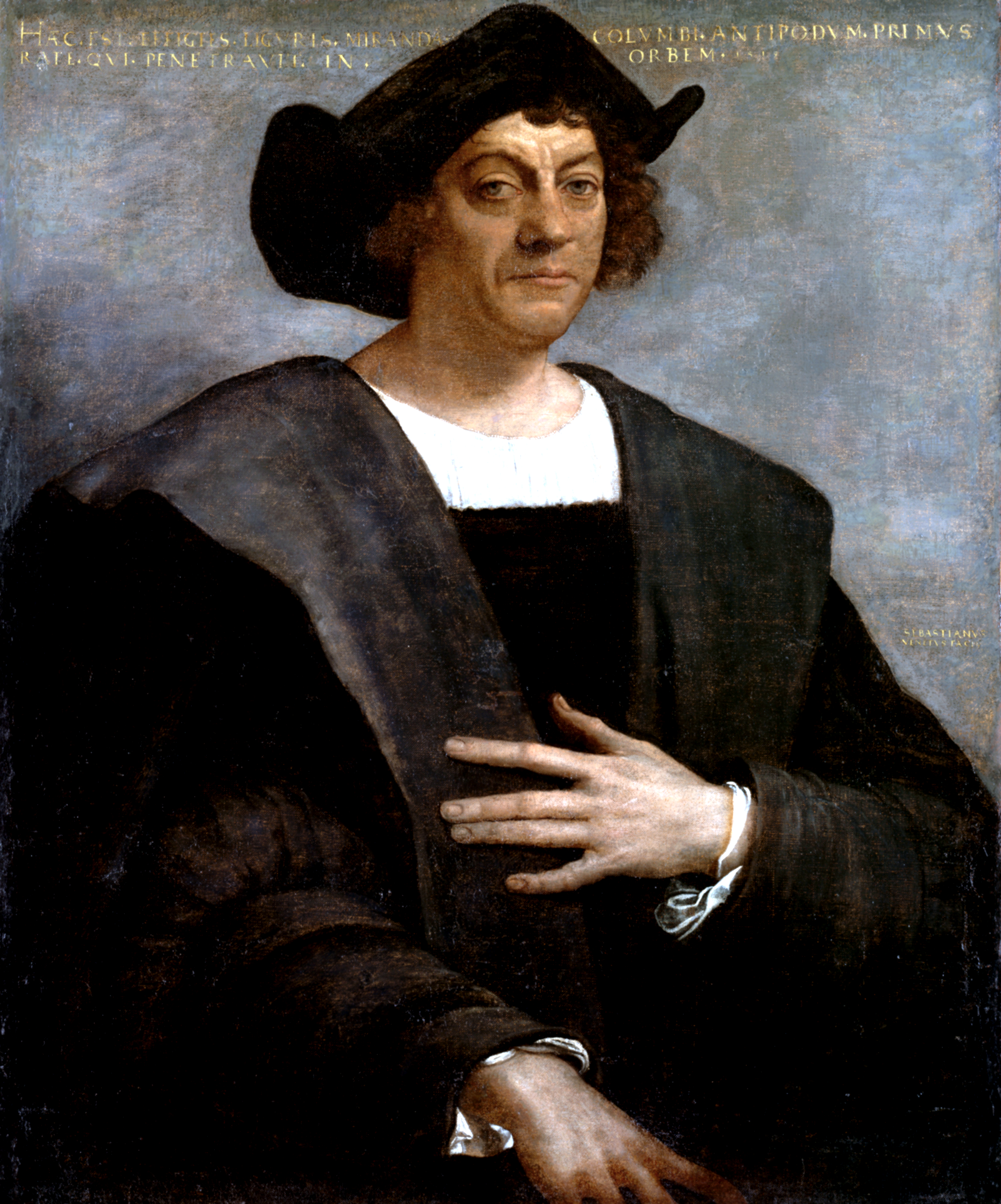
pedia/commons/5/5d/Christopher_Columbus.
PNG
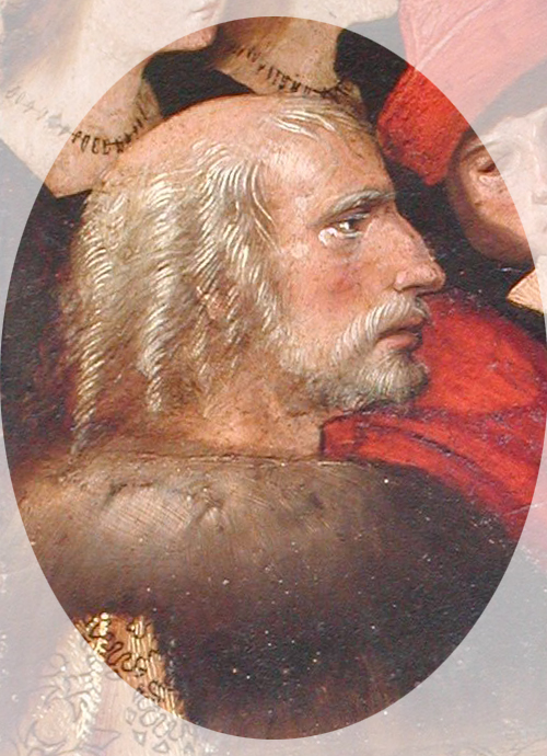
[2] Portrait of Christopher Columbus
from the painting Virgen de los
Navegantes (in the Sala de los
Almirantes, Royal Alcazar, Seville). A
painting by Alejo Fernández between
1505 and 1536. It is the only state
sponsored portrait of the First Admiral
of the Indias. Photo by a Columbus
historian, Manuel Rosa. More info
http://www.UnmaskingColumbus.com PD
source: http://en.wikipedia.org/wiki/Ima
ge:Christopher_Columbus_Face.jpg
[09/08/1522 AD]
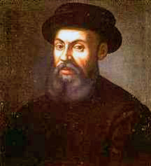
[1] An anonymous portrait of Ferdinand
Magellan, 16th or 17th century (The
Mariner's Museum Collection, Newport
News, VA) PD
source: http://en.wikipedia.org/wiki/Ima
ge:Ferdinand_Magellan.jpg
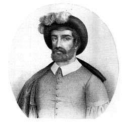
[2] Juan Sebastián
Elcano Litografía de J. Donon en
Historia de la Marina Real Española.
Madrid,
1854 http://marenostrum.org/bibliotecad
elmar/historia/pacifico/ PD
source: http://en.wikipedia.org/wiki/Ima
ge:Elcano.jpg
[1592 AD]

[1] Fig. 1. Galileo’s
thermoscope. from: David Sherry,
Thermoscopes, thermometers, and the
foundations of measurement, Studies In
History and Philosophy of Science Part
A, Volume 42, Issue 4, December 2011,
Pages 509-524, ISSN 0039-3681,
10.1016/j.shpsa.2011.07.001. (http://ww
w.sciencedirect.com/science/article/pii/
S0039368111000616) UNKNOWN
source: http://www.sciencedirect.com/cac
he/MiamiImageURL/1-s2.0-S003936811100061
6-gr1.jpg/0?wchp=dGLzVBA-zSkzS

[2] Thermoscope Instrument to
measure heat and cold invented by
Galileo Galilei (1564-1642) during his
stay in Padua. Santorio Santorio
(1561-1636) made a similar instrument
in Venice in 1612. A precursor of the
modern thermometer, the thermoscope
consists of a glass vessel with a long
neck. The vessel was heated with the
hands and partially immersed, in an
upright position, in a container full
of water. When the heat of the hands
was taken away, the water was observed
to rise in the thermoscope neck. The
experiment showed the changes in air
density produced by variations in
temperature. UNKNOWN
source: http://catalogue.museogalileo.it
/images/cat/approfondimenti_944/AF0020-5
1000_944.jpg
[1608 AD]
Netherlands)
[1] Hans Lippershey (1570-September
1619), Dutch lensmaker. PD
source: http://en.wikipedia.org/wiki/Ima
ge:Hans_Lippershey.jpg

[2] Description English: Early
depiction of a ‘Dutch telescope’
from the “Emblemata of zinne-werck”
(Middelburg, 1624) of the poet and
statesman Johan de Brune (1588-1658).
The print was engraved by Adriaen van
de Venne, who, together with his
brother Jan Pieters van de Venne,
printed books not far from the original
optical workshop of Hans
Lipperhey. Date 1624 Source
http://www.phys.uu.nl/~vgent/telesc
ope/telescopenl.htm Author Adriaen
Pietersz. van de Venne (1589–1662)
Link back to Creator infobox
template PD
source: http://upload.wikimedia.org/wiki
pedia/commons/5/51/Emblemata_1624.jpg
[1609 AD]
orbits is understood.
Stuttgart Region in the German state of
Baden-Württemberg, 30 km west of
Stuttgart's center)

[1] Johannes Kepler, ''Astronomia
nova'', 1609,
p267. http://www.e-rara.ch/zut/content/
titleinfo/162514 {Astronomia_nova_seu_p
hysica_coelestis_tradita_commentariis_de
_motibus_stellae_m.pdf} PD
AND Description English: Portrait
of Johannes Kepler. Date 8 March 2006
(original upload date) Source
Transferred from en.wikipedia Author
Original uploader was Brandmeister at
en.wikipedia Permission (Reusing this
file) PD-US; PD-ART. PD
source: http://www.e-rara.ch/zut/content
/titleinfo/162514http://upload.wikimedia
.org/wikipedia/commons/7/74/JKepler.jpg
[2] Johannes Kepler, ''Astronomia
nova'', 1609,
p267. http://www.e-rara.ch/zut/content/
titleinfo/162514 {Astronomia_nova_seu_p
hysica_coelestis_tradita_commentariis_de
_motibus_stellae_m.pdf} PD
source: http://www.e-rara.ch/zut/content
/titleinfo/162514
[01/??/1610 AD]
period determined.
Italy
[1] Galileo's Letter to Prince of
Venice PD
source: http://www2.jpl.nasa.gov/galileo
/ganymede/manuscript1.jpg
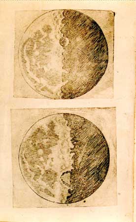
[2] Galileo's illustrations of the
Moon, from his Sidereus Nuncius (1610;
The Sidereal Messenger). Courtesy of
the Joseph Regenstein Library, The
University of Chicago PD
source: http://www.britannica.com/eb/art
-2914/Galileos-illustrations-of-the-Moon
-from-his-Sidereus-Nuncius?articleTypeId
=1
[1610 AD]
Netherlands)

[1] The microscope was first built in
1595 by Hans and Zacharias Jansen
(1588-1631) in Holland (see figure).
source: http://www.cerebromente.org.br/n
17/history/jansen-micro.JPG

[2] Description Portrait of Zacharias
Jansen Date 1655 Source Pierre
Borel, De vero telescopii
inventore Author Pierre Borel
source: http://upload.wikimedia.org/wiki
pedia/commons/3/3b/Zacharias.jpg
[1631 AD]

[1] Pierre Gassendi
(1592-1655). Peinture de Louis
Édouard Rioult. (Base Joconde du
Ministère de la Culture) PD
source: http://www.voltaire-integral.com
/Html/14/04CATALO_1_2.html

[2] Scientist: Gassendi, Pierre
(1592 - 1655) Discipline(s): Physics
; Astronomy Print Artist: Jacques
Lubin, 1637-1695 Medium: Engraving
Original Dimensions: Graphic: 17.6 x
14.1 cm / Sheet: 27.9 x 21.7 cm PD
source: http://www.sil.si.edu/digitalcol
lections/hst/scientific-identity/CF/by_n
ame_display_results.cfm?scientist=Gassen
di
[1635 AD]
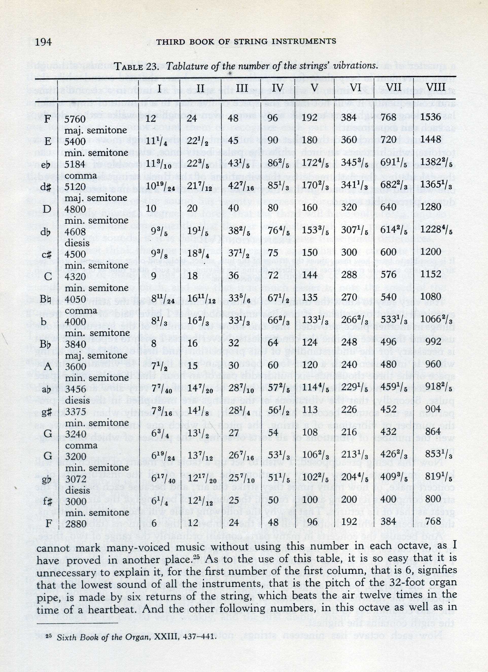
[1] Table of string
vibrations from: Marin Marsenne, tr:
R. E. Chapman, ''Harmonie
Universelle'', 1635, 1957,
p194. UNKNOWN
source: Marin Marsenne, tr: R. E.
Chapman, "Harmonie Universelle", 1635,
1957, p194.

[2] Ted Huntington adapted
from: http://upload.wikimedia.org/wikip
edia/en/math/6/c/8/6c88fce3e57d1eac8408b
abe264e1795.png GNU
source: http://upload.wikimedia.org/wiki
pedia/en/math/6/c/8/6c88fce3e57d1eac8408
babe264e1795.png
[1643 AD]
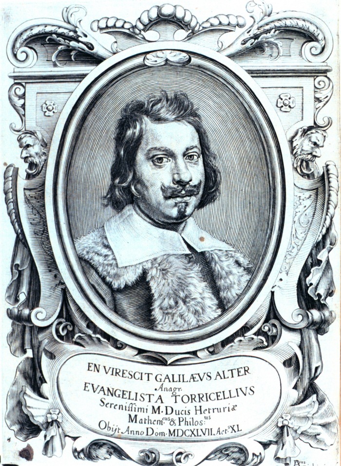
[1] Frontispiece to ''Lezioni
accademiche d'Evangelista
Torricelli....'', published in 1715.
Library Call Number Q155 .T69
1715. Image ID: libr0367, Treasures of
the NOAA Library Collection
Photographer: Archival Photograph by
Mr. Steve Nicklas, NOS, NGS Secondary
source: NOAA Central Library National
Oceanic & Atmospheric Adminstration
(NOAA), USA
http://www.photolib.noaa.gov/library/lib
r0367.htm PD
source: http://en.wikipedia.org/wiki/Ima
ge:Libr0367.jpg

[2] Frontispiece and title page to
''Lezioni accademiche d'Evangelista
Torricelli ....'', published in 1715.
Library Call Number Q155 .T69
1715. Image ID: libr0366, Treasures of
the NOAA Library Collection
Photographer: Archival Photograph by
Mr. Steve Nicklas, NOS, NGS Secondary
source: NOAA Central Library National
Oceanic & Atmospheric Adminstration
(NOAA),
USA http://www.photolib.noaa.gov/librar
y/libr0366.htm PD
source: http://commons.wikimedia.org/wik
i/Image:Libr0366.jpg
[1663 AD]
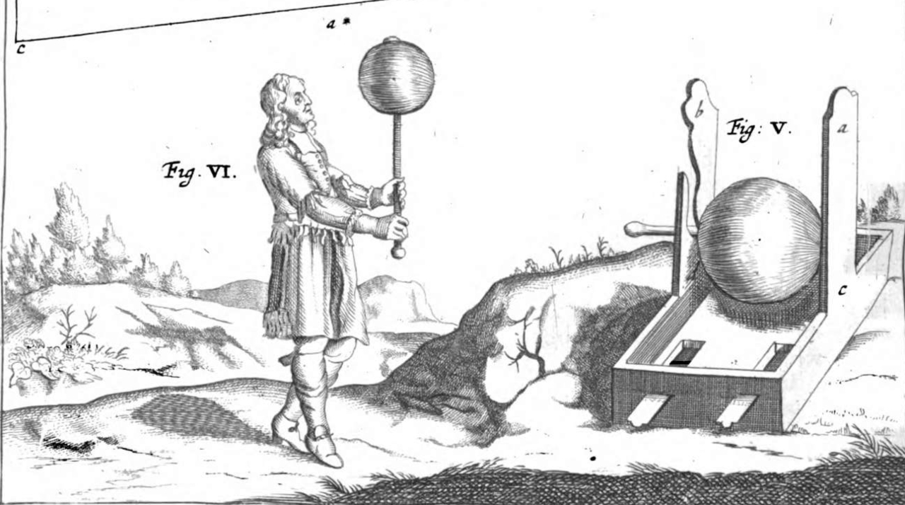
[1] Guericke's experiments with the
sulfur globe published 1672 PD
source: http://img.readtiger.com/wkp/en/
Guericke_Sulfur_globe.jpg

[2] Guericke's experiments with the
sulfur globe published 1672 PD
source: http://img.readtiger.com/wkp/en/
Guericke_Sulfur_globe.jpg
[1676 AD]
[1] ''Demonstration touchant le
mouvement de la lumiere trouvé par M.
Römer de l' Academie Royale des
Sciences'', Journal des sçavans,
December 7,
1676 http://books.google.com/books?id=5
scUAAAAQAAJ&pg=PA484 PD
source: http://books.google.com/books?id
=5scUAAAAQAAJ&pg=PA484

[2] Ole Rømer PD
source: http://www.rundetaarn.dk/dansk/o
bservatorium/grafik/roemer1.jpg
[1678 AD]
contracts muscle with electricity.
(presumably)
[1] One of Galvani’s decisive
experiments was to show that movement
could be induced by stroking an iron
plate against a brass hook inserted
into the frog’s spinal column, which
generated a small electric current. In
one version of Swammerdam’s nerve
muscle experiment, the nerve was
suspended in a brass hook, which was
then stroked with a silver
wire: PD/Corel
source: http://www.janswammerdam.net/Ima
ges/Fig4.jpg
[1687 AD]
attracts with a force that is the
product of their masses, and the
inverse of their distance squared.

[1] Sir Isaac Newton's own first
edition copy of his Philosophiae
Naturalis Principia Mathematica with
his handwritten corrections for the
second edition. The first edition was
published under the imprint of Samuel
Pepys who was president of the Royal
Society. By the time of the second
edition, Newton himself had become
president of the Royal Society, as
noted in his corrections. The book can
be seen in the Wren Library of Trinity
College, Cambridge. CC
source: http://en.wikipedia.org/wiki/Ima
ge:NewtonsPrincipia.jpg
[2] Description Isaac Newton Date
1689 Author Godfrey Kneller PD
source: http://en.wikipedia.org/wiki/Ima
ge:GodfreyKneller-IsaacNewton-1689.jpg
[11/04/1745 AD]
Germany(was for von Kleist: Pomerania?,
Prussia) (coast of Baltic Sea between
Germany and Poland)
[1]
http://books.google.com/books?id=ko9BAAA
AIAAJ&pg=PA71&dq=jar+%22von+Kleist%22&lr
=&as_brr=1&ei=aniTR_uCJ5HwsgOQ5bU4#PPA71
,M1 page with text and figure about
von Kleist's invention of the Leyden
jar Source Electricity in Every-day
Life Date 1905 Author Edwin J.
Houston PD
source: http://en.wikipedia.org/wiki/Ima
ge:Von_Kleist_Leyden_jar_1905.png

[2] Pieter van Musschenbroek aus:
http://20eeuwennederland.nl/actueel/1113
.htm PD
source: http://en.wikipedia.org/wiki/Ima
ge:Pieter_van_Musschenbroek.jpeg
[1769 AD]
[1] Nicolas-Joseph Cugnot's steam auto,
from 7 August, 1869 issue of Appleton's
Journal of Popular Literature, Science,
and Art. PD
source: http://en.wikipedia.org/wiki/Ima
ge:CugnotAppleton.jpg

[2] Fardier de Cugnot, modèle de
1771. Musée des Arts et Métiers,
Paris. 11 janvier 2005. (Note that
this is the second fardier, the
full-size one. It is not a 'model' (as
has been mis-translated
elsewhere)) Source : Photo et
photographisme © Roby 19:13, 12 Jan
2005 (UTC). Avec l'aimable permission
du Musée des Arts et Métiers, Paris.
GNU
source: http://upload.wikimedia.org/wiki
pedia/commons/5/56/FardierdeCugnot200501
11.jpg
[1791 AD]
contracted remotely by light
particles).

[1] Italian physicists Luigi
Galvani Source
http://www.museopalazzopoggi.unibo.it
//poggi_eng/palazzo/foto/prot Date
18-19 th century Author
Unknown PD
source: http://en.wikipedia.org/wiki/Ima
ge:Luigi_Galvani%2C_oil-painting.jpg
[2] The electrochemical behavior of
two dissimilar metals [(zinc (Z) and
copper (C)] in a bimetallic arch, in
contact with the electrolytes of
tissue, produces an electric
stimulating current that elicits
muscular contraction. [Malmivuo, J., &
Plonsey, R. (1995).
Bioelectromagnatism: Principles and
applications of bioelectric and
biomagnetic fields. New York: Oxford
University Press., Ch.1] URL:
http://butler.cc.tut.fi/~malmivuo/bem/be
mbook/01/01.htm Diagram of Luigi
Galvani's frog legs (~1770s) PD
source: http://en.wikipedia.org/wiki/Ima
ge:Galvani%27s_legs.gif
[03/20/1800 AD]

[1] Volta, ''On the Electricity excited
by the mere Contact of conducting
Substances of different Kinds.'',
Philosophical Magazine, September 1800,
p415. http://archive.org/download/lepid
opterarepor07winc/lepidopterarepor07winc
.pdf PD
source: http://archive.org/download/lepi
dopterarepor07winc/lepidopterarepor07win
c.pdf

[2] Description Alessandro Giuseppe
Antonio Anastasio Volta Source
http://www.anthroposophie.net/bibliot
hek/nawi/physik/volta/bib_volta.htm Dat
e 2006-03-02 (original upload
date) PD
source: http://en.wikipedia.org/wiki/Ima
ge:Alessandro_Volta.jpeg
[03/27/1800 AD]
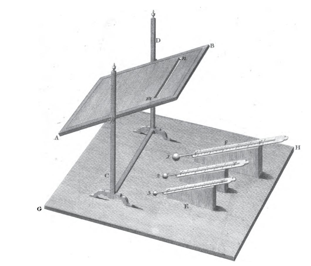
[1] William Herschel, ''Investigation
of the Powers of the Prismatic Colours
to Heat and Illuminate Objects; With
Remarks, That Prove the Different
Refrangibility of Radiant Heat. To
Which is Added, an Inquiry into the
Method of Viewing the Sun
Advantageously, with Telescopes of
Large Apertures and High Magnifying
Powers.'', Philosophical Transactions
of the Royal Society of London , Vol.
90, (1800), pp. 255-283.
books.google.com/books?id=dlFFAAAAcAAJ
&pg=PA255 PD
source: books.google.com/books?id=dlFFAA
AAcAAJ&pg=PA255
[2] Description Wilhelm Herschel,
German-British
astronomer. Date 1785 Source Nat
ional Portrait Gallery, London: NPG
98 Author Lemuel Francis Abbott PD
source: http://upload.wikimedia.org/wiki
pedia/commons/3/36/William_Herschel01.jp
g
[11/12/1801 AD]
[1] {ULSF: Table of light wavelengths
and frequencies calculated by Young
from Theory of Light and Colours
11/12/1801} The inch used in the table
is the French (Paris) inch of
27.07mm. PD/Corel AND Portrait of
Thomas Young in color PD
source: Young_Thomas_1802_on_the_theory_
of_light_and_colours.pdfhttp://ugadayki.
ru/images/sv/133163204157_full.jpg
[2] {ULSF: Table of light wavelengths
and frequencies calculated by Young
from Theory of Light and Colours
11/12/1801} The inch used in the table
is the French (Paris) inch of
27.07mm. PD/Corel
source: Young_Thomas_1802_on_the_theory_
of_light_and_colours.pdf
[1809 AD]

[1] Humphry Davy demonstrates his new
electric light for the members of the
Royal Institution of London. Power is
drawn from the banks of batteries in
the basement and rapidly used up by the
intense light. Electric light was then
only a scientific curiosity, practical
only when expense was no
object. Humphry Davy Demonstrating the
Arc Light, 1809 PD/COPYRIGHTED
source: http://people.clarkson.edu/%7Eek
atz/scientists/davy.htm
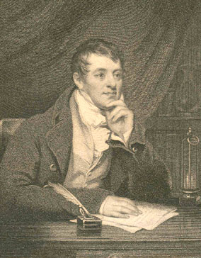
[2]
http://www.nndb.com/people/028/000083776
/humphry-davy-2-sized.jpg [left finger
1: ''left'' viewed as educated
intellectuals in 1800s England? just
coincidence?] PD
source: http://en.wikipedia.org/wiki/Ima
ge:Sir_Humphry_Davy2.jpg
[11/??/1815 AD]
multiples of hydrogen.

[1] William Prout (published
anonymously), On the Relation between
the Specific Gravities of Bodies in
their Gaseous State and the Weights of
their Atoms. Annals of Philosophy 6,
p321-330
(1815). books.google.com/books?id=tDowA
AAAYAAJ PD
source: http://books.google.com/books?id
=tDowAAAAYAA

[2] William Prout
(1785-1850) PD/COPYRIGHTED
source: http://www.uam.es/departamentos/
ciencias/qorg/docencia_red/qo/l0/1830.ht
ml
[04/21/1820 AD]
magnetism. First electromagnet.

[1] A younger Hans Christian Ørsted,
painted in the 19th century. PD
source: http://en.wikipedia.org/wiki/Ima
ge:%C3%98rsted.jpg
[2] Picture number :317 CD number
:9 Picture size :757x859[pixels],
66x75[mm] Date taken :0000-00-00
Date added
:2000-04-13 Fotographer/Owner :Engrave
d Location
:Denmark Description H.C. Oersted
(1777-1851). Danish physicist. Here as
a youngster. The picture was donated to
the Danish Polytech Institute,
Copenhagen, by his daughter Miss
Mathilde Oersted, April 19,
1905. PD/COPYRIGHTED
source: http://www.polytechphotos.dk/ind
ex.php?CHGLAN=2&CatID=286
[1820 AD]
England
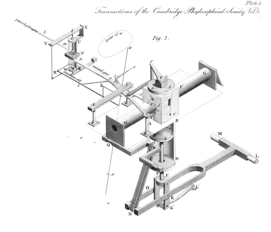
[1] W. Cecil's hydrogen combustion
vacuum engine PD/Corel
source: http://www.eng.cam.ac.uk/DesignO
ffice/projects/cecil/images/isometricalv
iew.jpg
[2] Cecil's figures PD/Corel
source: http://books.google.com/books?id
=hgYFAAAAQAAJ&printsec=frontcover&dq=edi
tions:0iE3HbhCd9wmSagF2t&as_brr=1#PPA230
,M1
[09/11/1821 AD]
England
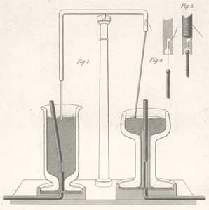
[1] The first electric motors - Michael
Faraday, 1821 From the Quarterly
Journal of Science, Vol XII, 1821 PD
source: http://www.sparkmuseum.com/MOTOR
S.HTM
[2] Description Michael Faraday,
oil, by Thomas Phillips Source
Thomas Phillips,1842 Date
1842 Author Thomas Phillips[3
wiki] The portrait shown here was
painted by Thomas Phillips (1770-1845),
oil on canvas, The National Portrait
Gallery, London.[7] PD
source: http://en.wikipedia.org/wiki/Ima
ge:M_Faraday_Th_Phillips_oil_1842.jpg
[1826 AD]

[1] English: By Nicéphore Niépce in
1826, entitled ''View from the Window
at Le Gras,'' captured on 20 × 25 cm
oil-treated bitumen. Due to the 8-hour
exposure, the buildings are illuminated
by the sun from both right and left.
This photo is generally considered the
first successful permanent
photograph. PD
source: http://en.wikipedia.org/wiki/Ima
ge:View_from_the_Window_at_Le_Gras%2C_Jo
seph_Nic%C3%A9phore_Ni%C3%A9pce.jpg

[2] Joseph-Nicéphore Niépce. ©
Bettmann/Corbis PD/COPYRIGHTED
source: http://concise.britannica.com/eb
c/art-59378/Joseph-Nicephore-Niepce
[03/27/1829 AD]
wire near a magnet.
[1] Francesco Zantedeschi PD/Corel
source: http://www.liceofoscarini.it/sto
ria/bio/zantedeschi.html
[2] Image of Francesco Zantedeschi
1797 to 1873 to illustrate that
article. Uploaded from
http://www.jergym.hiedu.cz/~canovm/objev
ite/objev4/zan.htm and
http://www.jergym.hiedu.cz/~canovm/objev
ite/objev4/zan2.htm (English
translation) This portrait of
Francesco Zantedeschi was published by
Stefano de Stefani, president of the
Academy of Agriculture, Arts and
Commerce of Verona, on March 21, 1875
to accompany his eulogy to Zantedeschi
on the occasion of the transport of his
ashes to the cemetery at Verona. Black
and white version PD
source: http://en.pedia.org//Image:Franc
esco_Zantedeschi_bw.jpg
[1830 AD]
Germany

[1] Wilhelm Eduard Weber
(1804-1891) PD
source: http://en.wikipedia.org/wiki/Ima
ge:Wilhelm_Eduard_Weber_II.jpg
[02/17/1831 AD]
England

[1] Michael Faraday, ''Experimental
Researches in Electricity'',
Philosophical Transactions of the Royal
Society of London (1776-1886), Volume
122, 1832. DOI 10.1098/rstl.1832.0006
{Faraday_1832_Experimental_Researches_
in_Electricity_1.pdf} PD
source: Faraday_1832_Experimental_Resear
ches_in_Electricity_1.pdf
[2] Description Michael Faraday,
oil, by Thomas Phillips Source
Thomas Phillips,1842 Date
1842 Author Thomas Phillips[3
wiki] The portrait shown here was
painted by Thomas Phillips (1770-1845),
oil on canvas, The National Portrait
Gallery, London.[7] PD
source: http://en.wikipedia.org/wiki/Ima
ge:M_Faraday_Th_Phillips_oil_1842.jpg
[09/??/1831 AD]
(constant current produced).
England
[1] Description Michael Faraday,
oil, by Thomas Phillips Source
Thomas Phillips,1842 Date
1842 Author Thomas Phillips[3
wiki] The portrait shown here was
painted by Thomas Phillips (1770-1845),
oil on canvas, The National Portrait
Gallery, London.[7] PD
source: http://en.pedia.org//Image:M_Far
aday_Th_Phillips_oil_1842.jpg
[2] Michael Faraday - Project
Gutenberg eText 13103 From The Project
Gutenberg eBook, Great Britain and Her
Queen, by Anne E.
Keeling http://www.gutenberg.org/etext/
13103 PD
source: http://en.pedia.org//Image:Micha
el_Faraday_-_Project_Gutenberg_eText_131
03.jpg
[1832 AD]
[1] Henri Braconnot, French
chemist H402/0577 Rights
Managed Credit: CCI ARCHIVES/SCIENCE
PHOTO LIBRARY Caption: Henri
Braconnot (1780-1855), French chemist
and pharmacist. At 13 Braconnot
undertook a two year apprenticeship in
a pharmacy in Nancy. As well as
pharmacology he also studied chemistry
and botany. He continued his education
in Strasbourg and Paris, before
returning to Nancy in 1802 to become
the chairman of the botanical garden.
His research lead to the discovery of
numerous plant compounds, including
acids and sugars, as well as
discovering chitin, the earliest known
polysaccharide, in mushrooms. Braconnot
was also the first chemist to create a
polymer when he added nitric acid to
wood or cotton to obtain
xyloidine. Release details: Model
and property releases are not available
PD
source: http://www.sciencephoto.com/imag
e/223788/large/H4020577-Henri_Braconnot,
_French_chemist-SPL.jpg
[2] Henri Braconnot, French
chemist H402/0577 Rights
Managed Credit: CCI ARCHIVES/SCIENCE
PHOTO LIBRARY Caption: Henri
Braconnot (1780-1855), French chemist
and pharmacist. At 13 Braconnot
undertook a two year apprenticeship in
a pharmacy in Nancy. As well as
pharmacology he also studied chemistry
and botany. He continued his education
in Strasbourg and Paris, before
returning to Nancy in 1802 to become
the chairman of the botanical garden.
His research lead to the discovery of
numerous plant compounds, including
acids and sugars, as well as
discovering chitin, the earliest known
polysaccharide, in mushrooms. Braconnot
was also the first chemist to create a
polymer when he added nitric acid to
wood or cotton to obtain
xyloidine. Release details: Model
and property releases are not available
PD
source:
[07/29/1839 AD]
(photoelectric effect).
France
[1] Becquerel, Alexandre-Edmond.
''Recherches sur les effets de la
radiation chimique de la lumière
solaire, au moyen des courants
électriques.'' CR Acad. Sci 9 (1839):
145-149. English: ''Studies of the
effect of actinitic radiation of
sunlight by means of electric
currents'' books.google.com/books?id=zm
ZFAAAAcAAJ&pg=PA145 PD AND [2]
Scientist: Becquerel, Alexandre Edmond
(1820 - 1891) Discipline(s):
Physics Print Artist: Charles
Jeremie Fuhr, b.1832 Medium:
Lithograph Original Artist: Pierre
Petit, 1832-1885 Original Dimensions:
Graphic: 25.5 x 19 cm / Sheet: 30.6 x
20.1 cm PD/Corel
source: books.google.com/books?id=zmZFAA
AAcAAJ&pg=PA145http://www.sil.si.edu/dig
italcollections/hst/scientific-identity/
fullsize/SIL14-B2-07a.jpg

[2] Becquerel, Alexandre-Edmond.
''Recherches sur les effets de la
radiation chimique de la lumière
solaire, au moyen des courants
électriques.'' CR Acad. Sci 9 (1839):
145-149. English: ''Studies of the
effect of actinitic radiation of
sunlight by means of electric
currents'' books.google.com/books?id=zm
ZFAAAAcAAJ&pg=PA145 PD
source: books.google.com/books?id=zmZFAA
AAcAAJ&pg=PA145
[04/??/1845 AD]
Ireland

[1] The Earl of Rosse. ''Observations
on the Nebulae.'' Philosophical
Transactions of the Royal Society of
London (1850):
499-514. http://www.jstor.org/stable/10
.2307/108449 PD
source: http://www.jstor.org/stable/10.2
307/108449
[2] Abb. 2 - Lord Rosse's drwaing of M
51 showing its spiral structure. [t
Notice that Parsons numbers stars which
appear to be part of the
galaxy] PD/Corel
source: http://www.klima-luft.de/steinic
ke/Artikel/birr/birr_e.htm
[09/23/1846 AD]
France)
[1] Galle, J. G., ''Account of the
discovery of Le Verrier's planet
Neptune, at Berlin, Sept. 23, 1846'',
Monthly Notices of the Royal
Astronomical Society, Vol. 7,
p.153. http://articles.adsabs.harvard.e
du/cgi-bin/nph-iarticle_query?1846MNRAS.
..7..153G&data_type=PDF_HIGH&whole_paper
=YES&type=PRINTER&filetype=.pdf PD
AND [1] Scientist: Le Verrier,
Urbain Jean Joseph (1811 -
1877) Discipline(s): Astronomy Print
Artist: Auguste Bry, 19th C. Medium:
Lithograph Original Dimensions:
Graphic: 12.5 x 10 cm / Sheet: 26.1 x
17 cm PD/Corel AND Galle, Johann
Gottfried (1812-1910) PD/Corel
source: http://articles.adsabs.harvard.e
du/cgi-bin/nph-iarticle_query?1846MNRAS.
..7..153G&data_type=PDF_HIGH&whole_paper
=YES&type=PRINTER&filetype=.pdfhttp://up
load.wikimedia.org/wikipedia/commons/8/8
9/Urbain_Le_Verrier.jpghttp://www.davidd
arling.info/images/Galle.jpg

[2] Scientist: Le Verrier, Urbain
Jean Joseph (1811 -
1877) Discipline(s): Astronomy Print
Artist: Auguste Bry, 19th C. Medium:
Lithograph Original Dimensions:
Graphic: 12.5 x 10 cm / Sheet: 26.1 x
17 cm PD/Corel
source: http://upload.wikimedia.org/wiki
pedia/commons/8/89/Urbain_Le_Verrier.jpg
[07/01/1858 AD]
understand their descent from a single
ancestor and the process of natural
selection.
England

[1] ''Charles Darwin, aged 51.''
Scanned from Karl Pearson, The Life,
Letters, and Labours of Francis Galton.
Photo originally from the 1859 or
1860. PD
source: http://upload.wikimedia.org/wiki
pedia/commons/4/42/Charles_Darwin_aged_5
1.jpg
[2] Charles Darwin as a 7-year old boy
in 1816 The seven-year-old Charles
Darwin in 1816, one year before his
mother's death. [t A rare smile, there
are not many photos of Darwin
smiling.] PD
source: http://upload.wikimedia.org/wiki
pedia/en/6/6c/Charles_Darwin_1816.jpg
[10/20/1859 AD]
spectra can be used to determine atomic
composition.
Germany

[1] Bunsen-Kirchhoff spectroscope with
the Bunsen burner (labeled D), from
Annalen der Physik (1860). Chemical
Heritage Foundation
Collections. PD/Corel AND Kirchhoff
(left) and Bunsen (right) PD
source: http://www.chemheritage.org/clas
sroom/chemach/images/lgfotos/04periodic/
bunsen-kirchhoff2.jpghttp://2.bp.blogspo
t.com/-DmiBHx0pAXc/TZGos9g4_yI/AAAAAAAAA
Wk/eFxlDSws9zI/s1600/kirchoff_bunsen.jpg
[2] Bunsen-Kirchhoff spectroscope with
the Bunsen burner (labeled D), from
Annalen der Physik (1860). Chemical
Heritage Foundation
Collections. PD/Corel
source: http://www.chemheritage.org/clas
sroom/chemach/images/lgfotos/04periodic/
bunsen-kirchhoff2.jpg
[10/26/1861 AD]
telephone. Sound converted to
electricity and back to sound again.
and cabinet in Garnier's Institute,
Friedrichsdorf, demonstrated before
Physical Society) Frankfort,
Germany

[1] Drawing of Philip Reiss telephone
used for 10/26/1861 demonstration
before Physical Society in Frankfort,
Germany. PD
source: http://books.google.com/books?id
=Fdpuup7RSrUC&pg=PA110&lpg=PA110&dq=%22g
alvanic+music%22&source=bl&ots=XSKEE-YQX
1&sig=LnqVekN9DrlsZbrt8uQvjga8znk&hl=en&
ei=ze-eSqviJYOgswPdgpSCDg&sa=X&oi=book_r
esult&ct=result&resnum=5#v=onepage&q=%22
galvanic%20music%22&f=false
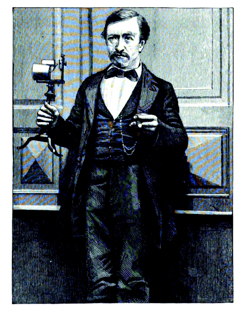
[2] portrait of Philip Reiss From
Silvanus Thompson: ''Reis is here
represented as holding in his hand the
telephone with which he had a few days
preceding (May 11, 1862) achieved such
success at his lecture before the
Freies Deutsches Hochstift (Free German
Institute) in Frankfort. '' PD
source: http://books.google.com/books?id
=YkHu_MiyFSkC&printsec=frontcover&dq=phi
lip+reis+inventor+of+the+telephone#v=one
page&q=&f=false
[08/28/1875 AD]
Electricity in nerve cells measured.

[1] Text of: Richard Caton, ''The
Electric Currents of the Brain'',
British Medical Journal, 1875, V2,
p278. http://www.bmj.com/content/2/765/
257.full.pdf+html {Caton_Richard_187508
28.pdf} PD
source: http://www.bmj.com/content/2/765
/257.full.pdf+html

[2]
http://books.google.com/books?id=_GMeW9E
1IB4C&pg=PA41 COPYRIGHTED
source: http://books.google.com/books?id
=_GMeW9E1IB4C&pg=PA41
[1880 AD]
Germany
[1] Figures 1 and 2 from: W. C.
Röntgen, ''Ueber die durch
Electricität bewirkten Form- und
Volumenänderungen von dielectrischen
Körpern'', Annalen der Physik, Volume
247, Issue 13, pages 771–786,
1880. http://onlinelibrary.wiley.com/do
i/10.1002/andp.18802471304/abstract {Ro
ntgen_Wilhelm_Conrad_188009xx.pdf}
English: ''About the changes in
shape and volume of dielectrics caused
by electricity'' PD
source: http://onlinelibrary.wiley.com/d
oi/10.1002/andp.18802471304/abstract

[2] English: Photo of Wilhelm Conrad
Röntgen. Cleaned up version of
http://images.google.com/hosted/life/l?i
mgurl=6b3da250c6b5560f Source
unknown source Date 1900 PD
source: http://upload.wikimedia.org/wiki
pedia/commons/7/71/Roentgen2.jpg
[03/24/1882 AD]
(radio).
Sommerville, Massachusetts, USA

[1] A. E. Dolbear, ''Mode of Electric
Communication'', Patent number: 350299,
Issue date: Oct 5,
1886. http://www.google.com/patents?id=
Pc9cAAAAEBAJ PD
source: http://www.google.com/patents?id
=Pc9cAAAAEBAJ
[2] From Scientific American
Supplement, December 11, 1886 PD/Corel
source: http://books.google.com/books?hl
=en&id=WE41AAAAMAAJ&dq=A+History+of+Wire
less+Telegraphy&printsec=frontcover&sour
ce=web&ots=08aQE8FQHe&sig=0AB8rC1DTmKfhh
sRE55cYSIq2PM&sa=X&oi=book_result&resnum
=2&ct=result#PPA98,M1
[06/21/1889 AD]

[1] The first (publicly known[t]) Films
Made on Celluloid (1889-1890) PD
source: Ray Allister, pseudonym for
Muriel Forth, "Friese-Greene: Close-up
of an Inventor", Marsland Publications,
1948.

[2] Description
Williamfriesegreen.jpg English:
William Friese-Greene photographed in
c.1890 Date c1890 PD
source: http://upload.wikimedia.org/wiki
pedia/commons/2/2d/Williamfriesegreen.jp
g
[11/05/1895 AD]
Germany

[1] English: Photo of Wilhelm Conrad
Röntgen. Cleaned up version of
http://images.google.com/hosted/life/l?i
mgurl=6b3da250c6b5560f Source
unknown source Date 1900 PD
AND Anna Berthe Roentgen.gif Print
of Wilhelm Röntgen's (1845-1923) first
x-ray, the hand of his wife Anna taken
on 1895-12-22, presented to Professor
Ludwig Zehnder of the Physik Institut,
University of Freiburg, on 1 January
1896. Source
http://en.wikipedia.org/wiki/Image:An
na_Berthe_Roentgen.gif Date 22
December 1895 (1895-12-22) Author
Wilhelm Röntgen PD
source: http://upload.wikimedia.org/wiki
pedia/commons/7/71/Roentgen2.jpghttp://u
pload.wikimedia.org/wikipedia/commons/6/
6e/Anna_Berthe_Roentgen.gif
[2] English: Photo of Wilhelm Conrad
Röntgen. Cleaned up version of
http://images.google.com/hosted/life/l?i
mgurl=6b3da250c6b5560f Source
unknown source Date 1900 PD
source: http://upload.wikimedia.org/wiki
pedia/commons/7/71/Roentgen2.jpg
[1898 AD]
data.
Copenhagen, Denmark
[1] Description Telegrafon
8154.jpg Magyar: Valdemar Poulsen
mágneses hangrögzítő készüléke
1898-ból. A Brede Værk ipari
múzeumban látható a dániai
Lingbyben. Saját felvétel. Dansk:
Valdemar Poulsen opfandt i i 1898 af en
magnetisk optageenhed der kaldes en
Telegrafon English: Magnetic wire
recorder, invented by Valdemar Poulsen,
1898. It is exhibited at Brede works
Industrial Museum, Lingby,
Danmark. Date 25 October
2009(2009-10-25) (original upload
date) Source Transferred from
hu.wikipedia; transferred to Commons by
User:Nico-dk using
CommonsHelper. Author Original
uploader was Bitman at
hu.wikipedia Permission (Reusing this
file) CC-BY-SA-2.5; Released under
the GNU Free Documentation
License. GNU
source: http://upload.wikimedia.org/wiki
pedia/commons/f/f9/Telegrafon_8154.jpg

[2] 1 Valdemar Poulsen (1869-1942),
der Erfinder der magnetischen
Schallaufzeichnung UNKNOWN
source: http://www.theimann.com/Analog/H
istory/100_Jahre/Bild1.jpg
[03/23/1903 AD]
USA

[1] Description First
flight2.jpg English: First successful
flight of the Wright Flyer, by the
Wright brothers. The machine traveled
120 ft (36.6 m) in 12 seconds at 10:35
a.m. at Kitty Hawk, North Carolina.
Orville Wright was at the controls of
the machine, lying prone on the lower
wing with his hips in the cradle which
operated the wing-warping mechanism.
Wilbur Wright ran alongside to balance
the machine, and just released his hold
on the forward upright of the right
wing in the photo. The starting rail,
the wing-rest, a coil box, and other
items needed for flight preparation are
visible behind the machine. This was
considered ''the first sustained and
controlled heavier-than-air, powered
flight'' by the Fédération
Aéronautique
Internationale. Français : L’un des
premier vols habités de l’histoire
dans un aéronef plus lourd que l’air
(36.6 mètres en 12 secondes), par les
frères Wright le 17 décembre 1903 à
10h35 sur la plage de Kitty Hawk en
Caroline du Nord. Orville est aux
commandes, allongé sur le ventre sur
l’aile basse et les hanches dans la
nacelle qui servait à contrôler le
mouvement des ailes ; Wilbur court le
long de l’appareil et vient de lacher
l’aile droite. Le rail de lancement,
des étais et d’autres équipements
nécessaires pour la préparation du
vol sont visibles. 日本語:
1903年12月17日、ライト兄弟が�
�類初の動力飛行機での有人飛
行に成功した時の写真。 Date
17 December 1903 PD
source: http://upload.wikimedia.org/wiki
pedia/commons/8/86/First_flight2.jpg
[2] * Description: Wilbur
Wright Background notes: Wright
brothers English: Early Wright
brother’s airplanes explored basic
principles of flight. The Wright
brothers are widely credited with
engineering the first aircraft capable
of sustained powered
flight. Commons-emblem-notice.svg
Wright brothers Wikipedia:
Asturianu Bosanski Català
Čeština Dansk Deutsch English
Esperanto Español Euskara Suomi
Français עברית Magyar Bahasa
Indonesia Italiano 日本語
한국어 Latina Lietuvių
Nederlands Norsk (Bokmål) Polski
Português Русский Slovenčina
Slovenščina Српски / Srpski
Svenska ไทย Türkçe Tiếng
Việt 中文 Other links: US
inventors *** Smithsonian Stories of
the Wright flights *** National Park
Service, Wright Brothers' Memorial ***
PBS Nova: The Wright Brothers' Flying
Machines * Source:
http://lcweb2.loc.gov/pp/wrihtml/wribac.
html * Photographer: unknwon PD
source: http://upload.wikimedia.org/wiki
pedia/commons/7/77/Wilbur_Wright.jpg
[12/21/1906 AD]
York City, New York, USA

[1] From De Forest 1907 Patent: Lee De
Forest, ''Space Telegraphy'', Patent
number: 879532, Filing date: Jan 29,
1907, Issue date: Feb 18,
1908 http://www.google.com/patents?id=6
i1vAAAAEBAJ&printsec=abstract&zoom=4&sou
rce=gbs_overview_r&cad=0#v=onepage&q&f=f
alse PD
source: http://www.google.com/patents?id
=6i1vAAAAEBAJ&printsec=abstract&zoom=4&s
ource=gbs_overview_r&cad=0#v=onepage&q&f
=false

[2] Description Lee De
Forest.jpg en:Lee De Forest,
published in the February 1904 issue of
The Electrical Age. PD
source: http://upload.wikimedia.org/wiki
pedia/commons/6/65/Lee_De_Forest.jpg
[05/??/1907 AD]
separated by mass.
England
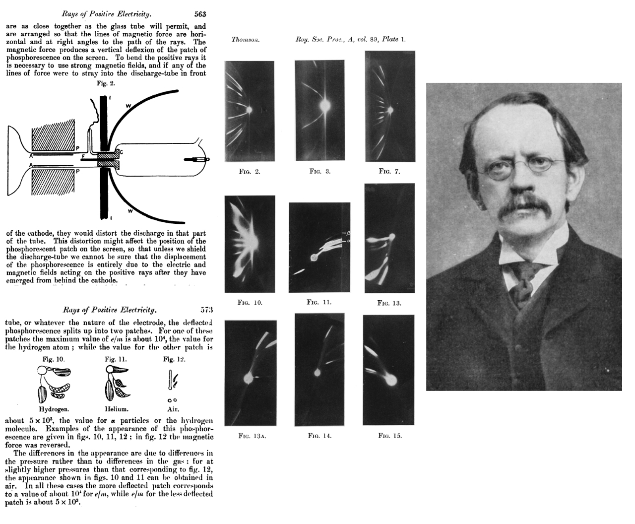
[1] fig 2 from: Thomson, J. J., ''On
Rays of Positive Electricity'', Phil.
Mag., S6, V13, N77, May 1907, p561. PD
AND figs 10-12 from: Thomson, J.
J., ''On Rays of Positive
Electricity'', Phil. Mag., S6, V13,
N77, May 1907, p561. PD AND [10]
figures 2-15 from: # Bakerian Lecture:
Rays of Positive Electricity # J. J.
Thomson # Proceedings of the Royal
Society of London. Series A, Containing
Papers of a Mathematical and Physical
Character, Vol. 89, No. 607 (Aug. 1,
1913), pp. 1-20 PD AND English
physicist J J Thomson Date GWS -
The Great War: The Standard History of
the All Europe Conflict (volume four)
edited by H. W. Wilson and J. A.
Hammerton (Amalgamated Press, London
1915) (So, it is taken before
1915) Source
http://www.firstworldwar.com/photos/g
raphics/gws_thomson_01.jpg First World
War.com PD
source: http://books.google.com/books?id
=vVjKOdktZhsC&printsec=frontcover&dq=edi
tions:UOM39015024088414#v=onepage&q=&f=f
alsehttp://www.jstor.org/stable/93452?&S
earch=yes&term=electricity&term=positive
&term=rays&list=hide&searchUri=%2Faction
%2FdoBasicSearch%3FQuery%3Drays%2Bof%2Bp
ositive%2Belectricity%26jc%3Dj100836%26w
c%3Don%26Search.x%3D0%26Search.y%3D0%26S
earch%3DSearch&item=1&ttl=262&returnArti
cleService=showArticlehttp://upload.wiki
media.org/wikipedia/commons/c/c1/J.J_Tho
mson.jpg
[2] fig 2 from: Thomson, J. J., ''On
Rays of Positive Electricity'', Phil.
Mag., S6, V13, N77, May 1907, p561. PD
source: http://books.google.com/books?id
=vVjKOdktZhsC&printsec=frontcover&dq=edi
tions:UOM39015024088414#v=onepage&q=&f=f
alse
[11/13/1907 AD]

[1] Paul Cornu's helicopter was the
first to achieve free flight while
carrying a passenger (1907). Credits
-National Air and Space
Museum, Smithsonian Institution (SI
Neg. No. 93-640) The French bicycle
maker and engineer Paul Cornu, born in
1881 in Lisieux, France, was the first
person to design and build a helicopter
that achieved free flight while
carrying a passenger. His twin-rotor
craft flew for about 20 seconds on
November 13, 1907, rising about one
foot (0.3 meter) off the ground. A
24-horsepower (18-kilowatt) engine
powered the helicopter, which had
counter-rotating rotors. The helicopter
had no effective means of control and
was abandoned after a few
flights. Cornu died in 1944. PD
source: http://www.centennialofflight.go
v/essay/Dictionary/Cornu/DI18G1.jpg
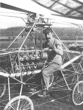
[2] Paul Cornu in his first helicopter
in 1907. Note that he is sitting
between the two rotors, which rotated
in opposite directions to cancel
torque. This helicopter was the first
flying machine to have risen from the
ground using rotor blades instead of
wings. Credits - © 2001 Smithsonian
Institution, National Air and Space
Museum, Videodisc. 2B 5847 PD
source: http://www.centennialofflight.go
v/essay/Rotary/early_20th_century/HE2G13
.jpg
[06/06/1908 AD]
[1] From top to bottom, left to
right Top: Plan View of Receiver
Showing Negative Received. Middle:
Plan View of Transmitter Showing
Traveling Carriage Carrying
Picture. Bottom Left: The Transmitting
Apparatus Bottom Middle: Photograph of
Edward VII. Transmitted by Wireless
Telegraphy. Bottom Right: The Receiver
Showing Relay to Which Recording Needle
is Connected. PD/Corel
source: KNUDSEN'S PROCESS OF
TRANSMITTING PICTURES BY WIRELESS
TELEGRAPHY. BY THE ENGLISH
CORREESPONDENT OF THE SCIENTIFIC
AMERICAN.. Scientific American
(1845-1908). New York: Jun 6, 1908.
Vol. Vol. XCVIII., Iss. No. 23.; p. 412
(1 page)
[06/21/1918 AD]
London, UK
[1] Image from: William Henry Eccles
and Frank Wilfred Jordan,
''Improvements in ionic relays''
British patent number: GB 148582
(filed: 21 June 1918; published: 5
August 1920).
http://worldwide.espacenet.com/publica
tionDetails/originalDocument?CC=GB&NR=14
8582&KC=&FT=E {Eccles_William_Henry_ele
ctronic_memory_GB148582A_19180621.pdf}
PD
source: http://worldwide.espacenet.com/p
ublicationDetails/originalDocument?CC=GB
&NR=148582&KC=&FT=E

[2] A simple yet powerful animation of
how an R-S flip-flop works. GNU
source: http://upload.wikimedia.org/wiki
pedia/commons/f/f4/R-S.gif
[04/??/1919 AD]
fusion; Nitrogen changed into Oxygen.
England
[1] Rutherford, Collision of α
Particles with Light Atoms, Phil. Mag.
June 1919, s6, 37, pp537-61. from:
Ernest Rutherford, ''The Collected
Papers of Lord Rutherford of Nelson'',
Vol 2, 1963,
p551. http://web.lemoyne.edu/~giunta/ru
therford.html {Rutherford_191904xx.pdf}
PD
source: Rutherford, Collision of α
Particles with Light Atoms, Phil. Mag.
June 1919, s6, 37, pp537-61. from:
Ernest Rutherford, "The Collected
Papers of Lord Rutherford of Nelson",
Vol 2, 1963, p551.
[2] Figure 1 from: Rutherford,
Collision of α Particles with Light
Atoms, Phil. Mag. June 1919, s6, 37,
pp537-61.
http://web.lemoyne.edu/~giunta/rutherf
ord.html {Rutherford_191904xx.pdf}
PD
source: Rutherford_191306xx003.pdf
[04/16/1932 AD]
University) Cambridge, England
[1] J. D. COCKCROFT & E. T. S. WALTON
, ''Disintegration of Lithium by Swift
Protons'', Nature 129, 649-649 (30
April
1932). http://www.nature.com/nature/jou
rnal/v129/n3261/abs/129649a0.html {Cock
croft_John_19320416.pdf}
COPYRIGHTED AND Enest S Walton,
Lord Rutherford and John Cockcroft:
1932 UNKNOWN
source: http://www.nature.com/nature/jou
rnal/v129/n3261/abs/129649a0.htmlhttp://
www.aip.org/history/exhibits/lawrence/im
ages/epa-12.jpg
[2] Enest S Walton, Lord Rutherford
and John Cockcroft: 1932 UNKNOWN
source: http://www.aip.org/history/exhib
its/lawrence/images/epa-12.jpg
[05/22/1937 AD]
Germany
[1] Figures 2-4 from: ''Fig 2.
Tungsten cathode (filament) [011] -
Direction in the middle. Fig 3.
Tungsten cathode [211] - Direction,
almost in the middle. Fig 4. Sphere
model with the lattice directions of a
cube-based emission tungsten cathode,
field of view as Fig 3.'' [2] Erwin W.
Müller, ''Elektronenmikroskopische
Beobachtungen von Feldkathoden'',
Zeitschrift für Physik A Hadrons and
Nuclei, Volume 106, Numbers 9-10,
541-550, DOI:
10.1007/BF01339895 http://www.springerl
ink.com/content/h425u71vqh66w886/ {Mull
er_Erwin_W_19370522.pdf}
English: ''Electron microscopic
observations of field cathode''
source: http://www.springerlink.com/cont
ent/h425u71vqh66w886/

[2] COPYRIGHTED
source: http://micro.magnet.fsu.edu/opti
cs/timeline/people/antiqueimages/mueller
.jpg
[06/22/1938 AD]
Gesellschaft/Berlin Medical Society)
Berlin, Germany
[1] (ubermikroskop) Ultramicroscope
image of the virus of ectromelia in the
point mouse. Infectious material from
the lymph of an infected paw. magnified
20,000x. Figure 1 from: B. v.
Borries, E. Ruska und H. Ruska,
''Bakterien und Virus in
übermikroskopischer
Aufnahme.'', Klin. Wochenschrift 17
(1938)
921-925. http://ernstruska.digilibrary.
de/bibliographie/q021/q021.html {Ruska_
Ernst_19380622.pdf} UNKNOWN
source: http://ernstruska.digilibrary.de
/bibliographie/q021/q021.html

[2] Ernst Ruska, 1939 UNKNOWN
source: http://www.siemens.com/history/p
ool/perseunlichkeiten/wissenschaftler/ru
ska_1939.jpg
[04/30/1939 AD]
Mansfield, Ohio, USA
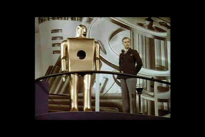
[1] Elektro at the 1939 World's
Fair UNKNOWN
source: http://img.youtube.com/vi/T35A3g
_GvSg/0.jpg

[2] Inside working of Westinghouse
Elektro walking robot UNKNOWN
source: http://davidszondy.com/future/ro
bot/elektro-interior.jpg
[04/02/1953 AD]
understood.
Cambridge) Cambridge, England
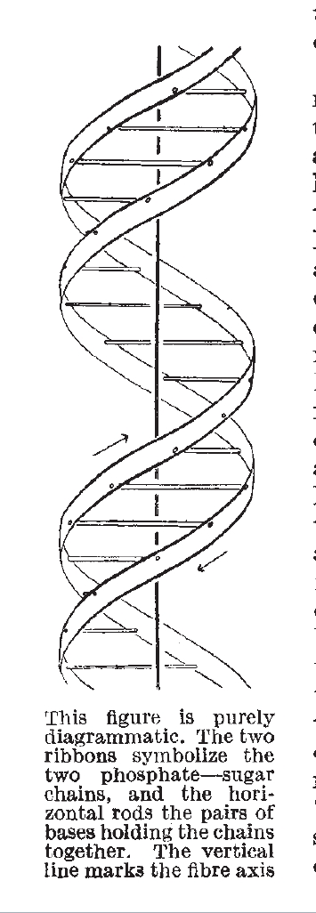
[1] Figure 1 from: J. D. WATSON & F.
H. C. CRICK, ''Molecular structure of
nucleic acids; a structure for
deoxyribose nucleic acid'', Nature,
(1953) volume: 171 issue: 4356 page:
737. http://www.nature.com/nature/journ
al/v171/n4356/abs/171737a0.html {Crick_
Francis_Harry_Compton_19530402.pdf} COP
YRIGHTED
source: http://www.nature.com/nature/jou
rnal/v171/n4356/abs/171737a0.html

[2] Francis Harry Compton Crick
UNKNOWN
source: http://scientistshowtell.wikispa
ces.com/file/view/FrancisHarryComptonCri
ck2.jpg/39149552/FrancisHarryComptonCric
k2.jpg
[05/05/1954 AD]
New York, USA
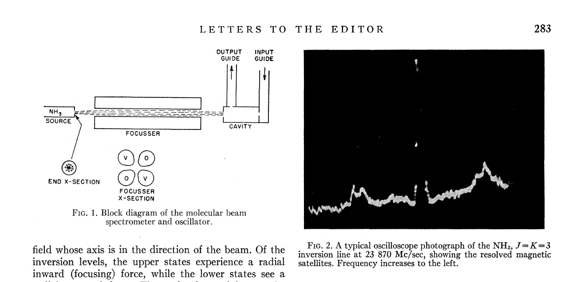
[1] Figures 1 and 2 from: J. P.
Gordon, H. J. Zeiger, and C. H. Townes,
''Molecular Microwave Oscillator and
New Hyperfine Structure in the
Microwave Spectrum of NH3'', Phys. Rev.
95, 282–284
(1954). http://prola.aps.org/abstract/P
R/v95/i1/p282_1 {Townes_Charles_Hard_19
540505.pdf} COPYRIGHTED
source: http://prola.aps.org/abstract/PR
/v95/i1/p282_1
[2] Charles Hard Townes Nobel Prize
photo COPYRIGHTED
source: http://nobelprize.org/nobel_priz
es/physics/laureates/1964/townes.jpg
[10/04/1957 AD]
Kazakhstan, U.S.S.R.
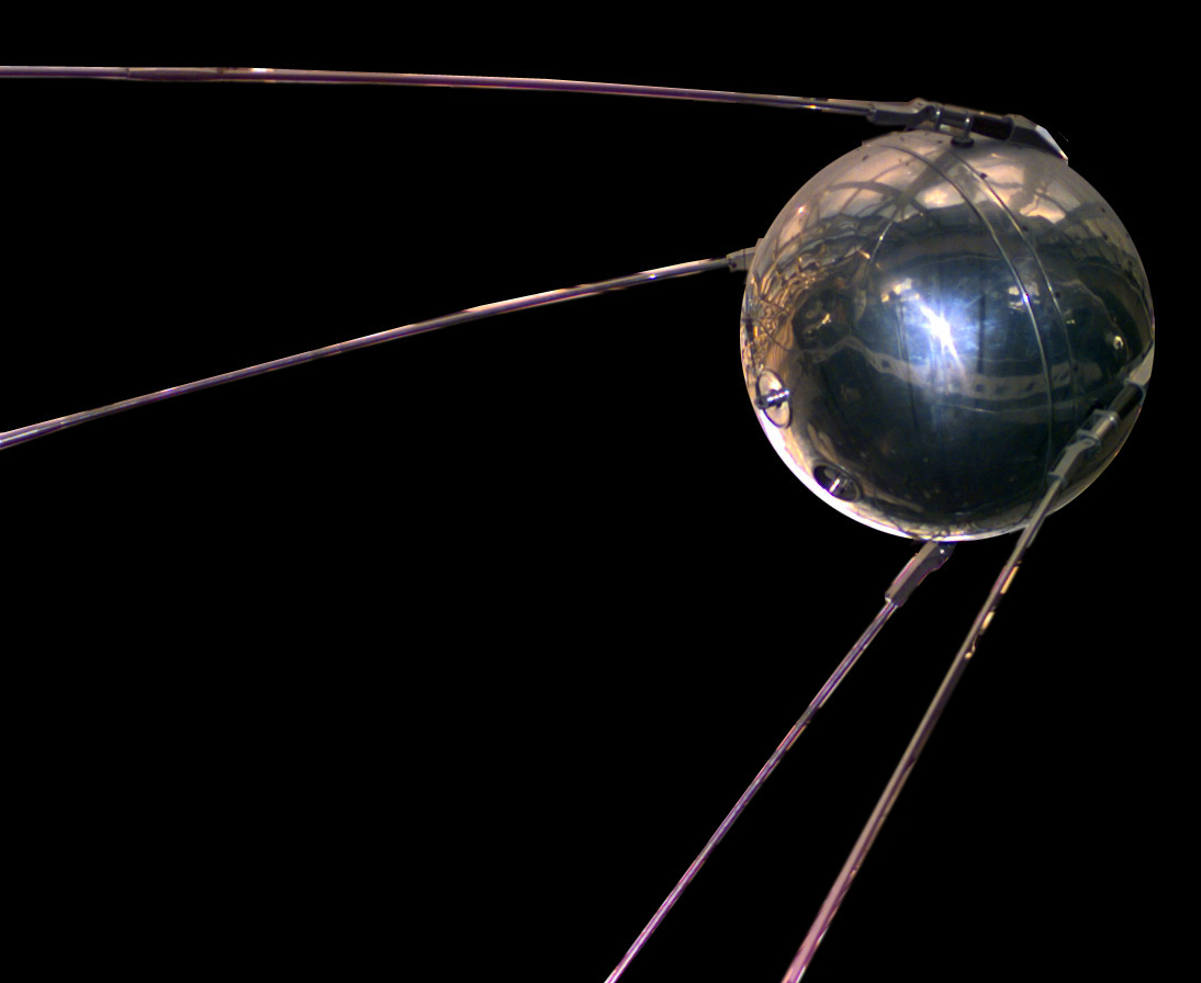
[1] Description Sputnik
asm.jpg English: A replica of Sputnik
1, the first artificial satellite in
the world to be put into outer space:
the replica is stored in the National
Air and Space Museum. فارسی:
مدل ماهواره
اسپوتنیک-۱، نخستین
ماهواره فضایی
جهان Suomi: Sputnik 1:n, maailman
ensimmäinen ihmisen laukaiseman Maata
kiertävän keinotekoisen satelliittin,
jäljennös. Date
2004(2004) Source
http://nssdc.gsfc.nasa.gov/database
/MasterCatalog?sc=1957-001B Author
NSSDC, NASA PD
source: http://upload.wikimedia.org/wiki
pedia/commons/b/be/Sputnik_asm.jpg
[09/14/1959 AD]
Kazakhstan, U.S.S.R.

[1] Luna 2 PD
source: http://nssdc.gsfc.nasa.gov/plane
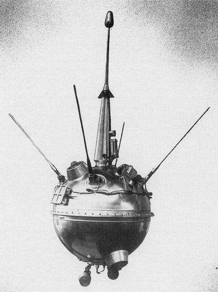
tary/image/luna_2.jpg
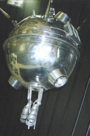
[2] Luna 1 PD
source: http://nssdc.gsfc.nasa.gov/image
/spacecraft/luna1_vsm.jpg
[04/22/1960 AD]
California

[1] Figure 1 from: Theodore H.
Mainman, ''Ruby Laser Systems'', Patent
number: 3353115, Filing date: Apr 13,
1961, Issue date: Nov 14,
1967 http://www.google.com/patents?id=b
-lUAAAAEBAJ&printsec=abstract&zoom=4&sou
rce=gbs_overview_r&cad=0#v=onepage&q&f=f
alse
{Maimon_Theodore_Harold_19610413.pdf}
PD
source: http://www.google.com/patents?id
=b-lUAAAAEBAJ&printsec=abstract&zoom=4&s
ource=gbs_overview_r&cad=0#v=onepage&q&f
=false

[2] Description Ted Maiman Holding
First Laser.jpg English: Theodore
Maiman holding his invention of the
world's first laser (invented May 16,
1960) Date 16 May
1983(1983-05-16) Source
Template:TRW Author
Kathleenfmaiman Permission (Reusi
ng this file) PD
source: http://upload.wikimedia.org/wiki
pedia/commons/d/df/Ted_Maiman_Holding_Fi
rst_Laser.jpg
[04/12/1961 AD]
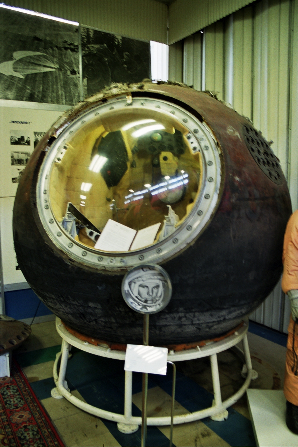
[1] The Vostok 1 capsule as recovered
after landing. Currently on display at
the RKK Energiya museum in Korolyov CC
source: http://upload.wikimedia.org/wiki
pedia/en/7/70/Vostok_1_after_landing.jpg

[2] Description Yuri Gagarin in
Vostok 1 Source Mission
photography Portion used
Sufficient to show the face of
Gagarin in his spacesuit within the
capsule Low resolution?
yes COPYRIGHTED
source: http://upload.wikimedia.org/wiki
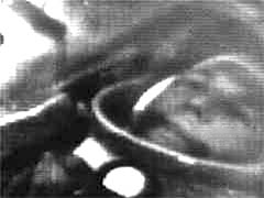
pedia/en/b/b1/Vostok1.jpg
[10/26/1962 AD]
data.
Angeles, California, USA

[1] From: Wayne R. Johnson, ''High
Speed, High Density, Optical Recording
System'', Patent number:
3154370 Filing date: Oct 26, 1962,
Issue date: Oct 27,
1964 http://www.google.com/patents?id=H
9x0AAAAEBAJ
source: http://www.google.com/patents?id
=H9x0AAAAEBAJ
[03/01/1966 AD]

[1] Venera 3 PD
source: http://nssdc.gsfc.nasa.gov/plane
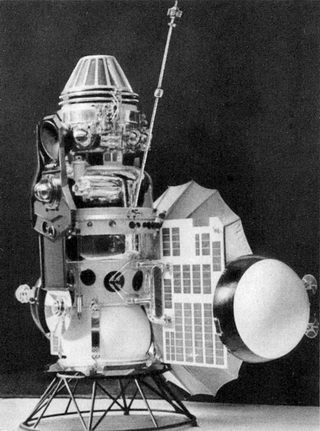
tary/image/venera_3.jpg
[07/21/1969 AD]
of Earth.

[1] ''That's one small step for man,
one giant leap for mankind.'' At 10:56
p.m. EDT on July 20, 1969, Neil
Armstrong became the first human to set
foot on the Moon. This image was taken
from the telecast of the event, watched
by over half a billion people around
the world. Armstrong composed the quote
after landing on the Moon, he had meant
to say, ''That's one small step for
aman ...''. The pictures were taken by
the Apollo lunar surface camera,
mounted on one of the LM legs. The
black bar running through the center of
the picture is an anomaly in the
Goldstone ground data system. (NASA
photo ID S69-42583) PD
source: http://nssdc.gsfc.nasa.gov/plane
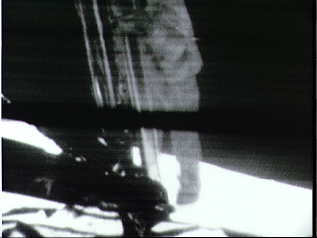
tary/lunar/images/a11tvarm.jpg
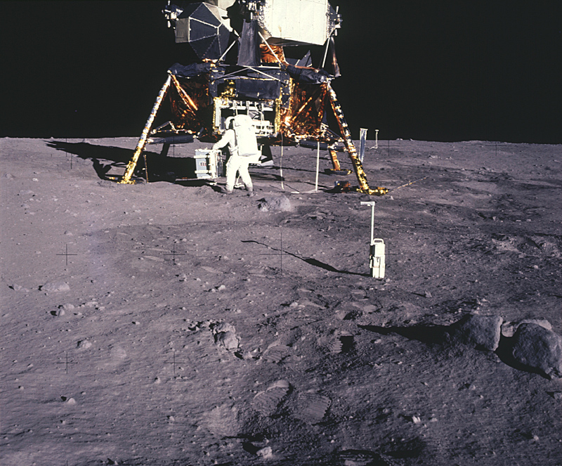
[2] Here Aldrin is unloading the
passive seismometer of the Early Apollo
Scientific Experiments Package (EASEP)
from the lunar module equipment bay.
The white apparatus in the foreground
is the 35 mm stereo close-up camera.
Beyond the right leg is the solar wind
experiment, and beyond that the lunar
surface TV camera. The LM legs are
wrapped in foil to provide thermal
insulation. There is a split rock in
the lower right of the frame which is
presumably ejecta from a nearby impact
crater. (NASA photo ID
AS11-40-5931) PD
source: http://nssdc.gsfc.nasa.gov/plane
tary/lunar/images/as11_40_5931.jpg
[11/14/1971 AD]
(Mars).

[1] Mariner 9 PD
source: http://nssdc.gsfc.nasa.gov/image
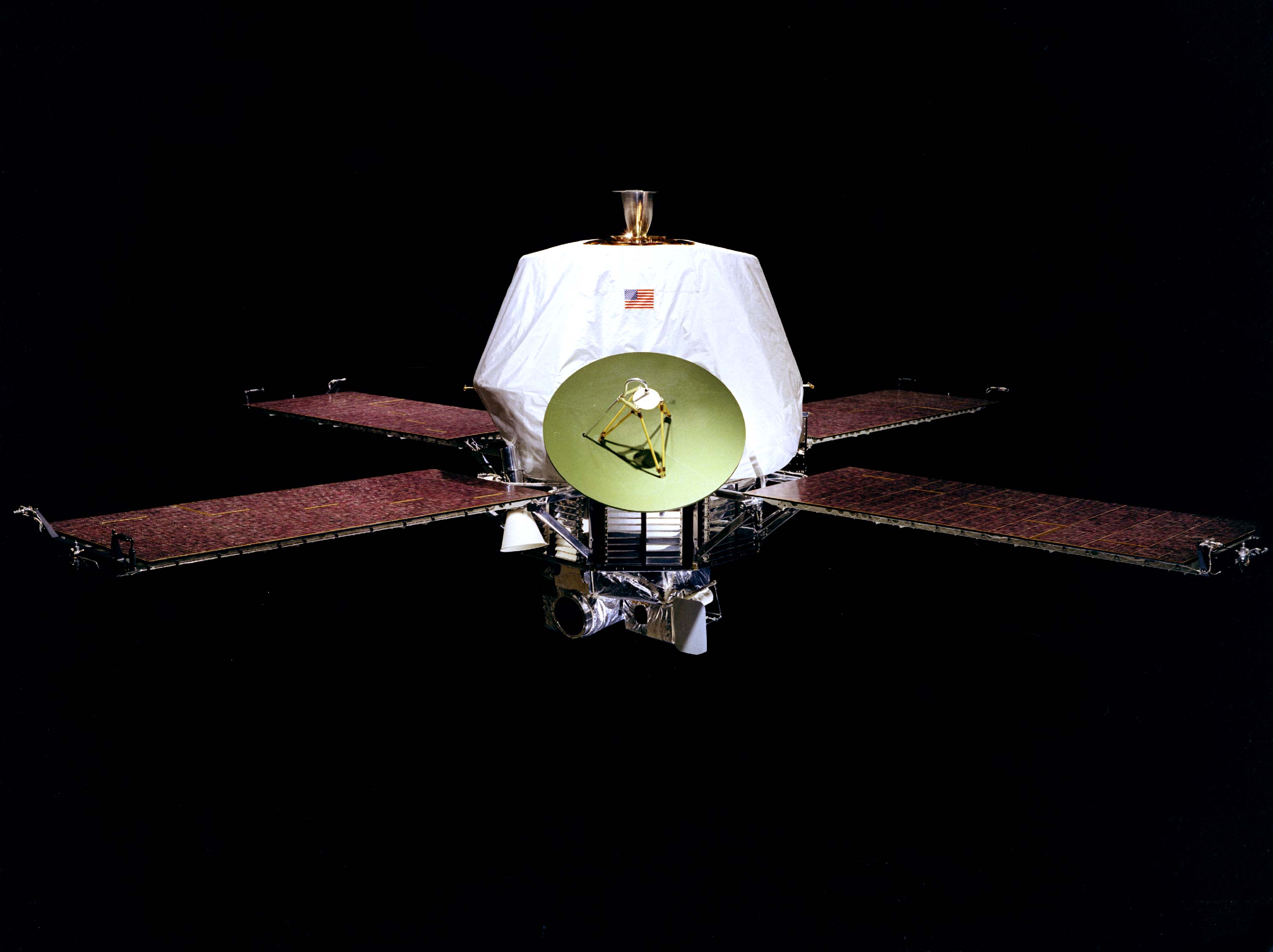
/spacecraft/mariner09.jpg
[2] Mariner 9 imagery of Olympus Mons
volcano on Mars compared to the eight
principal Hawaiian islands at the same
scale. (Mariner 9 image mosaic,
NASA/JPL) PD
source: http://pubs.usgs.gov/gip/volc/fi
g38.gif
[11/27/1971 AD]
[1] Mars 3 Lander PD
source: http://nssdc.gsfc.nasa.gov/image
/spacecraft/mars3_lander_vsm.jpg

[2] Description Mars3
iki.jpg English: The Mars 3
spacecraft Date Source
http://nssdc.gsfc.nasa.gov/image/sp
acecraft/mars3_iki.jpg Author
NASA PD
source: http://upload.wikimedia.org/wiki
pedia/commons/1/13/Mars3_iki.jpg
[12/02/1971 AD]
[1] Signal from mars-3 Lander UNKNOWN
source: http://www.mentallandscape.com/C
_Mars03_lander.jpg
[2] Mars 3 Lander PD
source: http://nssdc.gsfc.nasa.gov/image
/spacecraft/mars3_lander_vsm.jpg
[12/03/1973 AD]

[1] Description
http://history.nasa.gov/SP-349/p142.jpg
English: Pioneer 10 Jupiter
encounter. Date Source
http://history.nasa.gov/SP-349/ch8.
htm Author
NASA Permission (Reusing this
file) PD
source: http://history.nasa.gov/SP-349/p
142.jpg
[2] Pioneer 10 PD
source: http://nssdc.gsfc.nasa.gov/image
/spacecraft/pioneer10-11.jpg
[10/20/1975 AD]

[1] Image of the surface of Venus from
Venera 9 PD
source: http://nssdc.gsfc.nasa.gov/imgca
t/hires/v09_lander.gif

[2] Venera 9 Descent Craft PD
source: http://nssdc.gsfc.nasa.gov/plane
tary/image/venera_9_lander.jpg
[09/01/1979 AD]
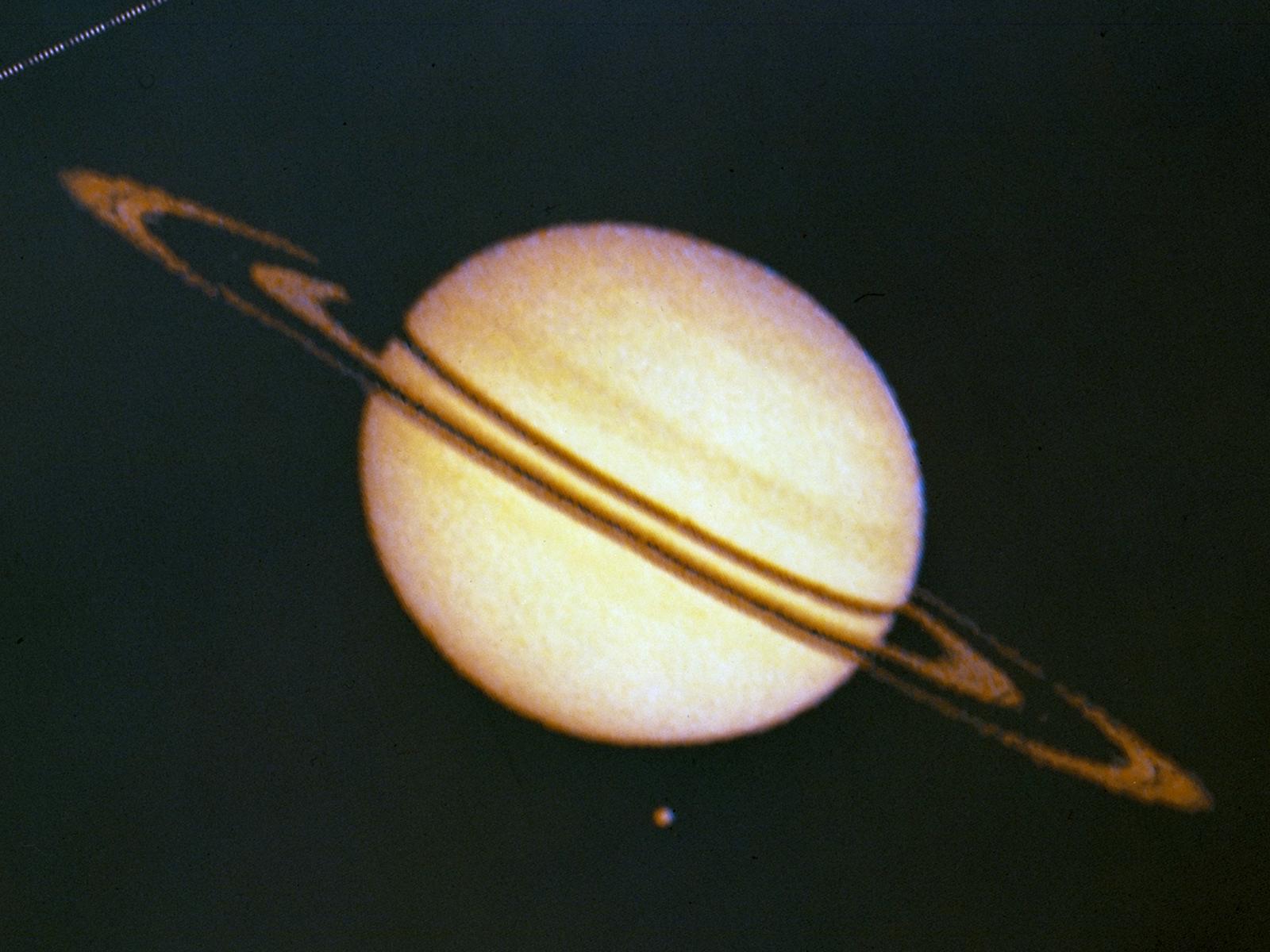
[1] Pioneer 11 Image of Saturn and Its
Moon Titan {ULSF: Titan is at the upper
left} The Pioneer 11 spacecraft
launched from Cape Canaveral forty
years ago, on April 5, 1973. Pioneer
11's path through Saturn's outer rings
took it within 21,000 km of the planet,
where it discovered two new moons
(almost smacking into one of them in
September 1979) and a new ''F'' ring.
The spacecraft also discovered and
charted the magnetosphere, magnetic
field and mapped the general structure
of Saturn's interior. The spacecraft's
instruments measured the heat radiation
from Saturn's interior and found that
its planet-sized moon, Titan, was too
cold to support life. This image
from Pioneer 11 shows Saturn and its
moon Titan. The irregularities in ring
silhouette and shadow are due to
technical anomalies in the preliminary
data later corrected. At the time this
image was taken, Pioneer was 2,846,000
km (1,768,422 miles) from
Saturn. › NASA Celebrates Four
Decades of Plucky Pioneer 11 Image
credit: NASA Ames PD
source: http://www.nasa.gov/images/conte
nt/739507main_739460main_AC79-9107.3_160
0-1200.jpg
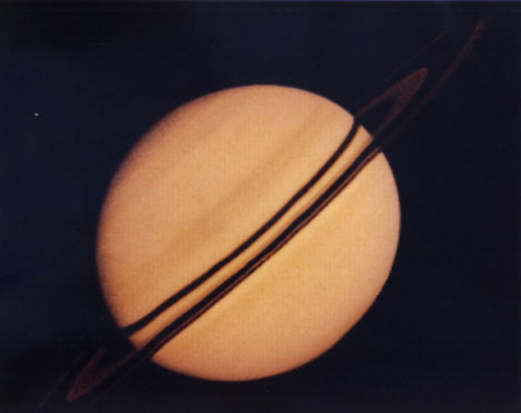
[2] Pioneer 10 PD
source: http://quest.nasa.gov/sso/cool/p
ioneer10/graphics/lasher/slide4.jpg
[01/24/1986 AD]

[1] Description Uranus.jpg English:
NASA photo of Uranus taken by Voyager
2. Caption: This pictures of Uranus was
compiled from images recorded by
Voyager 2 on January 10, 1986, when the
NASA spacecraft was 18 million
kilometers (11 million miles) from the
planet. The images were obtained by
Voyager's narrow-angle camera; the view
is toward the planet's pole of
rotation, which lies just left of
center. The picture has been processed
to show Uranus as human eyes would see
it from the vantage point of the
spacecraft. The dark shading of the
upper right edge of the disk is the
terminator, or day-night boundary. The
blue-green appearance of Uranus results
from methane in the atmosphere; this
gas absorbs red wavelengths from the
incoming sunlight, leaving the
predominant bluish color seen here.
Images shuttered through different
color filters were added and
manipulated by computer, greatly
enhancing the low-contrast details in
the original images. The planet reveals
a dark polar hood surrounded by a
series of progressively lighter
convective bands. The banded structure
is real, though exaggerated here. The
Voyager project is managed for NASA by
the Jet Propulsion Laboratory. Date
January 1986(1986-01) Source
http://photojournal.jpl.nasa.gov/ca
talog/PIA01360 Author NASA PD
source: http://upload.wikimedia.org/wiki
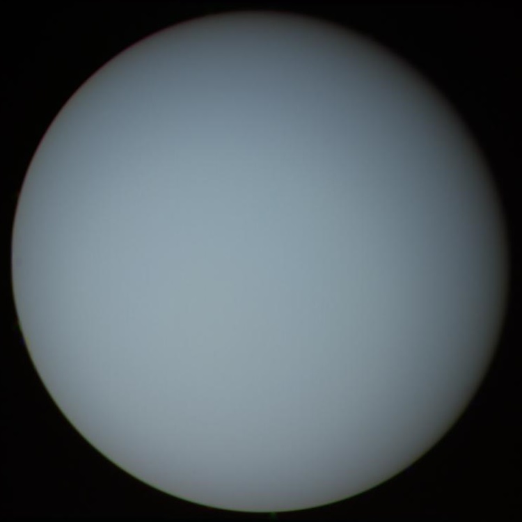
pedia/commons/b/bb/Uranus.jpg
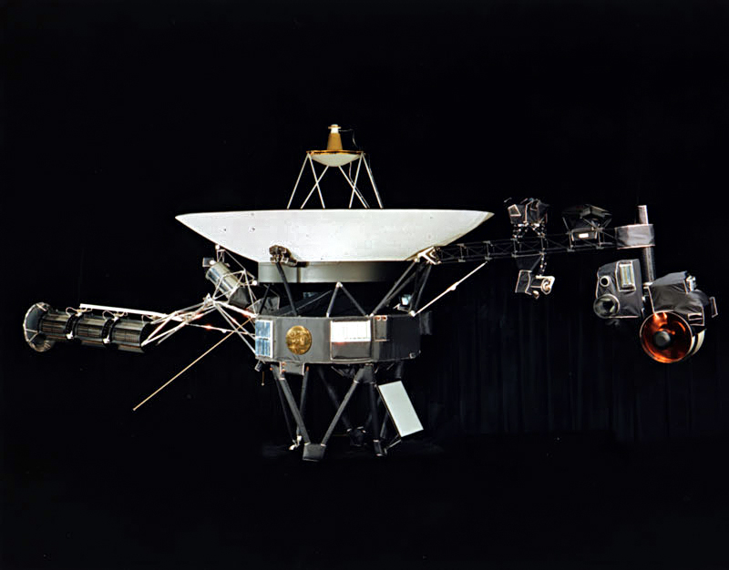
[2] Description
Voyager.jpg Voyager 1 / Voyager
2 English: NASA photograph of one of
the two identical Voyager space probes
Voyager 1 and Voyager 2 launched in
1977. The 3.7 metre diameter
high-gain antenna (HGA) is attached to
the hollow ten-sided polygonal body
housing the electronics, here seen in
profile. The Voyager Golden Record is
attached to one of the bus
sides. The angled square panel below
is the optical calibration target and
excess heat radiator. The three
radioisotope thermoelectric generators
(RTGs) are mounted end-to-end on the
left-extending boom. One of the two
planetary radio and plasma wave antenna
extends diagonally left and down, the
other extends to the rear, mostly
hidden here. The compact structure
between the RTGs and the HGA are the
high-field and low-field magnetometers
(MAG) in their stowed state; after
launch an Astromast boom extended to 13
metres to distance the low-field
magnetometers. The instrument boom
extending to the right holds, from left
to right: the cosmic ray subsystem
(CRS) above and Low-Energy Charged
Particle (LECP) detector below; the
Plasma Spectrometer (PLS) above; and
the scan platform that rotates about a
vertical axis. The scan platform
comprises: the Infrared Interferometer
Spectrometer (IRIS) (largest camera at
right); the Ultraviolet Spectrometer
(UVS) to the right of the UVS; the two
Imaging Science Subsystem (ISS) vidicon
cameras to the left of the UVS; and the
Photopolarimeter System (PPS) barely
visible under the ISS. Suggested for
English Wikipedia:alternative text for
images: A space probe with squat
cylindrical body topped by a large
parabolic radio antenna dish pointing
upwards, a three-element radioisotope
thermoelectric generator on a boom
extending left, and scientific
instruments on a boom extending right.
A golden disk is fixed to the
body. Date Source NASA
website http://voyager.jpl.nasa.gov/ima
ge/images/spacecraft/Voyager.jpg Author
NASA Permission (Reusing this
file) PD-NASA PD
source: http://upload.wikimedia.org/wiki
pedia/commons/d/d2/Voyager.jpg
[12/14/1988 AD]
Berkeley, California, USA
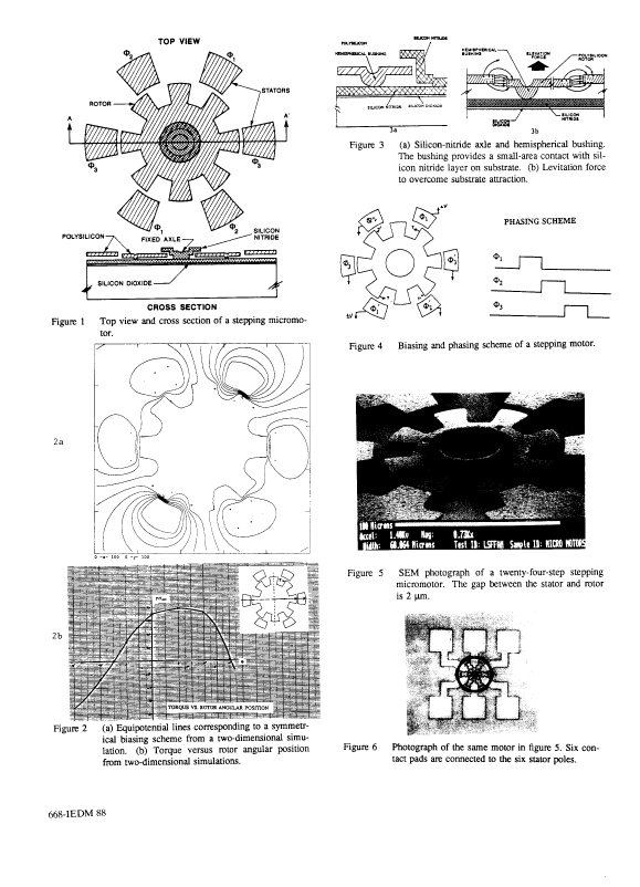
[1] Figures 1 from: Long-Sheng Fan;
Yu-Chong Tai; R.S. Muller; ,
''IC-processed electrostatic
micro-motors,'' Electron Devices
Meeting, 1988. IEDM '88. Technical
Digest., International , vol., no.,
pp.666-669, 1988 doi:
10.1109/IEDM.1988.32901 URL:
http://ieeexplore.ieee.org/stamp/stamp.j
sp?tp=&arnumber=32901&isnumber=1415 COP
YRIGHTED
source: URL:
http://ieeexplore.ieee.org/stamp/stamp.j
sp?tp=&arnumber=32901&isnumber=1415

[2] Figures 2 from: Long-Sheng Fan;
Yu-Chong Tai; R.S. Muller; ,
''IC-processed electrostatic
micro-motors,'' Electron Devices
Meeting, 1988. IEDM '88. Technical
Digest., International , vol., no.,
pp.666-669, 1988 doi:
10.1109/IEDM.1988.32901 URL:
http://ieeexplore.ieee.org/stamp/stamp.j
sp?tp=&arnumber=32901&isnumber=1415 COP
YRIGHTED
source: URL:
http://ieeexplore.ieee.org/stamp/stamp.j
sp?tp=&arnumber=32901&isnumber=1415
[08/25/1989 AD]

[1] A picture of Neptune taken by
Voyager 2, showing off the Great Dark
Spot which has since disappeared from
the planet's surface. Original
Caption Released with Image: During
August 16 and 17, 1989, the Voyager 2
narrow-angle camera was used to
photograph Neptune almost continuously,
recording approximately two and
one-half rotations of the planet. These
images represent the most complete set
of full disk Neptune images that the
spacecraft will acquire. This picture
from the sequence shows two of the four
cloud features which have been tracked
by the Voyager cameras during the past
two months. The large dark oval near
the western limb (the left edge) is at
a latitude of 22 degrees south and
circuits Neptune every 18.3 hours. The
bright clouds immediately to the south
and east of this oval are seen to
substantially change their appearances
in periods as short as four hours. The
second dark spot, at 54 degrees south
latitude near the terminator (lower
right edge), circuits Neptune every
16.1 hours. This image has been
processed to enhance the visibility of
small features, at some sacrifice of
color fidelity. The Voyager Mission is
conducted by JPL for NASA's Office of
Space Science and
Applications. Source:
http://photojournal.jpl.nasa.gov/catalog
/PIA00046 PD
source: http://upload.wikimedia.org/wiki
pedia/commons/0/06/Neptune.jpg
[2] Description
Voyager.jpg Voyager 1 / Voyager
2 English: NASA photograph of one of
the two identical Voyager space probes
Voyager 1 and Voyager 2 launched in
1977. The 3.7 metre diameter
high-gain antenna (HGA) is attached to
the hollow ten-sided polygonal body
housing the electronics, here seen in
profile. The Voyager Golden Record is
attached to one of the bus
sides. The angled square panel below
is the optical calibration target and
excess heat radiator. The three
radioisotope thermoelectric generators
(RTGs) are mounted end-to-end on the
left-extending boom. One of the two
planetary radio and plasma wave antenna
extends diagonally left and down, the
other extends to the rear, mostly
hidden here. The compact structure
between the RTGs and the HGA are the
high-field and low-field magnetometers
(MAG) in their stowed state; after
launch an Astromast boom extended to 13
metres to distance the low-field
magnetometers. The instrument boom
extending to the right holds, from left
to right: the cosmic ray subsystem
(CRS) above and Low-Energy Charged
Particle (LECP) detector below; the
Plasma Spectrometer (PLS) above; and
the scan platform that rotates about a
vertical axis. The scan platform
comprises: the Infrared Interferometer
Spectrometer (IRIS) (largest camera at
right); the Ultraviolet Spectrometer
(UVS) to the right of the UVS; the two
Imaging Science Subsystem (ISS) vidicon
cameras to the left of the UVS; and the
Photopolarimeter System (PPS) barely
visible under the ISS. Suggested for
English Wikipedia:alternative text for
images: A space probe with squat
cylindrical body topped by a large
parabolic radio antenna dish pointing
upwards, a three-element radioisotope
thermoelectric generator on a boom
extending left, and scientific
instruments on a boom extending right.
A golden disk is fixed to the
body. Date Source NASA
website http://voyager.jpl.nasa.gov/ima
ge/images/spacecraft/Voyager.jpg Author
NASA Permission (Reusing this
file) PD-NASA PD
source: http://upload.wikimedia.org/wiki
pedia/commons/d/d2/Voyager.jpg
[01/17/1990 AD]
Research Center) San Jose, California,
USA

[1] Figures 1 and 2 from: D. M. Eigler
& E. K. Schweizer, ''Positioning single
atoms with a scanning tunnelling
microscope'', Nature 344, 524 - 526 (05
April 1990);
doi:10.1038/344524a0 http://www.nature.
com/nature/journal/v344/n6266/abs/344524
a0.html COPYRIGHTED
source: http://www.nature.com/nature/jou
rnal/v344/n6266/abs/344524a0.html

[2] Figure 3 from: D. M. Eigler & E.
K. Schweizer, ''Positioning single
atoms with a scanning tunnelling
microscope'', Nature 344, 524 - 526 (05
April 1990);
doi:10.1038/344524a0 http://www.nature.
com/nature/journal/v344/n6266/abs/344524
a0.html COPYRIGHTED
source: http://www.nature.com/nature/jou
rnal/v344/n6266/abs/344524a0.html
[12/07/1995 AD]

[1] Drifting Galileo Date: 18 Oct
1989 Galileo spacecraft atop the
inertial upper stage drifts into the
blackness of space after deployment
from the Space Shuttle Atlantis payload
bay during mission STS-34 in October
1989. Image Credit: NASA Credit:
NASA PD
source: http://upload.wikimedia.org/wiki
pedia/commons/e/ea/Galileo_encounter_wit
h_Io.gif
[2] The Asteroid 243 Ida and Its Moon
Dactyl This color picture is made
from images taken from the Galileo
spacecraft about 14 minutes before its
closest approach to asteroid 243 Ida on
August 28, 1993. The range from the
spacecraft was about 10,500 kilometers
(6,500 miles). The images used are from
the sequence in which Ida's moon was
originally discovered; the tiny moon is
visible to the right of the asteroid.
The color is ''enhanced'' in the sense
that the CCD camera is sensitive to
near infrared wavelengths of light
beyond human vision; a ''natural''
color picture of this asteroid would
appear mostly gray. PD
source: http://solarsystem.nasa.gov/mult
imedia/gallery/STS34_10063774-browse.jpg
[06/28/2001 AD]

[1] Hitachi Develops a New RFID with
Embedded Antenna µ-Chip --Makes
Possible Wireless Links that Work Using
Nothing More Than a 0.4mm X 0.4mm Chip,
One of the World's Smallest ICs-- A
New RFID with Embedded Antenna
MU-Chip Tokyo, September 2,
2003-Hitachi, Ltd. (TSE: 6501) today
announced that it has developed a new
version of its RFID µ-Chip embedding
an antenna. When using Hitachi's
original µ-Chip, one of the world's
smallest RFID ICs measuring only 0.4mm
X 0.4mm, an external antenna must be
attached to the chip to allow external
devices to read the 128-bit ID number
stored in its ROM (Read-Only-Memory).
This newly developed version, however,
features an internal antenna, enabling
chips to employ the energy of incoming
electrical waves to wirelessly transmit
its ID number to a reader. The 0.4mm X
0.4mm chip can thus operate entirely on
its own, making it possible to use
µ-Chip as RFID IC tags without the
need to attach external devices. This
breakthrough opens the door to using
µ-Chips as RFID IC tags in extremely
minute and precise applications that
had been impractical until now. For
example, the new µ-Chip can be easily
embedded in bank notes, gift
certificates, documents and whole paper
media etc. The µ-Chip, announced by
Hitachi in July 2001, is one of the
world's smallest IC chips at 0.4mm X
0.4mm. The chip data is recorded in
read-only memory during the
semiconductor production process, and
therefore cannot be rewritten, thus
guaranteeing its authenticity.
Applications of the µ-Chip include a
system for managing the SCM materials
on sites, and entrance tickets for Expo
2005 Aichi Japan which opens on March
25, 2005. The primary features of
this revolutionary µ-Chip are as
follows. (1) A RFID IC chip measuring
only 0.4mm X 0.4mm with built-in
antenna Despite its extremely small
size, this µ-Chip has a built-in
antenna to permit contactless
communications (at very close
proximity) with other devices without
using an external antenna. (2) No need
for special manufacturing
equipment The antenna is formed using
bump-metalization technology (used to
create the electrical contacts of an
IC), a process already widely used by
semiconductor manufacturers, thus
eliminating any need for specialized
equipment. (3) Complete compatibility
with conventional µ-Chip With ID
numbers and support systems that are
fully compatible with those of existing
µ-Chip, the new chip is fully
compatible with all systems that use
current µ-Chip technology. Hitachi
plans to develop numerous markets for
this chip that take full advantage of
its outstanding features. Embedding the
chip in securities, identification and
other valuable documents such as
vouchers offers a highly sophisticated
means of preventing counterfeiting.
Another high-potential application is
agricultural products, where the chips
can help ensure the safety of food by
providing traceability of ingredients.
Additionally, the chips can be embedded
in business forms to automate logistics
systems and many other business
processes. UNKNOWN
source: http://www.hitachi.com/New/cnews
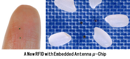
/030902_030902.jpg
[2] The world's smallest radio
frequency identification tags have been
unveiled by Japanese electronics firm
Hitachi. The minute devices measure
just 0.05mm by 0.05mm (0.002x0.002in)
and to the naked eye look like spots of
powder. Here the tiny tags can be
seen next to a human hair UNKNOWN
source: http://newsimg.bbc.co.uk/media/i
mages/42606000/jpg/_42606003_tag_203.jpg
[04/04/2003 AD]
Berkeley, California, USA

[1] Credit: Zettl Research Group LBNL,
University of California,
Berkley Electric Drives - Special
Purpose Motors (Description and
Applications) Motor
Construction Special purpose designs
have been developed to solve a wide
range of drive problems. Some common
examples are included here.
Integrated Starter Generator
(ISG) The electronically controlled
integrated starter generator used in
mild hybrid electric vehicles (HEVs)
combines the automotive starter and
alternator into a single machine. The
conventional starter is a low speed,
high current DC machine, while the
alternator is a variable speed 3 phase
AC machine. The ISG has four
important functions in a hybrid vehicle
application It enables the
''start-stop'' function, turning off
the engine when the vehicle is
stationary saving fuel. It
generates the electrical energy to
power all the electrical ancillaries.
It provides a power boost to assist
the engine when required, permitting
smaller engines for similar
performance. In some
configurations it recuperates energy
from regenerative braking. In a
typical implementation (below), the ISG
is a short axis, large diameter
''pancake'' shaped switched reluctance
machine mounted directly on the end of
the engine crankshaft between the
engine and the clutch in the gearbox
bell housing. Image source Long,
Schofield, Howe, Piron &
McClelland ''Design of a Switched
Reluctance Machine for Extended Speed
Operation'' IMEDC June 2003 The ISG
is a bi-directional energy converter
acting as a motor when powered by the
battery or a generator when driven by
the engine. The system voltage in a
mild HEV is 42 Volts which means that,
for the same cranking power as a 12
Volt machine, the starter current can
be reduced. Typical power throughput is
between 5kW and 15 kW with a possible
peak power of 70 kW for cold
cranking.. The brushless ISG design
eliminates one rotating machine
completely as well as the associated
commutator and brushes from the DC
machine and the sliprings and brushes
from the AC machine. The starter
solenoid, the Bendix ring (starter
gear) and the pulley or gear drive to
the alternator are also no longer
needed and because of the higher system
voltage, the diameter and weight of the
copper cabling is also reduced
substantially. The savings however
come at a cost. The system must be
integrated with several subsystems as
follows An AC/DC converter to
rectify the generator output voltage.
A DC/DC converter to supply the
vehicle's electrical power system
voltages. Power electronics and
software to control the ISG current,
voltage, speed, torque and temperature
as appropriate. An overall energy
management system integrated with the
vehicle's engine, battery and
brakes. Larger versions of this
construction are also used in full
hybrid electric vehicles. The
switched reluctance machine with its
simple rotor of inert iron is very
robust, able to operate at high speed
and to withstand the harsh operating
conditions in the engine
compartment. History
Outer Rotor Motors There are many
designs using this construction, mostly
for small sizes. Two examples of low
power motors are shown below. High
power versions are used for ''in
wheel'' automotive applications.
Inside Out Motor These are
permanent magnet motors with the moving
magnets arranged around the periphery
of a multi pole fixed stator carrying
the field windings.
Used for automotive drive
systems including in-wheel motors. Low
power versions used in small cooling
fans and direct drive record player
turntables. Toroidal Coil
Motor This is an ''inside out''
brushless permanent magnet motor with a
toroidal wound stator covered by a cup
shaped permanent magnet outer
rotor. Because
of the low inertia and friction free
rotor, the toroidal motor is capable of
speeds up to 25,000 RPM. Suitable for
low power applications it is used for
example to drive the polygonal rotating
mirrors which are mounted directly on
the rotor in laser printers.
Linear Motors In most cases the
linear motor can be considered as a
conventional rotary motor with both the
stator and the rotor split and rolled
out flat. The same electromagnetic
forces apply and these have been
employed in similar classes of AC and
DC machines. Except for traction motors
the travel of the motor armature is
usually quite short. Linear
Stepping Motors The most common
application is the stepping motor.
Stator poles are laid out along the
track and excited by windings fed from
a pulsed DC source. Permanent magnets
forming the armature are held in the
carriage. The carriage moves along the
track in response to pulses sent to the
the stator windings in much the same
way as the rotor turns in a brushless
DC motor. Closed loop control is
possible by mounting a position sensor
on the carriage. Despite the
elegance of the linear motor, linear
motion is more often provided by the
less expensive and more mundane method
of using a rotary stepping motor
driving a lead screw. Maglev
Traction Motors The principle of
the linear induction motor is used to
propel high speed Maglev (Magnetic
Levitation) trains which float on a
magnetic field created by
electromagnets in the trackbed under
the train . A separate set of trackside
guidance magnets is used to control the
lateral position of the train relative
to the track. Thus the maglev train
uses electromagnetic forces for three
different tasks, to suspend, to guide
and to propel the train. Maglev
trains have been developed in several
countries of the world using a variety
of configurations. Examples of the
essential features are described
below. Propulsion
The train has no onboard motor.
Electromagnets in the trackbed are
excited in sequence creating a linear
rather than a rotating field. By
transformer action, the trackbed coils
induce currents in coils on board the
train which are used to energise
powerful electromagnets. The Lorentz
force between the trackbed currents and
the the onboard electromagnets causes
the magnets to be propelled along by
the moving field. The
principles involved are very similar to
those of the induction motor but with
the static and moving parts
interchanged. See diagram below.
For illustrative
purposes the track can be likened to a
ladder formed by the unrolled squirrel
cage rotor of the induction motor. In
this case however it is fixed and it
supplies the moving field. Currents are
induced in the train's electromagnets
which are equivalent to the stator
poles of the induction motor but in
this case the magnets are free to move.
In practical designs the trackbed
currents are actually provided in a
series of individual coils laid along
the track. Levitation
Various levitation schemes are used.
The force holding the train aloft can
be created by the magnetic repulsion
between the same electromagnets on the
track and the onboard electromagnets in
the train which are used for
propulsion. The train's levitating
magnets are powered by direct current
supplied by a battery which is kept
charged by an induction generator
taking its power from the currents
induced by the trackbed coils in the
onboard generator coils. In
the diagram above, when the magnet is
directly above the current carrying
conductor as shown, the magnetic forces
(north and south poles) from the two
adjacent current loops cancel out and
there is no lift. If however the magnet
is moving very quickly over the coils,
it will reach a position over like,
repulsive, poles (north poles in the
diagram) which are displaced from the
attractive south poles so that the net
effect is a force repelling the magnet
away from the track. This is only
possible because the current in the
trackbed magnets lags the voltage due
to the inductance of the windings,
creating a delay in the build up of the
balanced field by which time the magnet
has moved into the adjacent region
where there is a net repulsive force.
This effect only happens when the
magnet on the train is moving at high
speed across the trackbed magnets. Thus
the train needs to be in motion for
this system to work and the train needs
wheels for support as it accelerates
from rest and when it is slowing to a
halt. Alternatively
levitation can be provided by separate
windings. The train's levitation
magnets protrude from the side of the
train and run between pairs of
vertically separated electromagnets in
guideways at each side of the train,
rather than in the trackbed. This
arrangement creates an attractive force
above the train's magnets combined with
a repulsive force beneath the train's
magnets to provide the levitating
force. Guidance For
guidance the train uses magnetic fields
provided by a separate set of weaker
magnets along each side of the train.
Similar in principle to the levitation
magnets they are used to control the
lateral position of the train relative
to the track.
Excitation of the trackside magnets is
arranged such that only the section
under the train is active. As the train
moves along the track between sections
the current to the previous section is
switched off and the current to the
next section is switched on pulling the
train along. This serves the dual
purpose of avoiding losses by
energising only the section of track
directly under the train and at the
same time, since the power to the rest
of the track is switched off, it
provides security against electric
shock to anybody near to the track and
avoids the possibility of accidentally
short circuiting the system by dropping
rubbish onto live conductors.
Very high armature currents of
thousands of amps or more are involved
and some designs use high temperature
superconductors ( HTS ) in the onboard
magnets, cooled with liquid nitrogen or
helium to minimise the resistive
losses. As might be expected some
sophisticated control systems are
needed to keep everything on track.
History Axial Field
Motors Axial field motors have been
developed for applications which
require short, flat, ''pancake''
construction. Printed Circuit
(PCB) or ''Pancake'' Motor The
printed circuit motor is an example of
an ironless or coreless motor with
several unique features. The pancake
construction uses an axial magnetic
field to achieve the short flat
construction. Radial field PCB motors
are also possible.
Construction The rotor windings
are printed, stamped or welded onto a
thin, disc shaped glass fibre circuit
board which rotates in the air gap
between pairs of permanent magnets
arranged around the periphery of the
disk. The windings fan out in a series
of radial loops around the surface of
the disk. The magnets are arranged
alternatively north and south so that
the magnetic fields in the air gaps of
adjacent magnet pairs are in opposite
directions. The magnets are held in
place by two iron end caps in a compact
''pancake'' shaped block to complete
the magnetic circuit. Current is fed to
the rotor windings via brushes through
precious metal commutator segments
printed on the disc. Operating
Principle Traditional electric
motors have a radial magnetic field or
flux with the rotor current flowing
axially along the length of the rotor.
In typical printed circuit motors the
construction is reversed. The magnetic
field is axial (oriented along the axis
of the machine) and the current flows
radially from the axis to the edge of
the disc and back again. A tangential
force on the disk is created by the
current passing through the magnetic
fields in the air gaps between the pole
pairs of the permanent magnets. So that
the return current does not cancel out
the effect of the outgoing current, the
return wire is physically separated or
displaced to one side from the outgoing
wire by the width of the magnet. In
this way it interacts with the magnetic
field of the adjacent magnet which is
in the opposite direction and thus
reinforces the tangential force on the
disk. In many ways it is similar
to Faraday's 1831disk or homopolar
motor which used a single magnet and
was driven by a unidirectional current
fed by brushes at the centre and on the
periphery of the disk.
Applications The printed circuit
motor is a very compact and light
weight design making it useful in
confined spaces. Since the rotor does
not have drag a lump of iron around, it
has very low inertia and can run up to
speed very quickly. Because of the many
commutator segments and the low current
capability of the windings, the PCB
motor is only suitable for low power
applications and is not suitable for
continuous operation. It is however
ideal for servo systems and industrial
controls and automotive applications
such as electric window winders.
Micro-motors
(Micro-ElectroMechanical Systems -
MEMS) Electrostatic Motor
The motor shown below is an example of
semiconductor manufacturing technology
used to fabricate very small mechanical
components. It measures 100 microns
across, or about the width of a human
hair. Similar in principle to a
reluctance motor, it depends on
electrostatic attraction, rather than
magnetic attraction, between the stator
and rotor poles. Because the dimensions
are so tiny, very high electric fields
can be built up with only a few volts
between the motor poles.
Fan Long-Shen, Tai Yu-Chong
and Richard S. Muller 1989
IC-processed electrostatic
micromotors Sensors Actuators 20
41-7 Fan L-S, Tai Y-C and R S
Muller 1988 Integrated moveable
micromechanical structures for sensors
and actuators IEEE Trans. Electron
Devices The motor is
not assembled from individual
components. Instead the components are
built up on a semiconductor substrate
by masking and etching and a mask-less
post-processing release step is
performed to etch away sacrificial
layers, allowing the structural layers
to move and rotate.
Micromachined micromotors can be
monolithically integrated together with
the necessary CMOS drive circuits,
containing oscillators, frequency
dividers and counters, and transistors
for the drive circuit all on one
silicon chip. Common uses
include defense/munitions applications,
computer hard drives, optics, sensors
and actuators.
History Nano-motors
(Nano-ElectroMechanical Systems -
NEMS) Electrostatic Motor
Even smaller motors have been made
using nanotechnology. An example is
shown below. It consists of a tiny gold
slab rotor, about 100 nm square,
mounted on concentric carbon nanotubes.
The outer tube carries the rotor,
driven by electrostatic electrodes,
rotating around an inner tube which
acts as a supporting shaft. By applying
voltage pulses of up to 5 Volts between
the rotor plate and stators, the
position, speed and direction of
rotation of the rotor can be
controlled. It measures about 500
nanometers across, 300 times smaller
than the diameter of a human hair.
UNKNOWN
source: http://www.mpoweruk.com/images/n
ems.gif
[2] Credit: Zettl Research
Group LBNL, University of California,
Berkley Electric Drives - Special
Purpose Motors (Description and
Applications) Motor
Construction Special purpose designs
have been developed to solve a wide
range of drive problems. Some common
examples are included here.
Integrated Starter Generator
(ISG) The electronically controlled
integrated starter generator used in
mild hybrid electric vehicles (HEVs)
combines the automotive starter and
alternator into a single machine. The
conventional starter is a low speed,
high current DC machine, while the
alternator is a variable speed 3 phase
AC machine. The ISG has four
important functions in a hybrid vehicle
application It enables the
''start-stop'' function, turning off
the engine when the vehicle is
stationary saving fuel. It
generates the electrical energy to
power all the electrical ancillaries.
It provides a power boost to assist
the engine when required, permitting
smaller engines for similar
performance. In some
configurations it recuperates energy
from regenerative braking. In a
typical implementation (below), the ISG
is a short axis, large diameter
''pancake'' shaped switched reluctance
machine mounted directly on the end of
the engine crankshaft between the
engine and the clutch in the gearbox
bell housing. Image source Long,
Schofield, Howe, Piron &
McClelland ''Design of a Switched
Reluctance Machine for Extended Speed
Operation'' IMEDC June 2003 The ISG
is a bi-directional energy converter
acting as a motor when powered by the
battery or a generator when driven by
the engine. The system voltage in a
mild HEV is 42 Volts which means that,
for the same cranking power as a 12
Volt machine, the starter current can
be reduced. Typical power throughput is
between 5kW and 15 kW with a possible
peak power of 70 kW for cold
cranking.. The brushless ISG design
eliminates one rotating machine
completely as well as the associated
commutator and brushes from the DC
machine and the sliprings and brushes
from the AC machine. The starter
solenoid, the Bendix ring (starter
gear) and the pulley or gear drive to
the alternator are also no longer
needed and because of the higher system
voltage, the diameter and weight of the
copper cabling is also reduced
substantially. The savings however
come at a cost. The system must be
integrated with several subsystems as
follows An AC/DC converter to
rectify the generator output voltage.
A DC/DC converter to supply the
vehicle's electrical power system
voltages. Power electronics and
software to control the ISG current,
voltage, speed, torque and temperature
as appropriate. An overall energy
management system integrated with the
vehicle's engine, battery and
brakes. Larger versions of this
construction are also used in full
hybrid electric vehicles. The
switched reluctance machine with its
simple rotor of inert iron is very
robust, able to operate at high speed
and to withstand the harsh operating
conditions in the engine
compartment. History
Outer Rotor Motors There are many
designs using this construction, mostly
for small sizes. Two examples of low
power motors are shown below. High
power versions are used for ''in
wheel'' automotive applications.
Inside Out Motor These are
permanent magnet motors with the moving
magnets arranged around the periphery
of a multi pole fixed stator carrying
the field windings.
Used for automotive drive
systems including in-wheel motors. Low
power versions used in small cooling
fans and direct drive record player
turntables. Toroidal Coil
Motor This is an ''inside out''
brushless permanent magnet motor with a
toroidal wound stator covered by a cup
shaped permanent magnet outer
rotor. Because
of the low inertia and friction free
rotor, the toroidal motor is capable of
speeds up to 25,000 RPM. Suitable for
low power applications it is used for
example to drive the polygonal rotating
mirrors which are mounted directly on
the rotor in laser printers.
Linear Motors In most cases the
linear motor can be considered as a
conventional rotary motor with both the
stator and the rotor split and rolled
out flat. The same electromagnetic
forces apply and these have been
employed in similar classes of AC and
DC machines. Except for traction motors
the travel of the motor armature is
usually quite short. Linear
Stepping Motors The most common
application is the stepping motor.
Stator poles are laid out along the
track and excited by windings fed from
a pulsed DC source. Permanent magnets
forming the armature are held in the
carriage. The carriage moves along the
track in response to pulses sent to the
the stator windings in much the same
way as the rotor turns in a brushless
DC motor. Closed loop control is
possible by mounting a position sensor
on the carriage. Despite the
elegance of the linear motor, linear
motion is more often provided by the
less expensive and more mundane method
of using a rotary stepping motor
driving a lead screw. Maglev
Traction Motors The principle of
the linear induction motor is used to
propel high speed Maglev (Magnetic
Levitation) trains which float on a
magnetic field created by
electromagnets in the trackbed under
the train . A separate set of trackside
guidance magnets is used to control the
lateral position of the train relative
to the track. Thus the maglev train
uses electromagnetic forces for three
different tasks, to suspend, to guide
and to propel the train. Maglev
trains have been developed in several
countries of the world using a variety
of configurations. Examples of the
essential features are described
below. Propulsion
The train has no onboard motor.
Electromagnets in the trackbed are
excited in sequence creating a linear
rather than a rotating field. By
transformer action, the trackbed coils
induce currents in coils on board the
train which are used to energise
powerful electromagnets. The Lorentz
force between the trackbed currents and
the the onboard electromagnets causes
the magnets to be propelled along by
the moving field. The
principles involved are very similar to
those of the induction motor but with
the static and moving parts
interchanged. See diagram below.
For illustrative
purposes the track can be likened to a
ladder formed by the unrolled squirrel
cage rotor of the induction motor. In
this case however it is fixed and it
supplies the moving field. Currents are
induced in the train's electromagnets
which are equivalent to the stator
poles of the induction motor but in
this case the magnets are free to move.
In practical designs the trackbed
currents are actually provided in a
series of individual coils laid along
the track. Levitation
Various levitation schemes are used.
The force holding the train aloft can
be created by the magnetic repulsion
between the same electromagnets on the
track and the onboard electromagnets in
the train which are used for
propulsion. The train's levitating
magnets are powered by direct current
supplied by a battery which is kept
charged by an induction generator
taking its power from the currents
induced by the trackbed coils in the
onboard generator coils. In
the diagram above, when the magnet is
directly above the current carrying
conductor as shown, the magnetic forces
(north and south poles) from the two
adjacent current loops cancel out and
there is no lift. If however the magnet
is moving very quickly over the coils,
it will reach a position over like,
repulsive, poles (north poles in the
diagram) which are displaced from the
attractive south poles so that the net
effect is a force repelling the magnet
away from the track. This is only
possible because the current in the
trackbed magnets lags the voltage due
to the inductance of the windings,
creating a delay in the build up of the
balanced field by which time the magnet
has moved into the adjacent region
where there is a net repulsive force.
This effect only happens when the
magnet on the train is moving at high
speed across the trackbed magnets. Thus
the train needs to be in motion for
this system to work and the train needs
wheels for support as it accelerates
from rest and when it is slowing to a
halt. Alternatively
levitation can be provided by separate
windings. The train's levitation
magnets protrude from the side of the
train and run between pairs of
vertically separated electromagnets in
guideways at each side of the train,
rather than in the trackbed. This
arrangement creates an attractive force
above the train's magnets combined with
a repulsive force beneath the train's
magnets to provide the levitating
force. Guidance For
guidance the train uses magnetic fields
provided by a separate set of weaker
magnets along each side of the train.
Similar in principle to the levitation
magnets they are used to control the
lateral position of the train relative
to the track.
Excitation of the trackside magnets is
arranged such that only the section
under the train is active. As the train
moves along the track between sections
the current to the previous section is
switched off and the current to the
next section is switched on pulling the
train along. This serves the dual
purpose of avoiding losses by
energising only the section of track
directly under the train and at the
same time, since the power to the rest
of the track is switched off, it
provides security against electric
shock to anybody near to the track and
avoids the possibility of accidentally
short circuiting the system by dropping
rubbish onto live conductors.
Very high armature currents of
thousands of amps or more are involved
and some designs use high temperature
superconductors ( HTS ) in the onboard
magnets, cooled with liquid nitrogen or
helium to minimise the resistive
losses. As might be expected some
sophisticated control systems are
needed to keep everything on track.
History Axial Field
Motors Axial field motors have been
developed for applications which
require short, flat, ''pancake''
construction. Printed Circuit
(PCB) or ''Pancake'' Motor The
printed circuit motor is an example of
an ironless or coreless motor with
several unique features. The pancake
construction uses an axial magnetic
field to achieve the short flat
construction. Radial field PCB motors
are also possible.
Construction The rotor windings
are printed, stamped or welded onto a
thin, disc shaped glass fibre circuit
board which rotates in the air gap
between pairs of permanent magnets
arranged around the periphery of the
disk. The windings fan out in a series
of radial loops around the surface of
the disk. The magnets are arranged
alternatively north and south so that
the magnetic fields in the air gaps of
adjacent magnet pairs are in opposite
directions. The magnets are held in
place by two iron end caps in a compact
''pancake'' shaped block to complete
the magnetic circuit. Current is fed to
the rotor windings via brushes through
precious metal commutator segments
printed on the disc. Operating
Principle Traditional electric
motors have a radial magnetic field or
flux with the rotor current flowing
axially along the length of the rotor.
In typical printed circuit motors the
construction is reversed. The magnetic
field is axial (oriented along the axis
of the machine) and the current flows
radially from the axis to the edge of
the disc and back again. A tangential
force on the disk is created by the
current passing through the magnetic
fields in the air gaps between the pole
pairs of the permanent magnets. So that
the return current does not cancel out
the effect of the outgoing current, the
return wire is physically separated or
displaced to one side from the outgoing
wire by the width of the magnet. In
this way it interacts with the magnetic
field of the adjacent magnet which is
in the opposite direction and thus
reinforces the tangential force on the
disk. In many ways it is similar
to Faraday's 1831disk or homopolar
motor which used a single magnet and
was driven by a unidirectional current
fed by brushes at the centre and on the
periphery of the disk.
Applications The printed circuit
motor is a very compact and light
weight design making it useful in
confined spaces. Since the rotor does
not have drag a lump of iron around, it
has very low inertia and can run up to
speed very quickly. Because of the many
commutator segments and the low current
capability of the windings, the PCB
motor is only suitable for low power
applications and is not suitable for
continuous operation. It is however
ideal for servo systems and industrial
controls and automotive applications
such as electric window winders.
Micro-motors
(Micro-ElectroMechanical Systems -
MEMS) Electrostatic Motor
The motor shown below is an example of
semiconductor manufacturing technology
used to fabricate very small mechanical
components. It measures 100 microns
across, or about the width of a human
hair. Similar in principle to a
reluctance motor, it depends on
electrostatic attraction, rather than
magnetic attraction, between the stator
and rotor poles. Because the dimensions
are so tiny, very high electric fields
can be built up with only a few volts
between the motor poles.
Fan Long-Shen, Tai Yu-Chong
and Richard S. Muller 1989
IC-processed electrostatic
micromotors Sensors Actuators 20
41-7 Fan L-S, Tai Y-C and R S
Muller 1988 Integrated moveable
micromechanical structures for sensors
and actuators IEEE Trans. Electron
Devices The motor is
not assembled from individual
components. Instead the components are
built up on a semiconductor substrate
by masking and etching and a mask-less
post-processing release step is
performed to etch away sacrificial
layers, allowing the structural layers
to move and rotate.
Micromachined micromotors can be
monolithically integrated together with
the necessary CMOS drive circuits,
containing oscillators, frequency
dividers and counters, and transistors
for the drive circuit all on one
silicon chip. Common uses
include defense/munitions applications,
computer hard drives, optics, sensors
and actuators.
History Nano-motors
(Nano-ElectroMechanical Systems -
NEMS) Electrostatic Motor
Even smaller motors have been made
using nanotechnology. An example is
shown below. It consists of a tiny gold
slab rotor, about 100 nm square,
mounted on concentric carbon nanotubes.
The outer tube carries the rotor,
driven by electrostatic electrodes,
rotating around an inner tube which
acts as a supporting shaft. By applying
voltage pulses of up to 5 Volts between
the rotor plate and stators, the
position, speed and direction of
rotation of the rotor can be
controlled. It measures about 500
nanometers across, 300 times smaller
than the diameter of a human hair.
UNKNOWN
source: http://www.mpoweruk.com/images/n
ems.gif
[07/01/2004 AD]

[1] * original caption: Jet Propulsion
Laboratory (JPL) workers use a
borescope to verify pressure relief
device bellows integrity on a
radioisotope thermoelectric generator
(RTG) which has been installed on the
Cassini spacecraft in the Payload
Hazardous Servicing Facility. The
activity is part of the mechanical and
electrical verification testing of RTGs
during prelaunch processing. RTGs use
heat from the natural decay of
plutonium to generate electric power.
The three RTGs on Cassini will enable
the spacecraft to operate far from the
Sun where solar power systems are not
feasible. They will provide electrical
power to Cassini on its 6.7-year trip
to the Saturnian system and during its
four-year mission at Saturn. The
Cassini mission is scheduled for an
Oct. 6 launch aboard a Titan
IVB/Centaur expendable launch vehicle.
Cassini is built and managed for NASA
by JPL. * date: 18. Dec 1997
* image ID: KSC-97PC-1070 *
source:
http://nix.ksc.nasa.gov/info;jsessionid=
1tplxxjif20rp?id=KSC-97PC-1070&orgid=5
PD
source: http://upload.wikimedia.org/wiki
pedia/commons/6/61/Cassini_assembly.jpg

[2] Original Caption Released with
Image: This is an artists concept of
Cassini during the Saturn Orbit
Insertion (SOI) maneuver, just after
the main engine has begun firing. The
spacecraft is moving out of the plane
of the page and to the right (firing to
reduce its spacecraft velocity with
respect to Saturn) and has just crossed
the ring plane. The SOI maneuver,
which is approximately 90 minutes long,
will allow Cassini to be captured by
Saturn's gravity into a five-month
orbit. Cassini's close proximity to the
planet after the maneuver offers a
unique opportunity to observe Saturn
and its rings at extremely high
resolution. Source:
http://photojournal.jpl.nasa.gov/catalog
/PIA03883 PD
source: http://upload.wikimedia.org/wiki
pedia/commons/b/b2/Cassini_Saturn_Orbit_
Insertion.jpg
[12/10/2008 AD]
what the eyes are seeing captured
remotely.
two Japanese Universities, two research
Institutes, and ATR Computational
Neuroscience Laboratories) Kyoto,
Japan
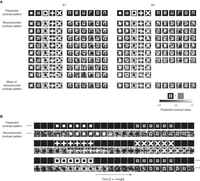
[1] Image from 12/10/2008 ''Neuron''
paper COPYRIGHTED
source: http://www.cell.com/neuron/image
/S0896-6273(08)00958-6?imageId=gr2&image
Type=large

[2] Image from 12/10/2008 ''Neuron''
paper COPYRIGHTED
source: http://www.cell.com/neuron/image
/S0896-6273(08)00958-6?imageId=gr1&image
Type=large
[2015 AD]
remotely.
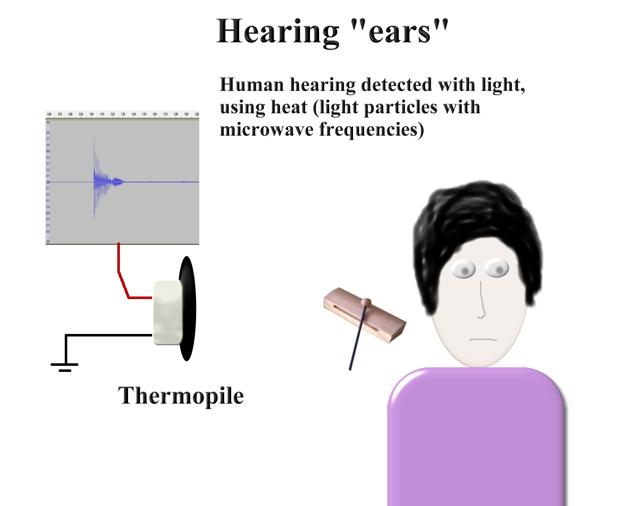
[1] Image by Ted Huntington GNU
source: Ted Huntington

[2] Storyboard image by Ted
Huntington GNU
source: Ted Huntington
[2018 AD]
organelle.

[1] Adapted from: Description
English: Drawing illustrating the
process of synaptic transmission in
neurons, cropped from original in an
NIA brochure. Date 2009-12-30,
first publication of original
unknown Source
http://www.nia.nih.gov/Alzheimers/P
ublications/UnravelingtheMystery/ Autho
r user:Looie496 created file, US
National Institutes of Health, National
Institute on Aging created
original Permission (Reusing this
file)
http://www.nia.nih.gov/Policies.htm Ot
her versions
http://en.wikipedia.org/wiki/File:Chemi
cal_synapse_schema.jpg PD
source: http://upload.wikimedia.org/wiki
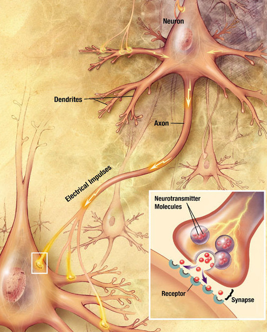
pedia/commons/3/30/Chemical_synapse_sche
ma_cropped.jpg

[2] Storyboard image by Ted
Huntington PD
source: Ted Huntington
[2020 AD]

[1] Drive Gear Side View Side view
of a microengine drive gear meshed with
another gear. PD
source: http://mems.sandia.gov/gallery/i
mages/rs1563_9.jpg

[2] Imaginary microscopic flying
camera on top of salt crystals Ted
Huntington PD
source: http://tedhuntington.com/saltcry
stal_127um.jpg
[2025 AD]
microscopic devices in neurons.

[1] Image of Remote neuron writing by
Ted Huntington PD
source: Ted Huntington
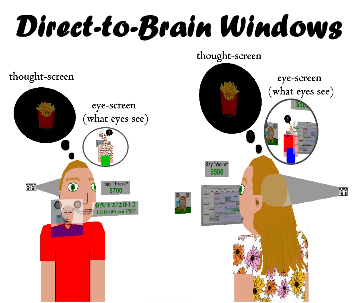
[2] Image of Remote neuron writing by
Ted Huntington PD
source: Ted Huntington
[2025 AD]

[1] Torsional Ratcheting Actuator A
high torque rotary electrostatic
actuator PD
source: http://www.mems.sandia.gov/image
s/mems5.jpg

[2] Storyboard image by Ted
Huntington PD
source: Ted Huntington
[2025 AD]
[1] Imaginary microscopic flying camera
on top of salt crystals Ted
Huntington PD
source: http://tedhuntington.com/saltcry
stal_127um.jpg

[2] Storyboard image by Ted
Huntington PD
source: Ted Huntington
[2030 AD]
out loud. Humans start to communicate
by thought-image and thought-sound
only.

[1] Hearing Thought by Ted
Huntington GNU
source: Ted Huntington
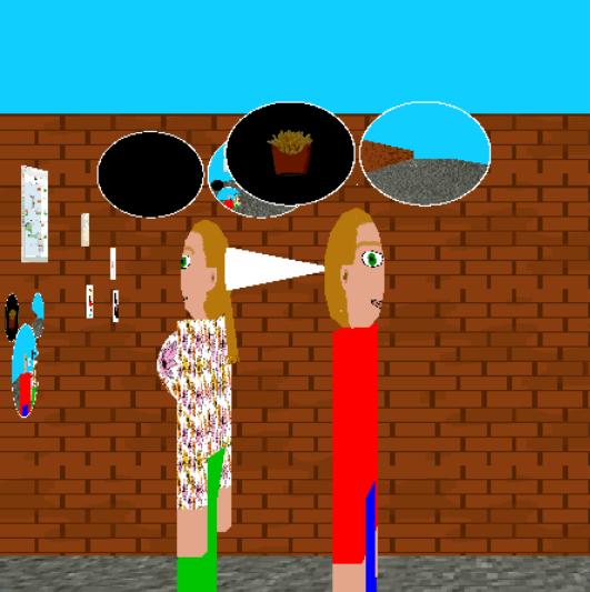
[2] Image of Direct-to-brain windows
by Ted Huntington GNU
source: http://www.tedhuntington.com/Mic
key_Mouse_eyes_thought_screens.jpg
[2030 AD]

[1] Muscular system P150/0098 Rights
Managed Credit: PASIEKA/SCIENCE
PHOTO LIBRARY Caption: Muscular
system. Computer artwork of a male
runner demonstrating the human muscular
system. These muscles, making up one of
the outer layers of the body, are
skeletal muscles, which are under the
conscious control of the brain. They
account for around 45% of the weight of
the body. Muscles are mainly composed
of proteins; in particular, the
proteins actin and myosin are involved
in making muscles contract. The muscles
then pull on tendons that are attached
to the bones of the skeleton, producing
movements such as running. Skin and fat
cover the muscles, while the internal
organs and bones lie
underneath. Release details: Model
and property releases are not available
UNKNOWN
source: http://www.sciencephoto.com/imag
e/302911/large/P1500098-Muscular_system-
SPL.jpg

[2] Muscular system P150/0098
Rights Managed Credit:
PASIEKA/SCIENCE PHOTO
LIBRARY Caption: Muscular system.
Computer artwork of a male runner
demonstrating the human muscular
system. These muscles, making up one of
the outer layers of the body, are
skeletal muscles, which are under the
conscious control of the brain. They
account for around 45% of the weight of
the body. Muscles are mainly composed
of proteins; in particular, the
proteins actin and myosin are involved
in making muscles contract. The muscles
then pull on tendons that are attached
to the bones of the skeleton, producing
movements such as running. Skin and fat
cover the muscles, while the internal
organs and bones lie
underneath. Release details: Model
and property releases are not available
UNKNOWN
source: http://www.sciencephoto.com/imag
e/302911/large/P1500098-Muscular_system-
SPL.jpg
[2030 AD]
publicly.
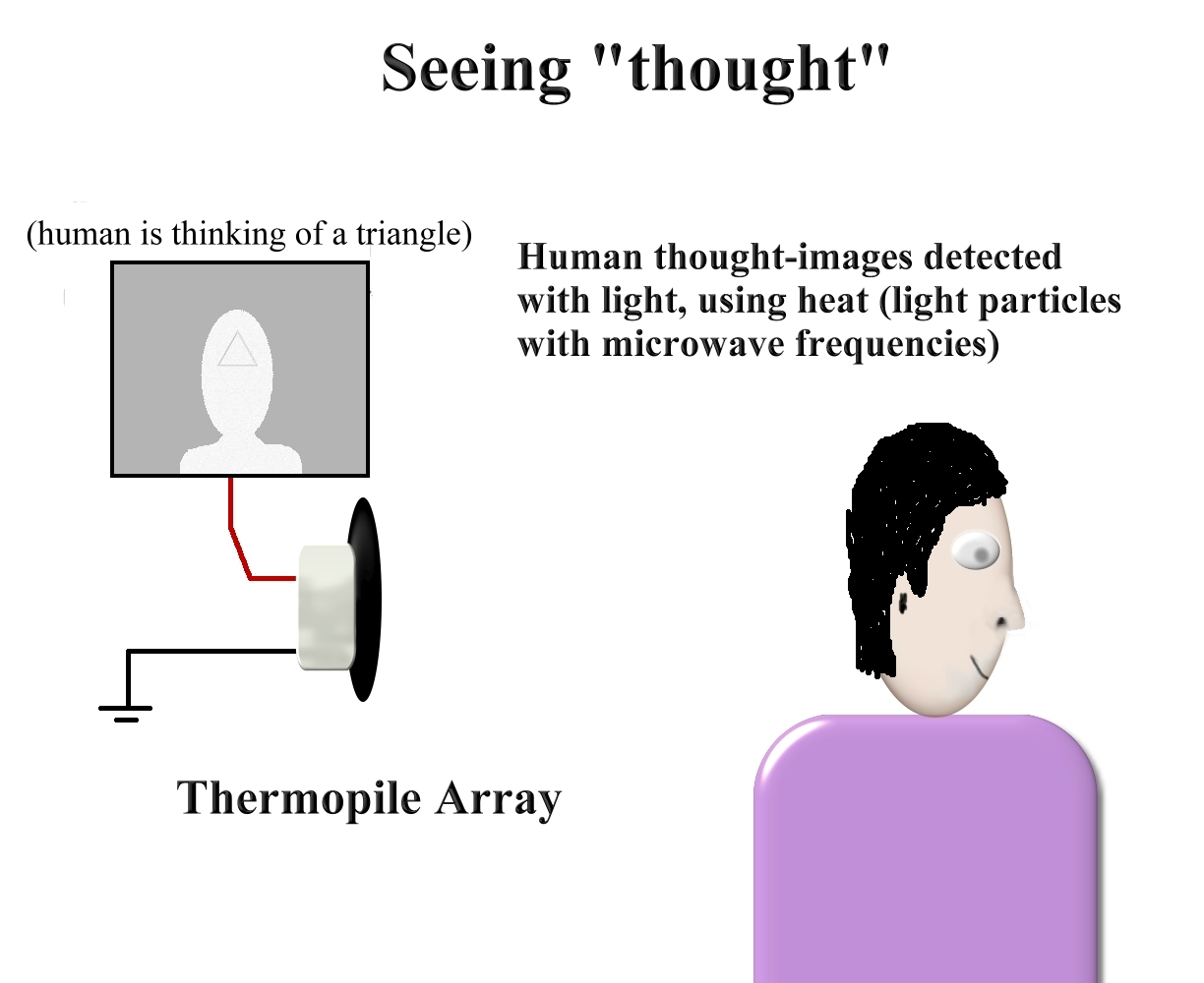
[1] Image of seeing thought by Ted
Huntington. PD
source: Ted Huntington
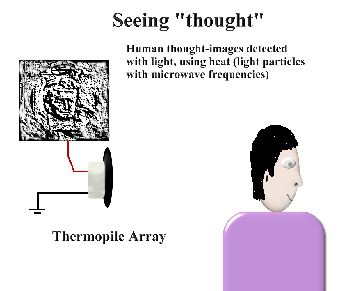
[2] Image of seeing thought by Ted
Huntington. Image of person
from: http://upload.wikimedia.org/wikip
edia/commons/3/30/Cassatt_Mary_Portrait_
of_a_Elderly_Lady_1883.jpg Title:
Portrait of a Elderly Lady Date:
1883 Mary Cassatt (1845–1926) Link
back to Creator infobox
template PD [1] Image of
Direct-to-brain windows by Ted
Huntington GNU
source: Ted
Huntingtonhttp://upload.wikimedia.org/wi
kipedia/commons/3/30/Cassatt_Mary_Portra
it_of_a_Elderly_Lady_1883.jpg
[2030 AD]
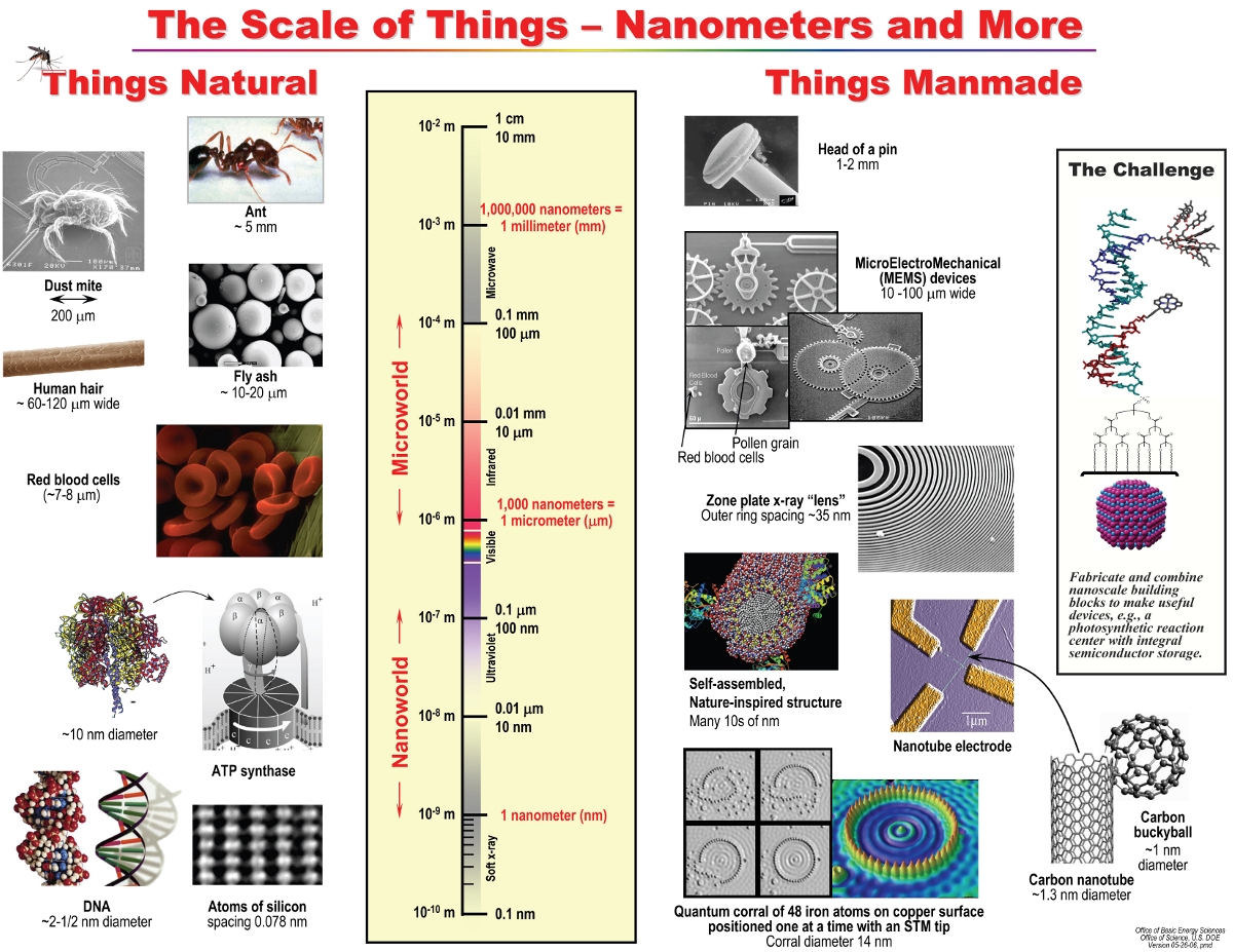
[1] The Scale of Things - Nanometers
and More the scale of things
graphic Chart also available in
Powerpoint file at:
http://science.energy.gov/bes/news-and-r
esources/scale-of-things-chart/. PD
source: http://science.energy.gov/~/medi
a/bes/images/scale-of-things-26may06.jpg

[2] The space between two carbon atoms
within a molecule is about one-tenth of
a nanometer. The DNA double helix has a
diameter of about two nanometers. The
smallest bacteria, on the other hand,
are much bigger: a few hundred
nanometers in length. PD
source: http://publications.nigms.nih.go
v/chemhealth/images/ch4_size.jpg
[2050 AD]
servants.

[1] Ted Huntington image of two humans
walking with robot servants. GNU
source: Ted Huntington
[2] Storyboard image by Ted
Huntington GNU
source: Ted Huntington
[2055 AD]
devices.

[1] Adapted from: Pictured is a breast
cancer cell, photographed by a scanning
electron microscope. This picture shows
the overall shape of the cell's surface
at a very high magnification. Cancer
cells are best identified by internal
details, but research with a scanning
electron microscope can show how cells
respond in changing environments and
can show mapping distribution of
binding sites of hormones and other
biological molecules. (National Cancer
Institute) UNKNOWN
source: http://cache.boston.com/universa
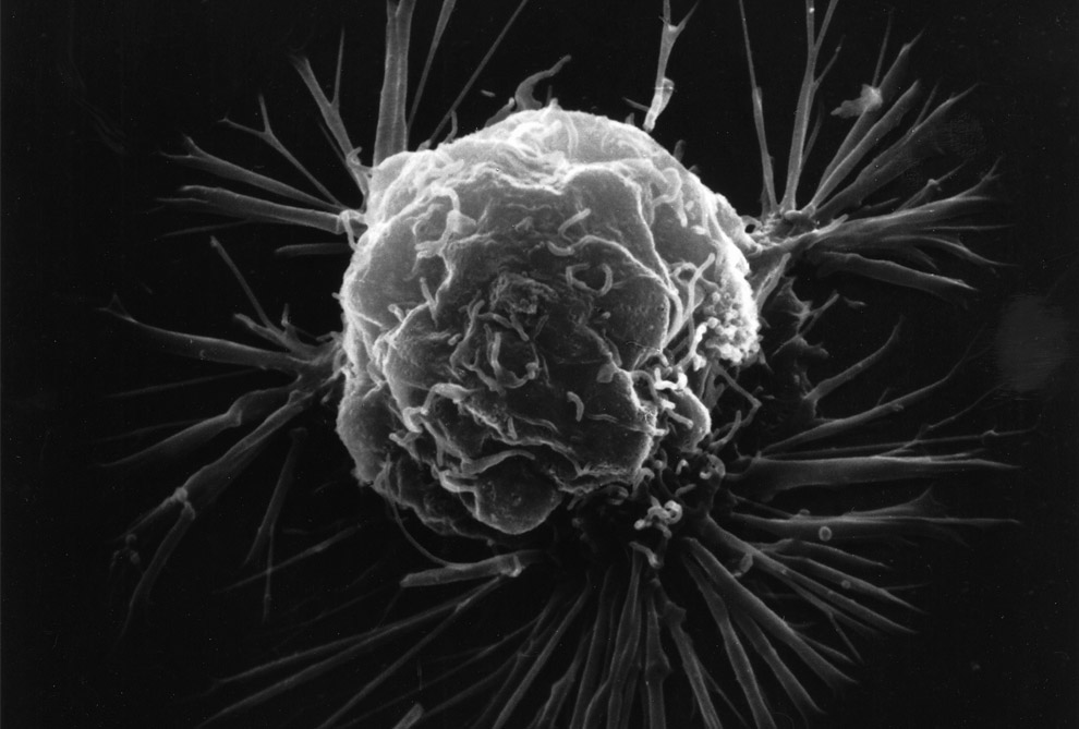
l/site_graphics/blogs/bigpicture/micro_1
1_14/m31_3b.jpg

[2] Lung Cancer Cells This image of
warped lung cancer cells is in stark
contrast to the healthy lung. UNKNOWN
source: http://1.bp.blogspot.com/_kGhJLc
78v60/TCytjueY3wI/AAAAAAAAA00/F8-TCWOsNq
4/s1600/Lung+cancer+cellsl.jpg
[2100 AD]
images and sounds of thought.
[1] Image of Direct-to-brain windows by
Ted Huntington GNU
source: http://www.tedhuntington.com/Mic
key_Mouse_eyes_thought_screens.jpg
[2] Image of Direct-to-brain windows
by Ted Huntington GNU
source: http://www.tedhuntington.com/dir
ect-to-brain_windows_002.jpg
[2100 AD]
of traffic above the streets.
[1] Image of single helicopter highway
by Ted Huntington GNU
source: Ted Huntington

[2] Image of double helicopter highway
by Ted Huntington Note that
helicopters are moving in wrong
way. GNU
source: Ted Huntington
[2100 AD]

[1] Three TDRS satellites, the
International Space Station (ISS) and
Hubble Space Telescope orbit a
blue-green Earth in this artist's
concept. The TDRS network facilitates
around the clock communication access
between ground stations and other
satellites and the ISS. Credit:
NASA/Goddard Space Flight Center ›
Larger image PD
source: http://www.nasa.gov/images/conte
nt/605410main_tdrsPlus.jpg

[2] Storyboard image by Ted
Huntington GNU
source: Ted Huntington
[2140 AD]
atoms like Iron converted into more
useful atoms like Hydrogen and Oxygen
using particle colliders.
[1] Image of Large Scale Atomic
Transmutation by Ted Huntington GNU
source: Ted Huntington
[2] Image of Large Scale Atomic
Transmutation by Ted Huntington GNU
source: Ted Huntington
[2150 AD]
microscopic devices.

[1] nanocaduceus Nanomedicine Art
Gallery Image 108 DNA Repair
Machines Image 108 Title:
DNA Repair Machines Date:
1989 Artist(s): Bryan Leister
Image Size: 45,592 bytes
[CLICK to download] Image Dimensions:
800 X 1,061 pixels Image
Palette: Color Image Size:
408,316 bytes [CLICK to
download] Image Dimensions: 1,170
X 1,552 pixels Image Palette:
Color Image Description:
Floating inside the nucleus of a human
cell, an assembler-built repair vessel
performs some genetic maintenance.
Stretching a supercoil of DNA between
its lower pair of robot arms, the
nanomachine gently pulls the unwound
strand through an opening in its prow
for analysis. Upper arms, meanwhile,
detach regulatory proteins from the
chain and place them in an intake port.
The molecular structures of both DNA
and proteins are compared to
information stored in the database of a
larger nanocomputer positioned outside
the nucleus and connected to the
cell-repair ship by a communications
link. Irregularities found in either
structure are corrected and the
proteins reattached to the DNA chain,
which re-coils into its original
form. With a diameter of only 50
nanometers, the repair vessel would be
smaller than most bacteria and viruses,
yet capable of therapies and cures well
beyond the reach of present-day
physicians. With trillions of these
machines coursing through a patient's
bloodstream, ''internal medicine''
would take on new significance. Disease
would be attacked at the molecular
level, and such maladies as cancer,
viral infections and arteriosclerosis
could be wiped out. Copyright Info:
© Copyright 1989 by Time-Life Books
Inc. For reprint permission, please
contact Time-Life Books Inc. at P.O.
Box C-32068, Richmond, VA 23261-2068,
Tel. 1-800-621-7026, URL:
http://www.timelife.com/. Print
Source(s): Time-Life Editors,
Alternative Computers, Time-Life Books,
Richmond VA, 1989, pp. 112-113. Online
Source(s): None COPYRIGHTED
source: http://www.foresight.org/Nanomed
icine/Gallery/Images/TimeLifeNM.jpeg
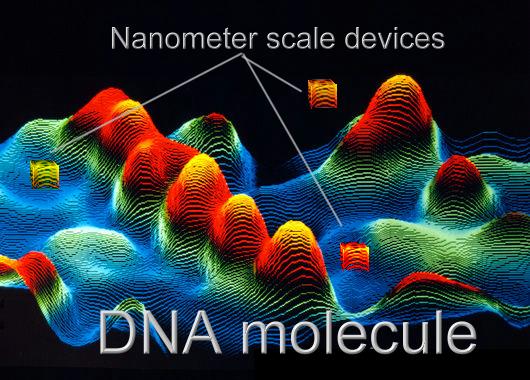
[2] Microscopic laser-machined
particle communication devices identify
and change nucleotides in a DNA
molecule as seen with an scanning
tunneling microscope.[t] Adapted
from: F/col STM image of
DNA G110/0150 Rights Managed Credit:
LAWRENCE LIVERMORE LABORATORY/SCIENCE
PHOTO LIBRARY Caption: False-colour
scanning tunnelling micrograph (STM) of
DNA. A sample of uncoated,
double-stranded DNA was dissolved in a
salt solution & deposited on graphite
prior to being imaged in air by the
STM. An STM image is formed by scanning
a fine point just above the specimen
surface & electronically recording the
height of the point as it moves. The
main feature of this image is a
right-handed, double-stranded DNA
molecule (a DNA duplex), which appears
as the row of orange/yellow peaks at
centre-left. These peaks correspond to
the ridges of the DNA double helix.
Magnification: x1,600,000 at 6x7cm
size. Release details: Model and
property releases are not
available UNKNOWN
source: http://www.sciencephoto.com/imag
e/209654/large/G1100150-F_col_STM_image_
of_DNA-SPL.jpg
[2180 AD]
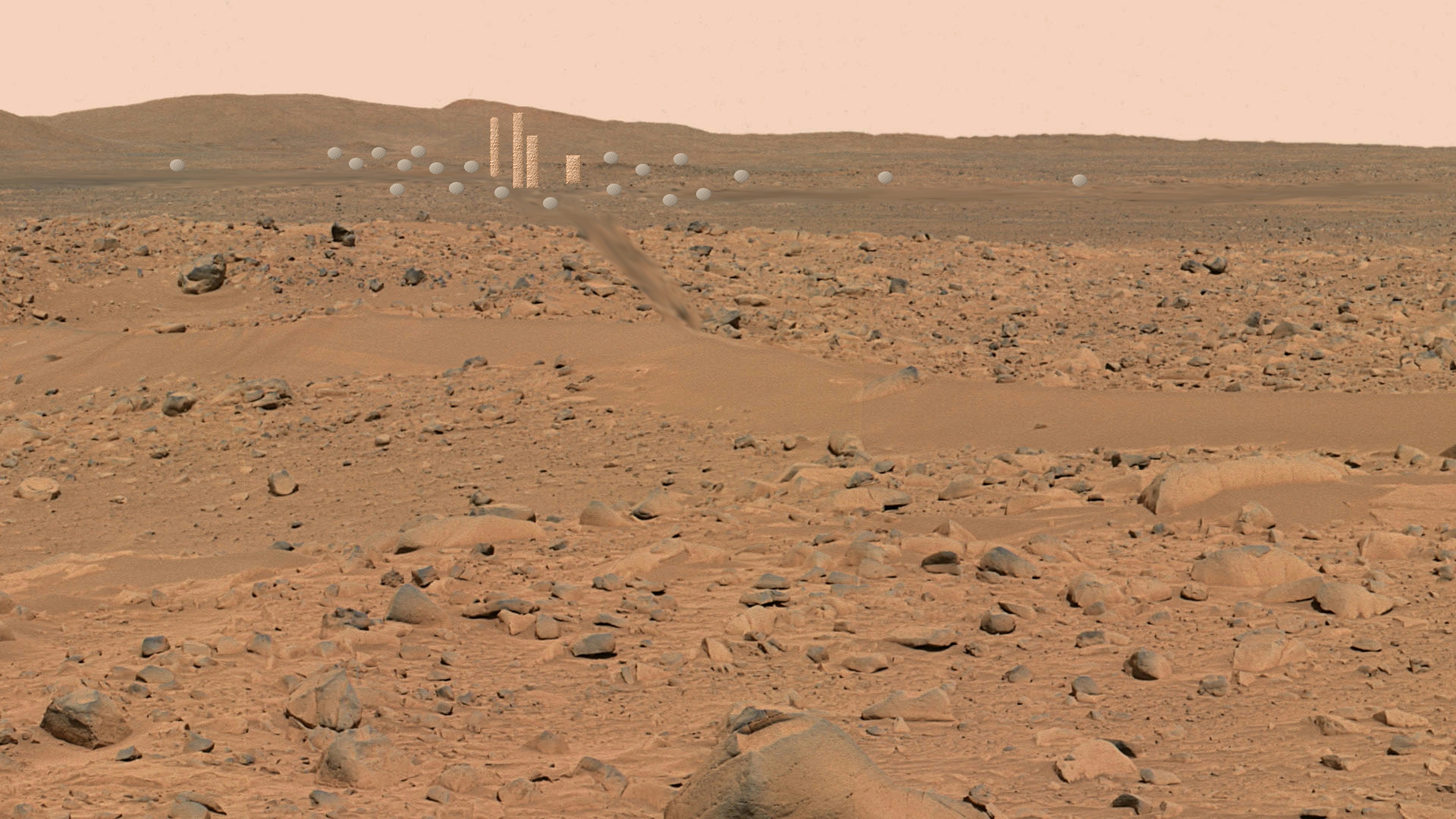
[1] Adapted from: Spirit's
Destination This image, cropped from
a larger panoramic image mosaic taken
by the Mars Exploration Rover Spirit
panoramic camera, shows the rover's
destination toward the hills nicknamed
the ''Columbia Hills.'' The rover is
currently positioned outside the view
of this image, on the right. This image
was taken on sols 68 and 69 of Spirit's
mission (March 12 and 13, 2004) from
the location the rover first reached on
the western rim of the crater. The
image is in approximate true color,
based on a scaling of data from the
red, green and blue (750 nanometers,
530 nanometers, and 480 nanometers)
filters. Image credit:
NASA/JPL/Cornell PD
source: http://marsrover.nasa.gov/galler
y/press/spirit/20040318a/10-JG-04-hills-
A074R1.jpg
[2] Storyboard image by Ted
Huntington GNU
source: Ted Huntington
[2200 AD]
most manual labor tasks (including
driving, cleaning, and food planting,
harvesting, preparing and serving).
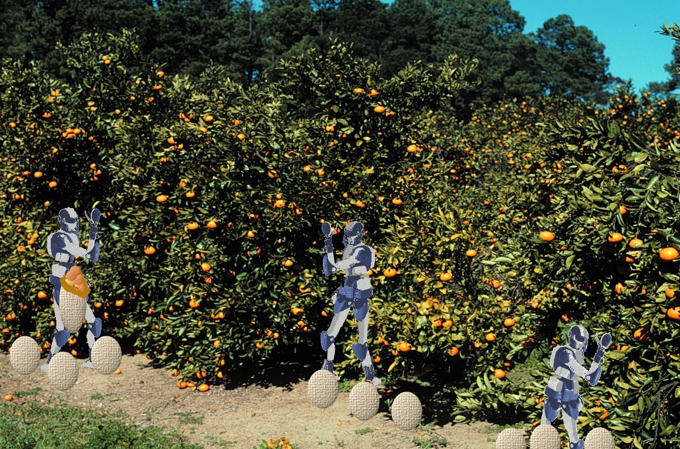
[1] Robots pick
oranges Viroids orange
trees Development of HRP-4, a Research
and Development Platform for Working
Humanoid Robots - Supply to external
research organizations from January
2011 - UNKNOWN and Orange
trees: Viroid diseases are
responsible for significant losses of
food and fiber. Our research goals are
to obtain fundamental knowledge about
the molecular interactions between
viroids and their host, to apply this
knowledge to the control of viroid
diseases, and to develop and test
viroid-induced dwarfing strategies to
increase citrus production efficiency.
Robert A. Owens PD UNKNOWN
source: http://www.aist.go.jp/aist_e/lat
est_research/2010/20101108/fig1.jpghttp:
//www.ars.usda.gov/sp2UserFiles/Place/12
752500/research/images/citrus.jpg
[2] Storyboard image by Ted
Huntington GNU
source: Ted Huntington
[2200 AD]
earth.

[1] Image adapted from from
http://www.nasa.gov/externalflash/gemini
_gallery/hi-resjpgs/10.jpg Cape
Canaveral, site of NASA's Kennedy Space
Center, from the Gemini V ... PD
source: http://www.nasa.gov/externalflas
h/gemini_gallery/hi-resjpgs/10.jpgTed
Huntington
[2] Storyboard image by Ted
Huntington GNU
source: Ted Huntington
[2200 AD]

[1] Adapted from image from NASA
Messenger ship PD
source: http://1.bp.blogspot.com/_qcuftp
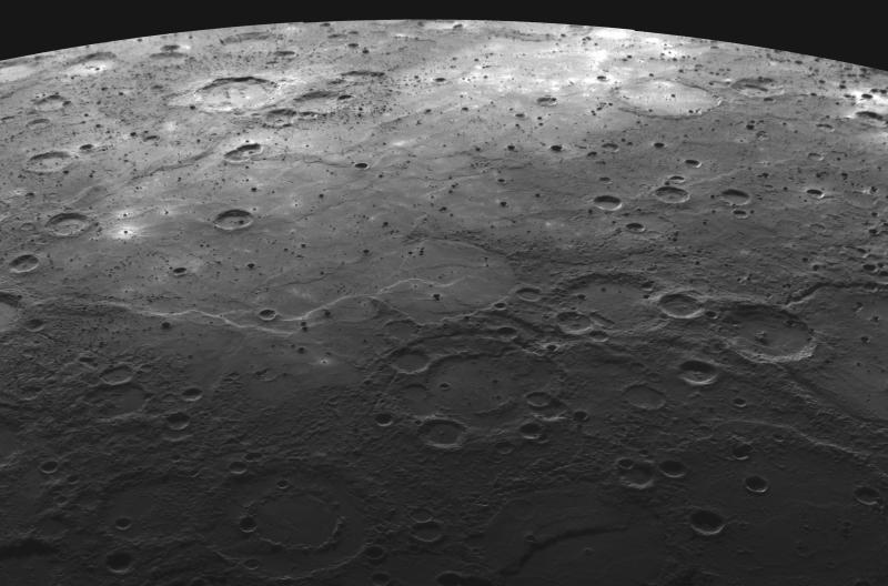
B9Hx8/TJOQmeFucWI/AAAAAAAACwg/Bl0M9a2_M1
0/s1600/Planet-Mercury.jpg
[2200 AD]
regrow, and reshape damaged cells.
[1] Skin cells, SEM C001/7939 Rights
Managed Credit: SCIENCE PHOTO
LIBRARY Caption: Skin cells.
Coloured scanning electron micrograph
(SEM) of squamous cells from the
surface of the skin. These are flat,
keratinised, dead cells that are
continuously sloughed off and replaced
with new cells from
below. AND Researchers are
developing a new class of tiny
mechanical devices containing
vibrating, hair-thin structures that
could be used to filter electronic
signals in cell phones and for other
more exotic applications. The work is
done inside a vacuum chamber sitting on
top of a special vibration-absorbing
platform critical to making the precise
measurements. A tiny prototype, roughly
comparable in size to a grain of sand,
is pictured on the monitor at right.
The device is an example of a
microelectromechanical system, or a
MEMS, which contains tiny moving parts.
(Birck Nanotechnology Center, Purdue
University) A publication-quality
photo is available at
http://news.uns.purdue.edu/images/+2009/
rhoads-mems2.jpg [1] A scanning
electron microscope (SEM) image of
nanowire-alginate composite scaffolds.
Star-shaped clusters of nanowires can
be seen in these images. Image
courtesy of the Disease Biophysics
Group, Harvard University UNKNOWN
source: http://www.sciencephoto.com/imag
e/82340/large/C0017939-Skin_cells,_SEM-S
PL.jpghttp://news.uns.purdue.edu/images/
+2009/rhoads-mems2.jpg

[2] Skin cells, SEM C001/7939
Rights Managed Credit: SCIENCE PHOTO
LIBRARY Caption: Skin cells.
Coloured scanning electron micrograph
(SEM) of squamous cells from the
surface of the skin. These are flat,
keratinised, dead cells that are
continuously sloughed off and replaced
with new cells from
below. AND Researchers are
developing a new class of tiny
mechanical devices containing
vibrating, hair-thin structures that
could be used to filter electronic
signals in cell phones and for other
more exotic applications. The work is
done inside a vacuum chamber sitting on
top of a special vibration-absorbing
platform critical to making the precise
measurements. A tiny prototype, roughly
comparable in size to a grain of sand,
is pictured on the monitor at right.
The device is an example of a
microelectromechanical system, or a
MEMS, which contains tiny moving parts.
(Birck Nanotechnology Center, Purdue
University) A publication-quality
photo is available at
http://news.uns.purdue.edu/images/+2009/
rhoads-mems2.jpg [1] A scanning
electron microscope (SEM) image of
nanowire-alginate composite scaffolds.
Star-shaped clusters of nanowires can
be seen in these images. Image
courtesy of the Disease Biophysics
Group, Harvard University UNKNOWN
source: http://www.sciencephoto.com/imag
e/82340/large/C0017939-Skin_cells,_SEM-S
PL.jpghttp://news.uns.purdue.edu/images/
+2009/rhoads-mems2.jpg
[2250 AD]
[1] The image show the Lander Falcon
skimming over one of the many ice
cravas of Jupiter’s moon Europa
looking for a suitable landing
place. COPYRIGHTED
source: http://api.ning.com/files/s7oIN4
97UMEE6dpA1xd*IhqzsZkYEn1zbiUE5*qsj*mBXD
EV7F1lGV*Qngn1qBdiZSdmNBsHbXquTTpGfoIHib
xxEsocyNr-/BB131FalconoverEuropaNR.jpg?w
idth=737&height=400

[2] Ganymede: Global Color View PD
source: http://solarsystem.nasa.gov/mult
imedia/gallery/gg1.jpg
[2280 AD]
[1] Saturn from the surface of
Dione. COPYRIGHTED
source: http://spaceart1.ning.com/photo/
saturn-from-dione/next?context=userhttp:
//microgravity.grc.nasa.gov/Advanced/Cap
abilities/ETDP/images/lunarlander.jpg
[2] Artist concept of Cassini at
Saturn. Image credit: NASA/JPL ›
Larger image PD
source: http://www.jpl.nasa.gov/images/c
assini/20090202/pia03883-full.jpg
[2300 AD]
[1] Adapted from: Uranus seen from
Oberon UNKNOWN
source: http://api.ning.com/files/DzXL-l
W6TdpjPVXja-k32xq4*PiPHvNiITlxVu5JoQ*XRl
Z72k*OlXD710b-zT2jIomp7im9tEUk0AzJ4HNiph
MGf2J-UCLg/Oberon.jpg?width=737&height=5
69http://microgravity.grc.nasa.gov/Advan
ced/Capabilities/ETDP/images/lunarlander
.jpg
[2350 AD]
[1] Intrepid-over-Proteus Neptune's
Moon Proteus The Lander Intrepid skims
the heavily cratered moon Proteus in
search for a landing area. Proteus is a
relatively large moon, similar in size
to Saturn’s moon Mimas, but was not
discovered until Voyager 2 flew by
because it is very dark and orbits very
close to Neptune. Like Mimas, it is
irregular in shape, heavily cratered,
and has no sign of internally generated
geologic activity in its
past. UNKNOWN
source: http://api.ning.com/files/n*cJoC
Qsunpuu6EpNQKC3KHkTJPnAZoABx8opILfQ7o_/I
ntrepidoverProteus.jpg?width=737&height=
469
[2350 AD]
Centauri). First close up pictures of
plane
ts of a different star.

[1] Storyboard image by Ted
Huntington GNU
source: Ted
Huntingtonhttp://upload.wikimedia.org/wi
kipedia/commons/0/08/Planet-alphacen1.pn
g

[2] Storyboard image by Ted
Huntington GNU
source: Ted
Huntingtonhttp://upload.wikimedia.org/wi
kipedia/commons/0/08/Planet-alphacen1.pn
g
[2370 AD]
another star identified (bacteria made
of DNA).

[1] Descripción English:
Filamentous cyanobacterium of a genus
Lyngbya, as collected in Baja
California, Mexico Česky: Vláknitá
sinice rodu Lyngbya; Baja California,
Mexico Fecha Fuente
http://microbes.arc.nasa.gov/images/con
tent/gallery/lightms/publication/lyngbya
.jpg Autor NASA PD
source: http://upload.wikimedia.org/wiki
pedia/commons/thumb/3/3e/Lyngbya.jpg/128
0px-Lyngbya.jpg
[2] Storyboard image by Ted
Huntington GNU
source: Ted Huntington
[2500 AD]
started.
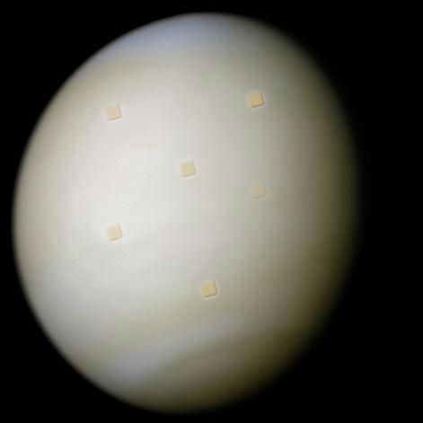
[1] Description Image of Venus in
real color processed from the clear and
blue filters (colors are probably
enhanced). Date 2006-09-16
(original upload
date) Source http://astrosurf.com/n
unes/explor/explor_m10.htm Author N
ASA/Ricardo Nunes PD
source: http://upload.wikimedia.org/wiki
pedia/commons/5/51/Venus-real.jpg

[2] Adapted from: A rover that could
survive the intense heat of Venus, seen
here in an artist's impression, could
revolutionise our understanding of the
planet. Cooled by a Stirling Cooler
with electronics at 200 °C and
external radiator at 500 °C. Since the
Venusian atmosphere is 'only' 450 °C
the radiator will lose
energy. Geoffrey Landis and Kenneth
Mellott from NASA's Glenn Research
Center in Ohio. PD
source: http://upload.wikimedia.org/wiki
pedia/commons/1/15/Venus_Rover.jpg
[2500 AD]

[1] Adapted by Ted Huntington
from: Solution structure of a
trans-opened (10S)-dA adduct of
+)-(7S,8R,9S,10R)-7,8-dihydroxy-9,10-epo
xy-7,8,9,10-tetrahydrobenzo[a]pyrene in
a DNA duplex. GNU AND Multiple Gear
Speed Reduction Unit Top view of
gear reduction unit. PD PD
source: http://upload.wikimedia.org/wiki
pedia/commons/d/d8/Benzopyrene_DNA_adduc
t_1JDG.pnghttp://mems.sandia.gov/gallery
/images/tg8.jpgTed Huntington

[2] Storyboard image by Ted
Huntington GNU
source: Ted Huntington
[2600 AD]
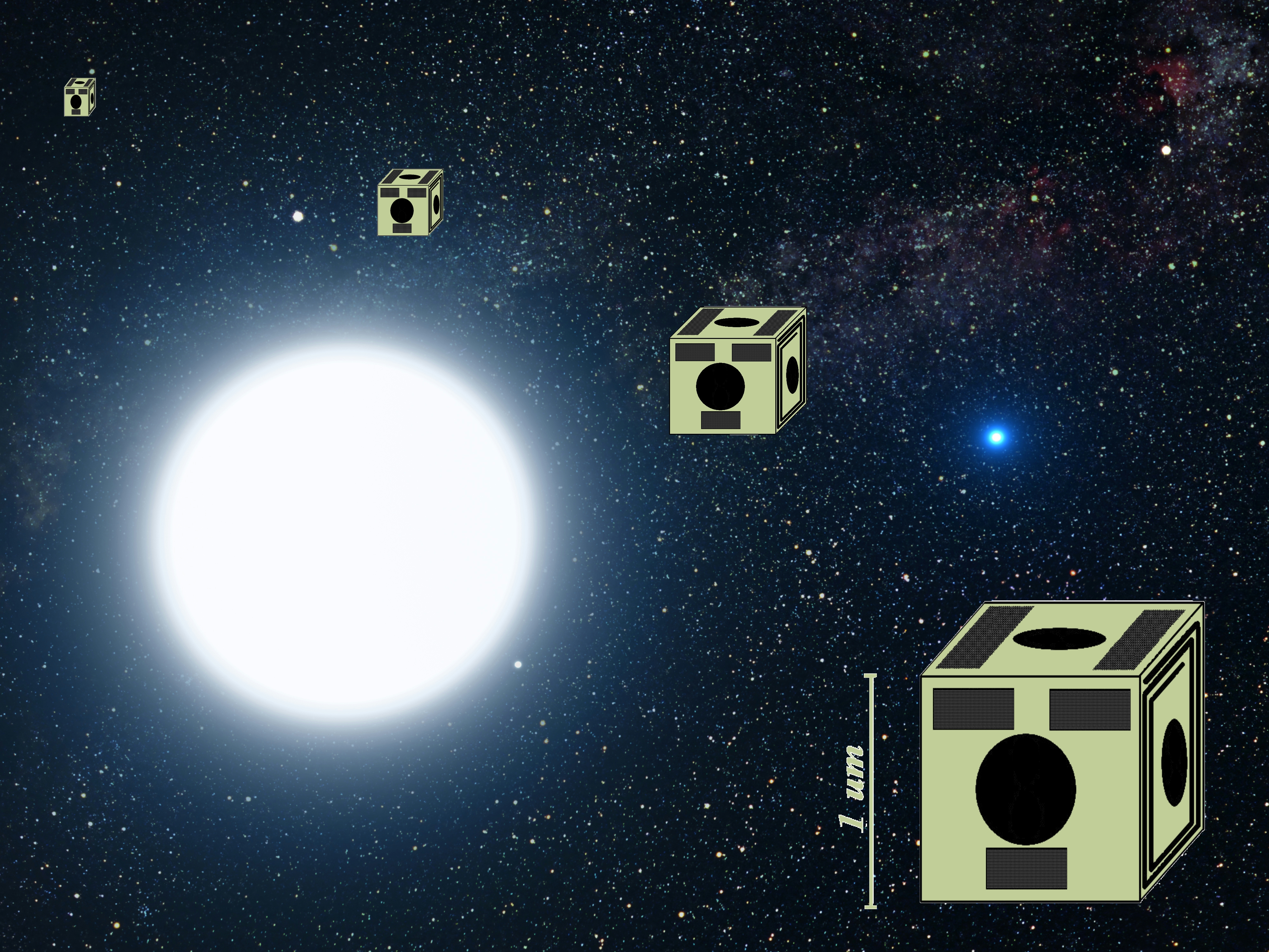
[1] {ULSF: adapted by Ted Huntington
from below:} Description This
picture is an artist's impression
showing how the binary star system of
Sirius A and its diminutive blue
companion, Sirius B, might appear to an
interstellar visitor. The large,
bluish-white star Sirius A dominates
the scene, while Sirius B is the small
but very hot and blue white-dwarf star
on the right. The two stars revolve
around each other every 50 years. White
dwarfs are the leftover remnants of
stars similar to our Sun. The Sirius
system, only 8.6 light-years from
Earth, is the fifth closest stellar
system known. Sirius B is faint because
of its tiny size. Its diameter is only
7,500 miles (about 12 thousand
kilometres), slightly smaller than the
size of our Earth. The Sirius system is
so close to Earth that most of the
familiar constellations would have
nearly the same appearance as in our
own sky. In this rendition, we see in
the background the three bright stars
that make up the Summer Triangle:
Altair, Deneb, and Vega. Altair is the
white dot above Sirius A; Deneb is the
dot to the upper right; and Vega lies
below Sirius B. But there is one
unfamiliar addition to the
constellations: our own Sun is the
second-magnitude star, shown as a small
dot just below and to the right of
Sirius
A. Date Source http://www.spacete
lescope.org/images/html/heic0516b.html
Author NASA, ESA Credit: G. Bacon
(STScI) PD
source: http://upload.wikimedia.org/wiki
pedia/commons/c/c9/Sirius_A_and_B_artwor
k.jpg

[2] Adapted from: Description This
picture is an artist's impression
showing how the binary star system of
Sirius A and its diminutive blue
companion, Sirius B, might appear to an
interstellar visitor. The large,
bluish-white star Sirius A dominates
the scene, while Sirius B is the small
but very hot and blue white-dwarf star
on the right. The two stars revolve
around each other every 50 years. White
dwarfs are the leftover remnants of
stars similar to our Sun. The Sirius
system, only 8.6 light-years from
Earth, is the fifth closest stellar
system known. Sirius B is faint because
of its tiny size. Its diameter is only
7,500 miles (about 12 thousand
kilometres), slightly smaller than the
size of our Earth. The Sirius system is
so close to Earth that most of the
familiar constellations would have
nearly the same appearance as in our
own sky. In this rendition, we see in
the background the three bright stars
that make up the Summer Triangle:
Altair, Deneb, and Vega. Altair is the
white dot above Sirius A; Deneb is the
dot to the upper right; and Vega lies
below Sirius B. But there is one
unfamiliar addition to the
constellations: our own Sun is the
second-magnitude star, shown as a small
dot just below and to the right of
Sirius
A. Date Source http://www.spacete
lescope.org/images/html/heic0516b.html
Author NASA, ESA Credit: G. Bacon
(STScI) PD
source: http://upload.wikimedia.org/wiki
pedia/commons/c/c9/Sirius_A_and_B_artwor
k.jpghttp://aetd.gsfc.nasa.gov/code540/5
40/new_images/MLAS.jpg
[2650 AD]
particles (photon fusion).

[1] Humans create atoms from light
particles Ted Huntington PD
source: Ted Huntington

[2] Storyboard image by Ted
Huntington GNU
source: Ted Huntington
[2800 AD]

[1] Adapted from: The Missing Craters
of Asteroid Itokawa Credit &
Copyright: ISAS, JAXA Explanation:
Where are the craters on asteroid
Itokawa? No one knows. The Japanese
robot probe Hayabusa recently
approached the Earth-crossing asteroid
and is returning pictures showing a
surface unlike any other Solar System
body yet photographed -- a surface
possibly devoid of craters. One
possibility for the lack of common
circular indentations is that asteroid
Itokawa is a rubble pile -- a bunch of
rocks and ice chunks only loosely held
together by a small amount of gravity.
If so, craters might be filled in
whenever the asteroid gets jiggled by a
passing planet -- Earth in this case.
Alternatively, surface particles may
become electrically charged by the Sun,
levitate in the microgravity field, and
move to fill in craters. Over the
weekend, Hayabusa lowered itself to the
surface of the strange asteroid in an
effort to study the unusual body and
collect surface samples that could be
returned to Earth in 2007. PD
source: http://apod.nasa.gov/apod/image/
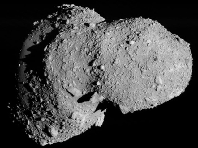
0511/itokawa05_hayabusa.jpg
[2800 AD]

[1] Summary A rover that could
survive the intense heat of Venus, seen
here in an artist's impression, could
revolutionise our understanding of the
planet. Cooled by a Stirling Cooler
with electronics at 200 °C and
external radiator at 500 °C. Since the
Venusian atmosphere is 'only' 450 °C
the radiator will lose
energy. Geoffrey Landis and Kenneth
Mellott from NASA's Glenn Research
Center in Ohio. PD
source: http://upload.wikimedia.org/wiki
pedia/commons/1/15/Venus_Rover.jpg
[2] Storyboard image by Ted
Huntington GNU
source: Ted Huntington
[2800 AD]
moon.
[1] A map of Io This map of Io was
created by taking three relatively low
resolution Voyager 2 maps of Io and
composing them into a color map. These
maps were taken from this FTP site.
Each of the three original maps was
created from images taken through
differently colored filters. I then
replaced the luminosity channel of the
color map with a very high resolution
grayscale map from the above site. That
map was created from Voyager 1 photos.
The result was impressive despite the
fact that Io changed somewhat between
the two Voyager flybys (it is after all
the most volcanically active body in
the solar system !). I then removed the
most prominent seams and color
discontinuities. Some seams still
remain (I was more lazy when creating
this map than e.g. the Europa map ;)
but remarkably they are usually not
visible in 3D renderings except for
renderings showing Io from a close
range. I may remove these defects some
day. Finally I replaced small, black
areas near the poles (where no imagery
is available) with fictional data
created by cloning adjacent
areas. This map is aesthetically
better than the ones at David Seal's
site and also of higher resolution but
the color is probably less realistic
than in his ''yellowish'' map. His maps
are also more realistic in the sense
that there is no fictional data at the
poles, they are simply blank. Click
the map below to view the 2880x1440
pixel (442 KB JPG) full size map.
Actually my original map is 5760x2880
pixels but I do not have space for such
a monster here. UNKNOWN
source: http://www.mmedia.is/~bjj/data/i
o/io.jpg

[2] Storyboard image by Ted
Huntington GNU
source: Ted Huntington
[2850 AD]
planet.

[1] Image of ships surrounding Earth in
the future by Ted Huntington Source of
Texture map for Earth unknown GNU
source: Ted Huntington
[2] Storyboard image by Ted
Huntington GNU
source: Ted Huntington
[2900 AD]
Jupiter. First image of the surface of
Jupiter.

[1] Ted Huntington GNU
source: Ted Huntington

[2] This close-up of the northwestern
region of the Sun shows a filament
erupting. Credit: NASA/SDO PD
source: http://www.nasa.gov/images/conte
nt/480276main_20100904-fulldisk.jpg
[3150 AD]
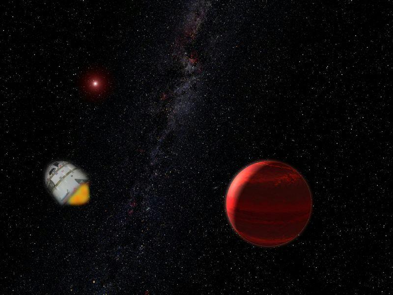
[1] Adapted
from: Description English: Artist's
conception of a the red dwarf star CHRX
73 A and its companion object CHRX 73
B. The companion object is around 12
Jupiter masses, and may either be a
planet, a failed star or a brown
dwarf Date 2006-09-02 Source Sel
f-made JPEG version of original TIFF
image at Hubble
website Author NASA, ESA and G.
Bacon (STScI) PD
source: http://upload.wikimedia.org/wiki
pedia/commons/thumb/6/68/RedDwarfPlanet.
jpg/800px-RedDwarfPlanet.jpghttp://aetd.
gsfc.nasa.gov/code540/540/new_images/MLA
S.jpg
[3200 AD]
with objects.
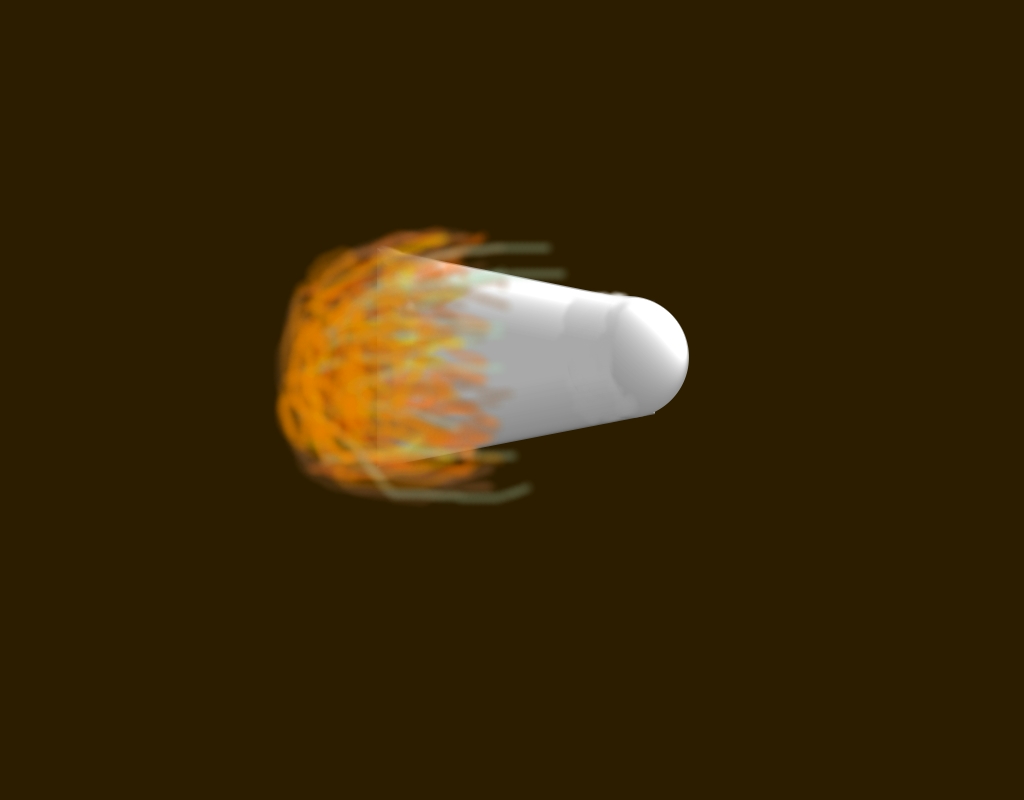
[1] Ship from Centauri reaches
Earth PD
source: Ted Huntington
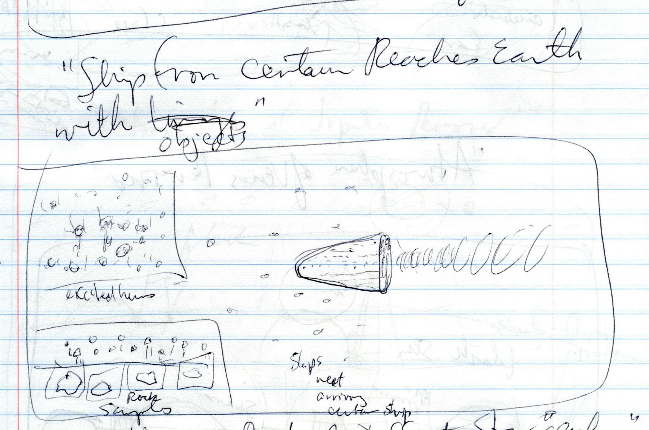
[2] Storyboard image by Ted
Huntington GNU
source: Ted Huntington
[3200 AD]
Centauri.
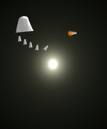
[1] Humans reach Alpha
Centauri adapted by Ted Huntington
from: http://upload.wikimedia.org/wikip
edia/commons/0/08/Planet-alphacen1.png
CC
source: http://upload.wikimedia.org/wiki
pedia/commons/0/08/Planet-alphacen1.png

[2] Storyboard image by Ted
Huntington GNU
source: Ted Huntington
[3500 AD]
removed.
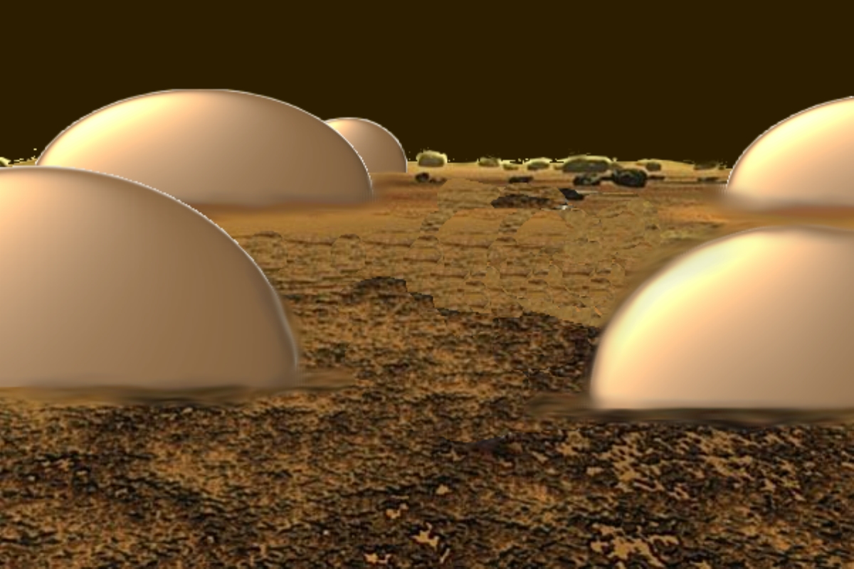
[1] Atmosphere of Venus completely
removed adapted by Ted Huntington
from: Summary A rover that could
survive the intense heat of Venus, seen
here in an artist's impression, could
revolutionise our understanding of the
planet. Cooled by a Stirling Cooler
with electronics at 200 °C and
external radiator at 500 °C. Since the
Venusian atmosphere is 'only' 450 °C
the radiator will lose
energy. Geoffrey Landis and Kenneth
Mellott from NASA's Glenn Research
Center in Ohio. PD
source: http://upload.wikimedia.org/wiki
pedia/commons/1/15/Venus_Rover.jpg

[2] Venus with no atmosphere adapted
from Mars texture map UNKNOWN
source:
[4000 AD]

[1] Jupiter after atmosphere is
consumed. By Ted Huntington, adapted
from: http://volcanoes.usgs.gov/Imgs/Jp
g/Photoglossary/aa_large.jpg GNU
source: http://volcanoes.usgs.gov/Imgs/J
pg/Photoglossary/aa_large.jpg
[2] Storyboard image by Ted
Huntington GNU
source: Ted Huntington
[4000 AD]
systems.
[1] Description English: Praesepe:
the open cluster Messier 44 Date
Source 2MASS Atlas Image Gallery:
The Messier Catalog Author Two Micron
All Sky Survey (2MASS), a joint project
of the University of Massachusetts and
the Infrared Processing and Analysis
Center/California Institute of
Technology, funded by the National
Aeronautics and Space Administration
and the National Science
Foundation. Licensing Public domain
This image is from the Two Micron All
Sky Survey (2MASS) project. The images
from this project have been released
into the public domain. 2MASS kindly
requests acknowledgement in one of the
following forms, the longer of which is
preferred. Atlas Image [or Atlas
Image mosaic] obtained as part of the
Two Micron All Sky Survey (2MASS), a
joint project of the University of
Massachusetts and the Infrared
Processing and Analysis
Center/California Institute of
Technology, funded by the National
Aeronautics and Space Administration
and the National Science Foundation.
Atlas Image [or Atlas Image mosaic]
courtesy of
2MASS/UMass/IPAC-Caltech/NASA/NSF. PD
source: http://upload.wikimedia.org/wiki
pedia/commons/3/3a/Messier_044_2MASS.jpg

[2] Description English: Praesepe:
the open cluster Messier 44 Date
Source 2MASS Atlas Image Gallery:
The Messier Catalog Author Two Micron
All Sky Survey (2MASS), a joint project
of the University of Massachusetts and
the Infrared Processing and Analysis
Center/California Institute of
Technology, funded by the National
Aeronautics and Space Administration
and the National Science
Foundation. Licensing Public domain
This image is from the Two Micron All
Sky Survey (2MASS) project. The images
from this project have been released
into the public domain. 2MASS kindly
requests acknowledgement in one of the
following forms, the longer of which is
preferred. Atlas Image [or Atlas
Image mosaic] obtained as part of the
Two Micron All Sky Survey (2MASS), a
joint project of the University of
Massachusetts and the Infrared
Processing and Analysis
Center/California Institute of
Technology, funded by the National
Aeronautics and Space Administration
and the National Science Foundation.
Atlas Image [or Atlas Image mosaic]
courtesy of
2MASS/UMass/IPAC-Caltech/NASA/NSF. PD
source: http://upload.wikimedia.org/wiki
pedia/commons/3/3a/Messier_044_2MASS.jpg
[4500 AD]

[1] International Space Station crew
members are trained to observe and
document dynamic events on the
Earth’s surface, such as hurricanes,
forest fires, and volcanic eruptions.
Their observations provide scientists
and the general public a different
perspective on these events. Earlier
this week, astronauts in the crew of
the ISS-5 mission were able to observe
Mt. Etna’s spectacular eruption, and
photograph the details of the eruption
plume as well as smoke from fires
triggered by the lava as it flowed down
the 11,000-foot mountain. This image is
looking obliquely to the southeast over
the island of Sicily. A wider view
(ISS005-E-19016) shows the ash plume
curving out toward the horizon, caught
first by low-level winds blowing to the
southeast, and to the south toward
Africa at higher altitudes. Ashfall was
reported in Libya, more than 350 miles
away. The lighter-colored plumes
downslope and north of the summit (see
detailed view, ISS005-E-19024) are
produced by gas emissions from a line
of vents on the mountain’s north
flank. The detailed image provides a
three-dimensional profile of the
eruption plume. This was one of
Etna’s most vigorous eruptions in
years. The eruption was triggered by a
series of earthquakes on October 27.
These images were taken on October 30,
2002. Sicilans have learned to live
with Etna’s eruptions. Although
schools were closed and air traffic was
diverted because of the ash, no towns
or villages were threatened by the lava
flow. Astronaut photographs
ISS005-E-19016 and ISS005-E-19024 were
taken on October 30, 2002, at about
11:30 GMT and are provided by the Earth
Sciences and Image Analysis Laboratory
at Johnson Space Center. Additional
images taken by astronauts and
cosmonauts can be viewed at the
NASA-JSC Gateway to Astronaut
Photography of Earth. Instrument:
ISS - Digital Camera PD
source: http://eoimages.gsfc.nasa.gov/im
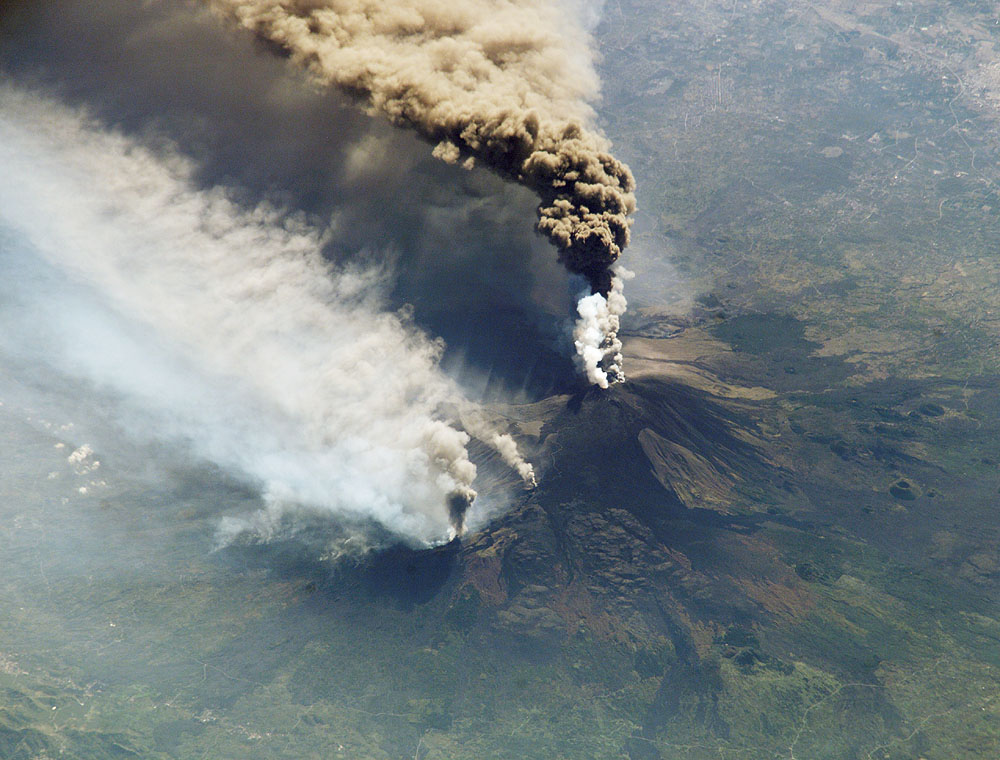
ages/imagerecords/2000/2923/etna2_ISS200
2303_lrg.jpg
[4500 AD]
[1] Lava flows on Krafla in
Iceland Licensing: This image
was created by Chris 73. The image is
licensed under a dual license; please
choose either of the two licenses below
as desired. Attribution to Wikipedia or
another project of the Wikimedia
foundation is required for both
licenses if the image is used outside
of projects of the Wikimedia
foundation. Attribution to me is not
required. GNU head Es ist erlaubt,
die Datei unter den Bedingungen der
GNU-Lizenz für freie Dokumentation,
Version 1.3 oder einer späteren
Version, veröffentlicht von der Free
Software Foundation, zu kopieren, zu
verbreiten und/oder zu modifizieren; es
gibt keine unveränderlichen
Abschnitte, keinen vorderen und keinen
hinteren Umschlagtext. Der
vollständige Text der Lizenz ist im
Kapitel GNU-Lizenz für freie
Dokumentation verfügbar. GNU
source: http://upload.wikimedia.org/wiki
pedia/commons/d/d6/Lava_at_Vulkan_Krafla
_Iceland_1.JPG
[2] Volle resolusie (3 888 × 2 592
pixels, lêergrootte: 5,96 MG, MIME
type: image/jpeg) Hierdie lêer kom
vanaf Wikimedia Commons en kan ook in
ander projekte gebruik word. Die
beskrywing op die lêer se
inligtingsblad word hieronder
weergegee. Beskrywing English: Lava
flow at The Big Island of Hawaii. The
lava flow is due to July 21 fissure
eruption. The picture was taken from a
helicopter. Datum 09/10/07 Bron
Created and originally uploaded to the
English Wikipedia by Brocken
Inaglory. Outeur Brocken
Inaglory Toestemming (Hergebruik van
die lêer)
CC-BY-SA-3.0,2.5,2.0,1.0; Released
under the GNU Free Documentation
License. Kameraligging 19°
23' 11.0'' N, 155° 6' 18.0''
W Hierdie en ander beelde in: Google
Maps - Google Earth - OpenStreetMa CC
source: http://upload.wikimedia.org/wiki
pedia/commons/6/62/Hawaiian_lava_flow.jp
g
[4500 AD]
control.
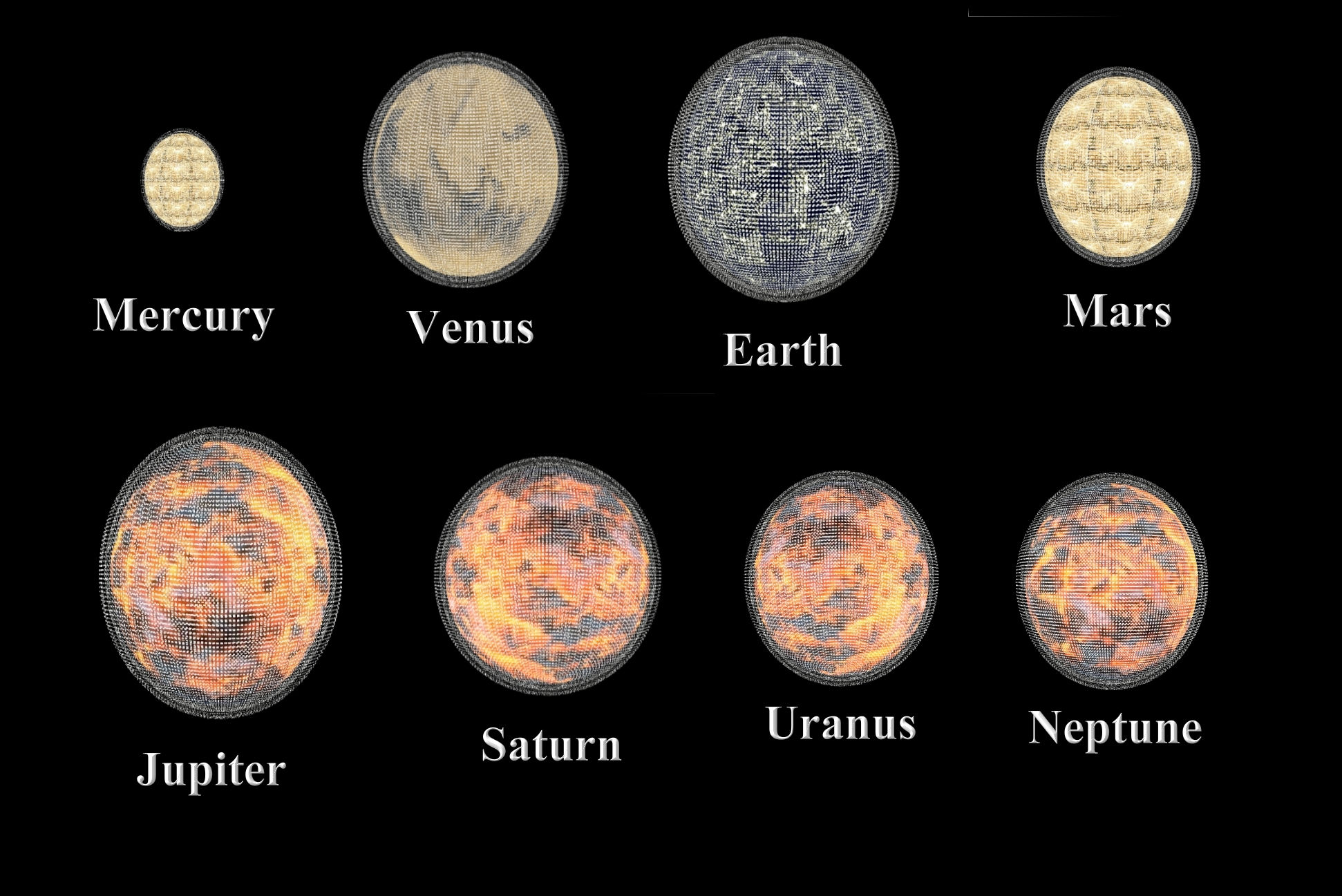
[1] Various planetary texture maps and
public domain lava texture map mapped
onto 3D sphere and rendered two spheres
of ships added by Ted Huntington PD
source: Various planetary texture maps
and public domain lava texture map
mapped onto 3D sphere and rendered two
spheres of ships added by Ted
Huntington

[2] Storyboard image by Ted
Huntington GNU
source: Ted Huntington
[5100 AD]
living objects that evolved around a
different star.
[1] Life of another star by Ted
Huntington with background
image: 说明 English: Open Star
Cluster NGC663 in Cassiopeia 日期
2010年11月9日 来源
自己的作品 作者
Hewholooks CC
source: Ted
Huntingtonhttp://upload.wikimedia.org/wi
kipedia/commons/1/12/NGC663HunterWilson.
jpg
[2] Living object from other star by
Ted Huntington with image in
background: Description English:
This impressive image of the open
cluster known as Trumpler 14 was
obtained with the Multi-conjugate
Adaptive optics Demonstrator (MAD)
mounted on ESO’s Very Large
Telescope. The cluster, which is found
to be only 500 000 years old — a
blink of an eye in the Universe’s
history — resides at the outskirts of
the central region of the Carina
Nebula, located some 8000 light-years
away towards the constellation of
Carina (the Keel). Trumpler 14 is not
only the youngest, but also one of the
most populous clusters within the
nebula. Astronomers counted about 2000
stars in the very central parts of this
cluster. The MAD instrument allows
astronomers to obtain very sharp images
over a wide area and this image is the
adaptive optics image that so far
covers the widest area on the sky. The
field of view is about 2 arcminutes
across and the image is based on data
obtained through two different filters
(K and H). Date 03-12-2009 Source
http://www.eso.org/public/images/eso094
7a/ Author ESO/H. Sana CC
source: Ted
Huntingtonhttp://upload.wikimedia.org/wi
kipedia/commons/5/57/ESO-Trumpler14-clus
ter.jpg
[5500 AD]
of Earth moved in direction of
Centauri.
[1] Humans move star by Ted
Huntington PD
source: Ted Huntington
[2] Storyboard image by Ted
Huntington GNU
source: Ted Huntington
[6000 AD]
objects that evolved around a different
star.

[1] Humans touch advanced life of
another star by Ted Huntington with
background image of: Double Cluster
in Perseus Credit & Copyright: Roth
Ritter (Dark
Atmospheres) Explanation: Some 7,000
light-years away, this pair of open or
galactic star clusters is an easy
binocular target, a lovely starfield in
the northern constellation Perseus.
Also visible to the unaided eye from
dark sky areas, it was cataloged in 130
BC by Greek astronomer Hipparchus. Now
known as h and chi Persei, or NGC
869(above right) and NGC 884, the
clusters themselves are separated by
only a few hundred light-years and
contain stars much younger and hotter
than the Sun. In addition to being
physically close together, the
clusters' ages based on their
individual stars are similar - evidence
that both clusters were likely a
product of the same star-forming
region. PD
source: Ted
Huntingtonhttp://apod.nasa.gov/apod/imag
e/0901/clusters_2_ritter.jpg

[2] Storyboard image by Ted
Huntington GNU
source: Ted Huntington
[7000 AD]
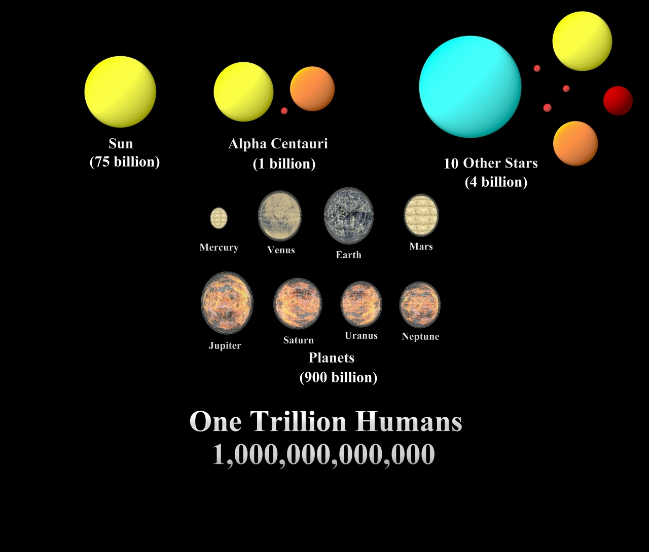
[1] Image summarizing population of
humans around planets and stars by Ted
Huntington PD
source: Ted Huntington
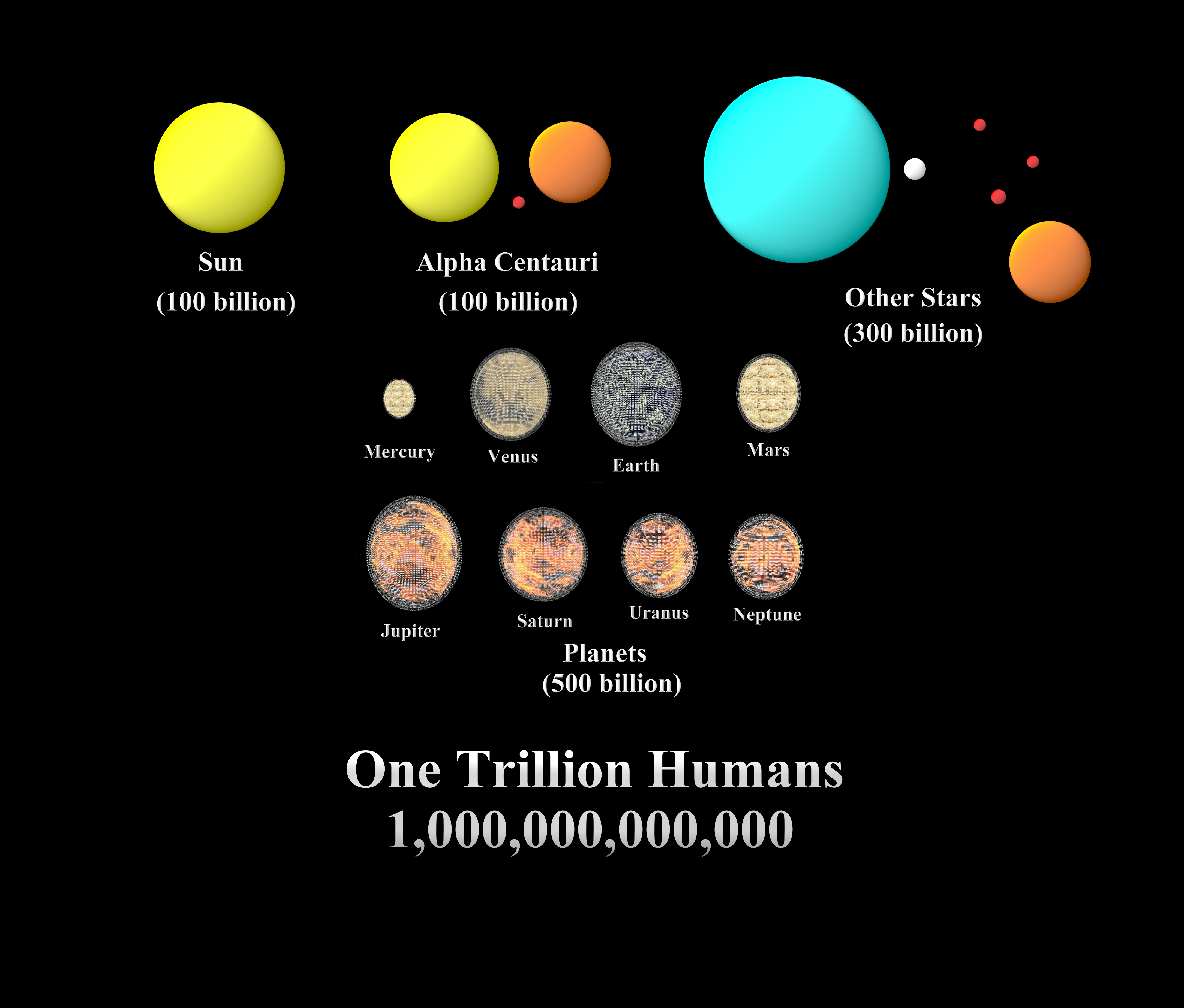
[2] Image summarizing population of
humans around planets and stars by Ted
Huntington PD
source: Ted Huntington
[27000 AD]
a globular cluster of 10 stars.

[1] Hyades for the Holidays Image
Credit & Copyright: Jerry Lodriguss
(Catching the Light) Explanation:
Recognized since antiquity and depicted
on the shield of Achilles according to
Homer, stars of the Hyades cluster form
the head of the constellation Taurus
the Bull. Their general V-shape is
anchored by Aldebaran, the eye of the
Bull and by far the constellation's
brightest star. Yellowish in
appearance, red giant Aldebaran is not
a Hyades cluster member, though. Modern
astronomy puts the Hyades cluster 151
light-years away making it the nearest
established open star cluster, while
Aldebaran lies at less than half that
distance, along the same line-of-sight.
Along with colorful Hyades stars, this
stellar holiday portrait locates
Aldebaran just below center, as well as
another open star cluster in Taurus,
NGC 1647 at the left, some 2,000
light-years or more in the background.
Just slide your cursor over the image
to identify the stars. The central
Hyades stars are spread out over about
15 light-years. Formed some 800 million
years ago, the Hyades star cluster may
share a common origin with M44
(Praesepe), a naked-eye open star
cluster in Cancer, based on M44's
motion through space and remarkably
similar age. UNKNOWN
source: http://apod.nasa.gov/apod/image/
1212/HyadesLodriguss.jpg

[2] Hyades for the Holidays Image
Credit & Copyright: Jerry Lodriguss
(Catching the Light) Explanation:
Recognized since antiquity and depicted
on the shield of Achilles according to
Homer, stars of the Hyades cluster form
the head of the constellation Taurus
the Bull. Their general V-shape is
anchored by Aldebaran, the eye of the
Bull and by far the constellation's
brightest star. Yellowish in
appearance, red giant Aldebaran is not
a Hyades cluster member, though. Modern
astronomy puts the Hyades cluster 151
light-years away making it the nearest
established open star cluster, while
Aldebaran lies at less than half that
distance, along the same line-of-sight.
Along with colorful Hyades stars, this
stellar holiday portrait locates
Aldebaran just below center, as well as
another open star cluster in Taurus,
NGC 1647 at the left, some 2,000
light-years or more in the background.
Just slide your cursor over the image
to identify the stars. The central
Hyades stars are spread out over about
15 light-years. Formed some 800 million
years ago, the Hyades star cluster may
share a common origin with M44
(Praesepe), a naked-eye open star
cluster in Cancer, based on M44's
motion through space and remarkably
similar age. UNKNOWN
source: http://www.cfht.hawaii.edu/Scien
ce/Astros/Imageofweek/ciw-image/050600-2
.jpg
[47000 AD]
form a globular cluster of 100 stars.
[1] The CFHT Open Cluster Survey : NGC
2099 Credit: Image courtesy of
Harvey Richer1, Patrick Durrell1,
Gregory Fahlman2, J. Kalirai1, F.
D'Antona3 & G. Marconi3 1 University
of British Columbia, Vancouver,
Canada 2 Canada-France-Hawaii
Telescope Corporation, Hawaii, USA 3
Osservatorio Astronomico di Roma, Italy
COPYRIGHTED
source: http://www.cfht.hawaii.edu/Scien
ce/Astros/Imageofweek/ciw-image/050600-2
.jpg
[2] Hyades for the Holidays Image
Credit & Copyright: Jerry Lodriguss
(Catching the Light) Explanation:
Recognized since antiquity and depicted
on the shield of Achilles according to
Homer, stars of the Hyades cluster form
the head of the constellation Taurus
the Bull. Their general V-shape is
anchored by Aldebaran, the eye of the
Bull and by far the constellation's
brightest star. Yellowish in
appearance, red giant Aldebaran is not
a Hyades cluster member, though. Modern
astronomy puts the Hyades cluster 151
light-years away making it the nearest
established open star cluster, while
Aldebaran lies at less than half that
distance, along the same line-of-sight.
Along with colorful Hyades stars, this
stellar holiday portrait locates
Aldebaran just below center, as well as
another open star cluster in Taurus,
NGC 1647 at the left, some 2,000
light-years or more in the background.
Just slide your cursor over the image
to identify the stars. The central
Hyades stars are spread out over about
15 light-years. Formed some 800 million
years ago, the Hyades star cluster may
share a common origin with M44
(Praesepe), a naked-eye open star
cluster in Cancer, based on M44's
motion through space and remarkably
similar age. UNKNOWN
source: http://apod.nasa.gov/apod/image/
1212/HyadesLodriguss.jpg
[65000 AD]
Earth.
[1] by Ted Huntington PD
source: Ted Huntington

[2] Adapted from: The Death Star is
the size of a small moon. See more Star
Wars pictures. Photo courtesy ©
Lucasfilm Ltd. & TM. All Rights
Reserved. COPYRIGHTED
source: http://static.ddmcdn.com/gif/dea
th-star-1.jpg
[67000 AD]
living objects.
[1] Earth is completely filled by Ted
Huntington PD
source: Ted Huntington

[2] Storyboard image by Ted
Huntington GNU
source: Ted Huntington
[72000 AD]
form a globular cluster of 1,000 stars.
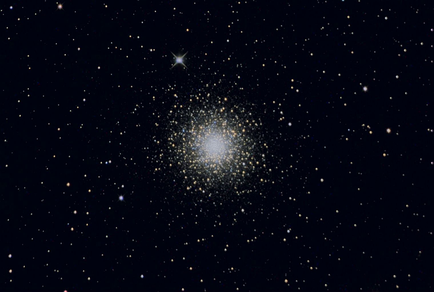
[1] M15 Second attempt for a star
cluster. L 12x2min, RGB each 5x2min,
Dark no Flat. 1 click on the picture
(1024x690, 115 KB) Distance: 35000
Ly UNKNOWN
source: http://www.luluobservatorium.de/
Big%20Pictures/M15.jpg

[2] Storyboard image by Ted
Huntington GNU
source: Ted Huntington
[92000 AD]
10,000 stars leaves the plane of the
Milky Way Galaxy.
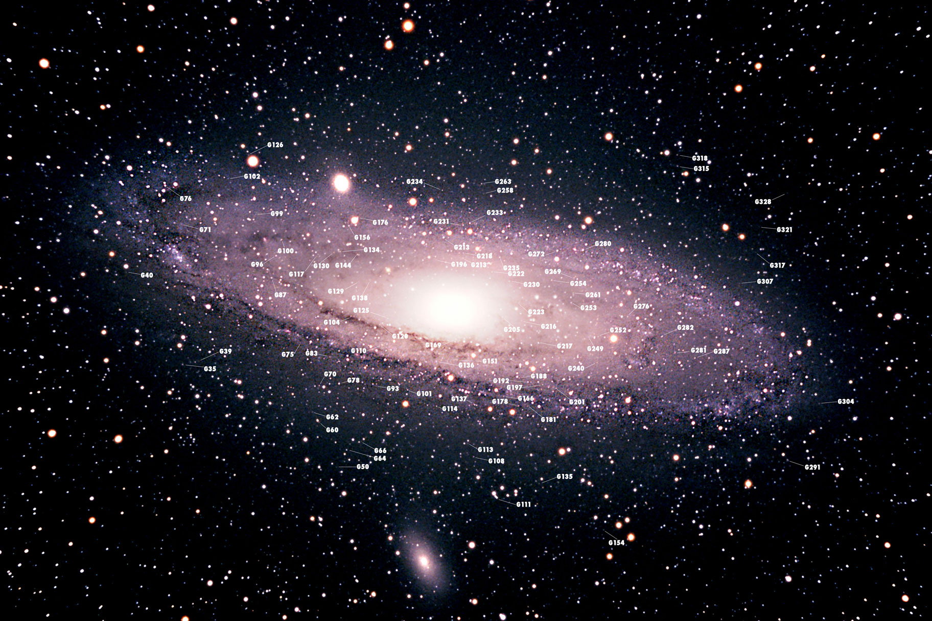
[1] M31 with some globular clusters
marked [t Note that each globular
cluster can be imagined to be formed by
some intelligent living
objects.] Image by Peter
Kennett UNKNOWN
source: http://www.petesastrophotography
.com/m31globs.jpg

[2] M31, Southwest Arm, NGC
206 copyright Robert Gendler
2005 UNKNOWN
source: http://www.robgendlerastropics.c
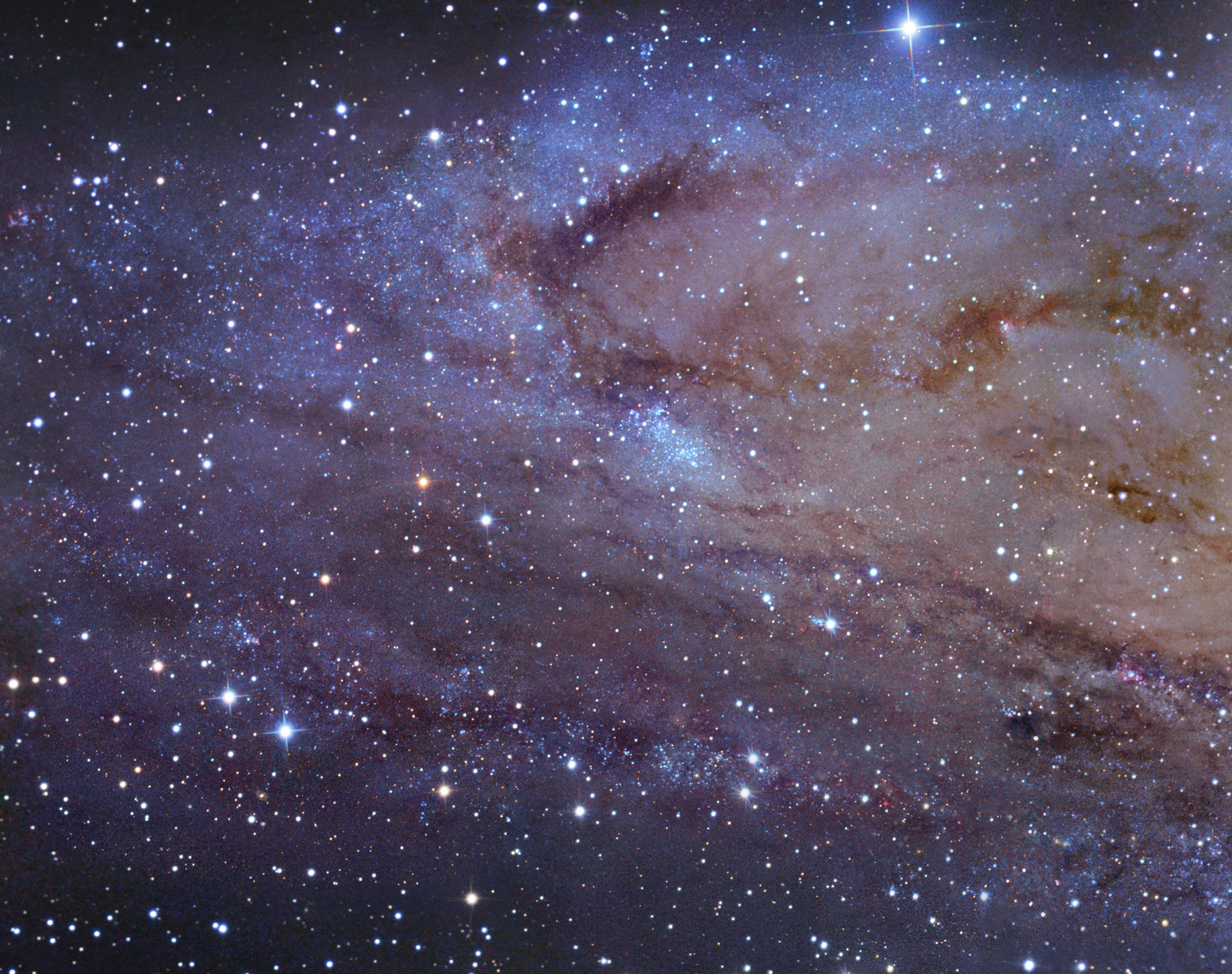
om/M31NMmosaicSW.jpg
consumed.

[1] Star with many ships around
it. Adapted from: English: The Sun
photographed by the Atmospheric Imaging
Assembly (AIA 304) of NASA's Solar
Dynamics Observatory (SDO). This is a
false color image of the sun observed
in the extreme ultraviolet region of
the spectrum. For example, similar
image. Date 2010-08-19T00:32:21Z
(ISO 8601) Source NASA/SDO
(AIA). Author NASA/SDO (AIA). PD
source: http://upload.wikimedia.org/wiki
pedia/commons/thumb/b/b4/The_Sun_by_the_
Atmospheric_Imaging_Assembly_of_NASA%27s
_Solar_Dynamics_Observatory_-_20100819.j
pg/628px-The_Sun_by_the_Atmospheric_Imag
ing_Assembly_of_NASA%27s_Solar_Dynamics_
Observatory_-_20100819.jpg

[2] Globular Star Cluster 47
Tuc Image Credit & Copyright: Dieter
Willasch
(Astro-Cabinet) Explanation:
Globular star cluster 47 Tucanae is a
jewel of the southern sky. Also known
as NGC 104, it roams the halo of our
Milky Way Galaxy along with some 200
other globular star clusters. The
second brightest globular cluster
(after Omega Centauri) as seen from
planet Earth, it lies about 13,000
light-years away and can be spotted
naked-eye near the Small Magellanic
Cloud in the constellation of the
Toucan. The dense cluster is made up of
several million stars in a volume only
about 120 light-years across. Red giant
stars on the outskirts of the cluster
are easy to pick out as yellowish stars
in this sharp telescopic portrait.
Globular cluster 47 Tuc is also home to
exotic x-ray binary star systems. PD
source: http://apod.nasa.gov/apod/image/
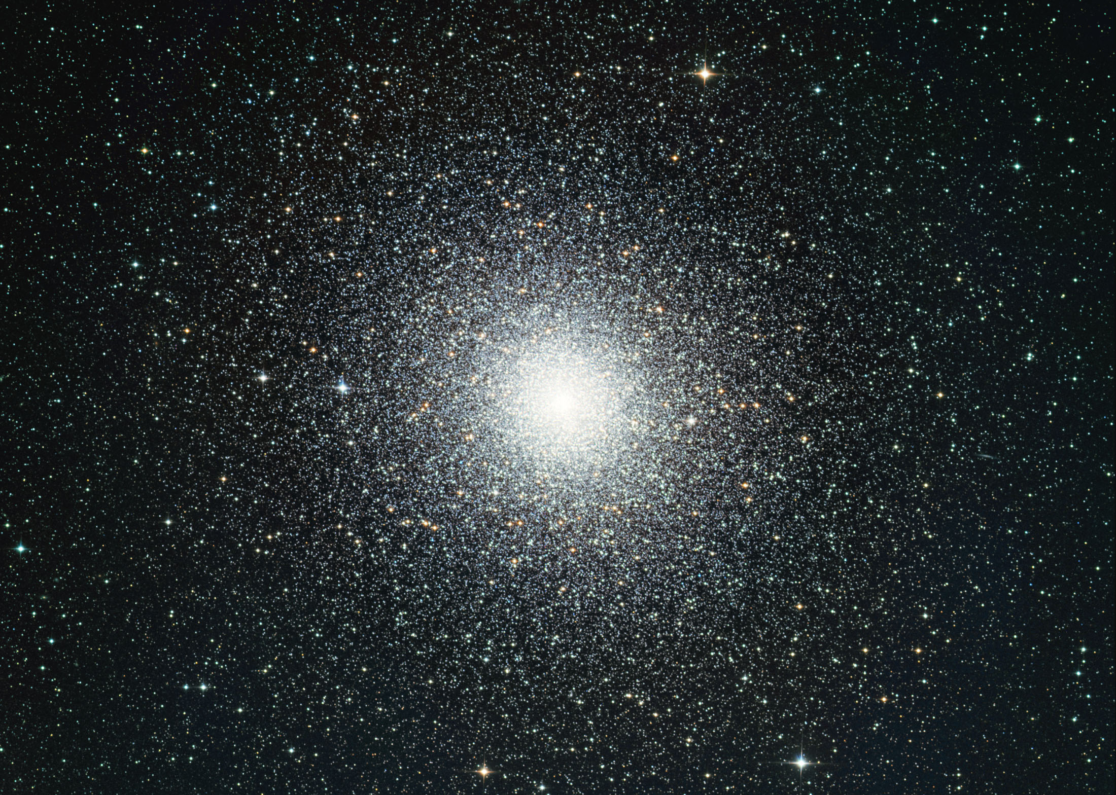
1101/47Tuc_DW.jpg
[1] Star with many ships around
it. Adapted from: English: The Sun
photographed by the Atmospheric Imaging
Assembly (AIA 304) of NASA's Solar
Dynamics Observatory (SDO). This is a
false color image of the sun observed
in the extreme ultraviolet region of
the spectrum. For example, similar
image. Date 2010-08-19T00:32:21Z
(ISO 8601) Source NASA/SDO
(AIA). Author NASA/SDO (AIA). PD
source: http://upload.wikimedia.org/wiki
pedia/commons/thumb/b/b4/The_Sun_by_the_
Atmospheric_Imaging_Assembly_of_NASA%27s
_Solar_Dynamics_Observatory_-_20100819.j
pg/628px-The_Sun_by_the_Atmospheric_Imag
ing_Assembly_of_NASA%27s_Solar_Dynamics_
Observatory_-_20100819.jpg
[2] Globular Star Cluster 47
Tuc Image Credit & Copyright: Dieter
Willasch
(Astro-Cabinet) Explanation:
Globular star cluster 47 Tucanae is a
jewel of the southern sky. Also known
as NGC 104, it roams the halo of our
Milky Way Galaxy along with some 200
other globular star clusters. The
second brightest globular cluster
(after Omega Centauri) as seen from
planet Earth, it lies about 13,000
light-years away and can be spotted
naked-eye near the Small Magellanic
Cloud in the constellation of the
Toucan. The dense cluster is made up of
several million stars in a volume only
about 120 light-years across. Red giant
stars on the outskirts of the cluster
are easy to pick out as yellowish stars
in this sharp telescopic portrait.
Globular cluster 47 Tuc is also home to
exotic x-ray binary star systems. PD
source: http://apod.nasa.gov/apod/image/
1101/47Tuc_DW.jpg
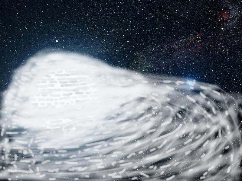
[1] Adapted from: Description This
picture is an artist's impression
showing how the binary star system of
Sirius A and its diminutive blue
companion, Sirius B, might appear to an
interstellar visitor. The large,
bluish-white star Sirius A dominates
the scene, while Sirius B is the small
but very hot and blue white-dwarf star
on the right. The two stars revolve
around each other every 50 years. White
dwarfs are the leftover remnants of
stars similar to our Sun. The Sirius
system, only 8.6 light-years from
Earth, is the fifth closest stellar
system known. Sirius B is faint because
of its tiny size. Its diameter is only
7,500 miles (about 12 thousand
kilometres), slightly smaller than the
size of our Earth. The Sirius system is
so close to Earth that most of the
familiar constellations would have
nearly the same appearance as in our
own sky. In this rendition, we see in
the background the three bright stars
that make up the Summer Triangle:
Altair, Deneb, and Vega. Altair is the
white dot above Sirius A; Deneb is the
dot to the upper right; and Vega lies
below Sirius B. But there is one
unfamiliar addition to the
constellations: our own Sun is the
second-magnitude star, shown as a small
dot just below and to the right of
Sirius
A. Date Source http://www.spacete
lescope.org/images/html/heic0516b.html
Author NASA, ESA Credit: G. Bacon
(STScI) PD
source: http://upload.wikimedia.org/wiki
pedia/commons/thumb/c/c9/Sirius_A_and_B_
artwork.jpg/800px-Sirius_A_and_B_artwork
.jpg
[2] Adapted from: Description This
picture is an artist's impression
showing how the binary star system of
Sirius A and its diminutive blue
companion, Sirius B, might appear to an
interstellar visitor. The large,
bluish-white star Sirius A dominates
the scene, while Sirius B is the small
but very hot and blue white-dwarf star
on the right. The two stars revolve
around each other every 50 years. White
dwarfs are the leftover remnants of
stars similar to our Sun. The Sirius
system, only 8.6 light-years from
Earth, is the fifth closest stellar
system known. Sirius B is faint because
of its tiny size. Its diameter is only
7,500 miles (about 12 thousand
kilometres), slightly smaller than the
size of our Earth. The Sirius system is
so close to Earth that most of the
familiar constellations would have
nearly the same appearance as in our
own sky. In this rendition, we see in
the background the three bright stars
that make up the Summer Triangle:
Altair, Deneb, and Vega. Altair is the
white dot above Sirius A; Deneb is the
dot to the upper right; and Vega lies
below Sirius B. But there is one
unfamiliar addition to the
constellations: our own Sun is the
second-magnitude star, shown as a small
dot just below and to the right of
Sirius
A. Date Source http://www.spacete
lescope.org/images/html/heic0516b.html
Author NASA, ESA Credit: G. Bacon
(STScI) PD
source: http://upload.wikimedia.org/wiki
pedia/commons/thumb/c/c9/Sirius_A_and_B_
artwork.jpg/800px-Sirius_A_and_B_artwork
.jpg
[1] Globular Star Cluster 47 Tuc Image
Credit & Copyright: Dieter Willasch
(Astro-Cabinet) Explanation:
Globular star cluster 47 Tucanae is a
jewel of the southern sky. Also known
as NGC 104, it roams the halo of our
Milky Way Galaxy along with some 200
other globular star clusters. The
second brightest globular cluster
(after Omega Centauri) as seen from
planet Earth, it lies about 13,000
light-years away and can be spotted
naked-eye near the Small Magellanic
Cloud in the constellation of the
Toucan. The dense cluster is made up of
several million stars in a volume only
about 120 light-years across. Red giant
stars on the outskirts of the cluster
are easy to pick out as yellowish stars
in this sharp telescopic portrait.
Globular cluster 47 Tuc is also home to
exotic x-ray binary star systems. PD
source: http://apod.nasa.gov/apod/image/
1101/47Tuc_DW.jpg
[2] Storyboard image by Ted
Huntington GNU
source: Ted Huntington
integrates with the Magellanic Cloud
galaxies.
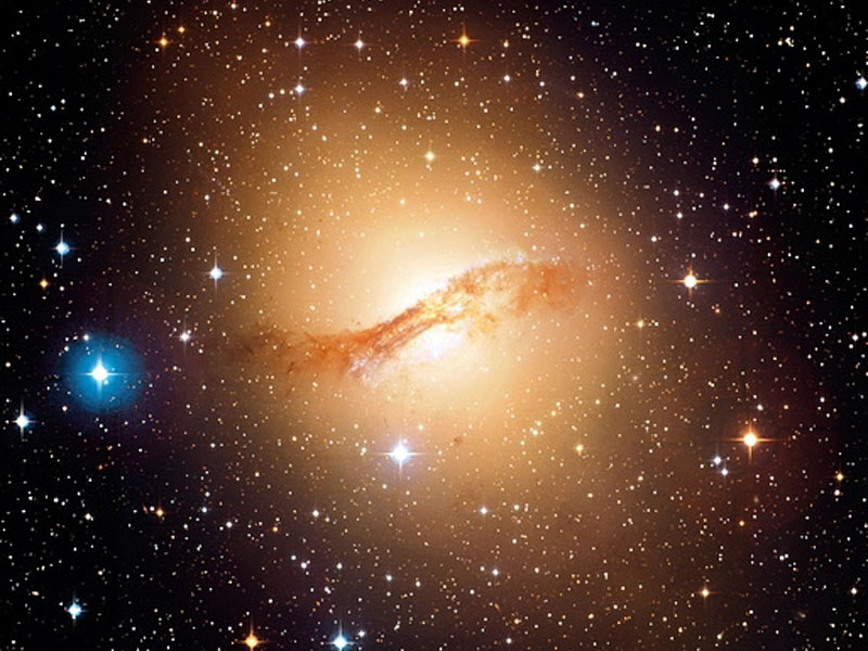
[1] Elliptical Galaxy Centaurus A from
CFHT Credit & Copyright: Jean-Charles
Cuillandre (CFHT) & Giovanni Anselmi
(Coelum Astronomia), Hawaiian
Starlight Explanation: Why is
peculiar galaxy Centaurus A so dusty?
Dramatic dust lanes that run across the
galaxy's center mark Cen A. These dust
lanes are so thick they almost
completely obscure the galaxy's center
in visible light. This is particularly
unusual as Cen A's red stars and round
shape are characteristic of a giant
elliptical galaxy, a galaxy type
usually low in dark dust. Cen A, also
known as NGC 5128, is also unusual
compared to an average elliptical
galaxy because it contains a higher
proportion of young blue stars and is a
very strong source of radio emission.
Evidence indicates that Cen A is likely
the result of the collision of two
normal galaxies. During the collision,
many young stars were formed, but
details of the creation of Cen A's
unusual dust belts are still being
researched. Cen A lies only 13 million
light years away, making it the closest
active galaxy. Cen A, pictured above,
spans 60,000 light years and can be
seen with binoculars toward the
constellation of Centaurus. PD
source: http://apod.nasa.gov/apod/image/
0607/cenA_cfht.jpg

[2] Storyboard image by Ted
Huntington GNU
source: Ted Huntington
globular galaxies join.
Galaxy
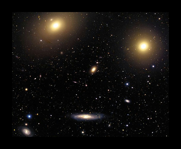
[1] See Explanation. Clicking on the
picture will download the highest
resolution version available. In the
Center of the Virgo Cluster Credit &
Copyright: Jean-Charles Cuillandre
(CFHT), Hawaiian Starlight,
CFHT Explanation: The Virgo Cluster
of Galaxies is the closest cluster of
galaxies to our Milky Way Galaxy. The
Virgo Cluster is so close that it spans
more than 5 degrees on the sky - about
10 times the angle made by a full Moon.
It contains over 100 galaxies of many
types - including spiral, elliptical,
and irregular galaxies. The Virgo
Cluster is so massive that it is
noticeably pulling our Galaxy toward
it. The cluster contains not only
galaxies filled with stars but also gas
so hot it glows in X-rays. Motions of
galaxies in and around clusters
indicate that they contain more dark
matter than any visible matter we can
see. Pictured above, the center of the
Virgo cluster might appear to some as a
human face, and includes bright Messier
galaxies M86 at the top, M84 on the far
right, NGC 4388 at the bottom, and NGC
4387 in the middle. PD
source: http://apod.nasa.gov/apod/image/
0308/virgocenter_cfht.jpg
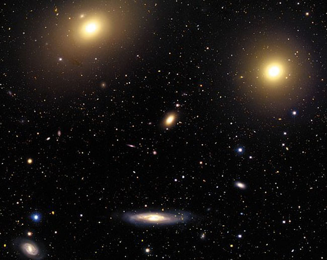
[2] See Explanation. Clicking on the
picture will download the highest
resolution version available. In the
Center of the Virgo Cluster Credit &
Copyright: Jean-Charles Cuillandre
(CFHT), Hawaiian Starlight,
CFHT Explanation: The Virgo Cluster
of Galaxies is the closest cluster of
galaxies to our Milky Way Galaxy. The
Virgo Cluster is so close that it spans
more than 5 degrees on the sky - about
10 times the angle made by a full Moon.
It contains over 100 galaxies of many
types - including spiral, elliptical,
and irregular galaxies. The Virgo
Cluster is so massive that it is
noticeably pulling our Galaxy toward
it. The cluster contains not only
galaxies filled with stars but also gas
so hot it glows in X-rays. Motions of
galaxies in and around clusters
indicate that they contain more dark
matter than any visible matter we can
see. Pictured above, the center of the
Virgo cluster might appear to some as a
human face, and includes bright Messier
galaxies M86 at the top, M84 on the far
right, NGC 4388 at the bottom, and NGC
4387 in the middle. PD
source: http://apod.nasa.gov/apod/image/
0308/virgocenter_cfht.jpg